www.jpis.org
pISSN 2093-2278 eISSN 2093-2286 Copyright © 2012 Korean Academy of PeriodontologyThis is an Open Access article distributed under the terms of the Creative Commons Attribution Non-Commercial License (http://creativecommons.org/licenses/by-nc/3.0/).
Influence of crown-to-implant ratio on periimplant marginal bone loss in the posterior region: a five-year retrospective study
Kyung-Jin Lee, Yong-Gun Kim, Jin-Woo Park, Jae-Mok Lee, Jo-Young Suh* Department of Periodontology, Kyungpook National University School of Dentistry, Daegu, Korea
Purpose: The aim of this study was to evaluate the influence of the crown-to-implant (C/I) ratio on the change in marginal bone level around the implant and to determine the site-related factors influencing the relationship between the C/I ratio and periimplant marginal bone loss.
Methods: A total of 259 implants from 175 patients were evaluated at a mean follow-up of five years. Implants were divided into two groups according to their C/I ratios: ≤1, and >1. Site-related factors having an influence on the relationship between C/I ratio and periimplant marginal bone loss were analyzed according to the implant location, implant diameter, implant manufacturer, prosthesis type, and guided bone regeneration (GBR) procedure.
Results: It was found that 1) implants with a C/I ratio below 1 exhibited greater periimplant marginal bone loss than implants with a C/I ratio more than 1, 2) site-related factors had an effect on periimplant marginal bone loss, except for the implant sys- tem used, 3) the C/I ratio was the factor having more dominant influence on periimplant marginal bone loss, compared with implant diameter, prosthesis type, implant location, and GBR procedure, 4) implants with a C/I ratio below 1 showed greater periimplant marginal bone loss than implants with a C/I ratio greater than 1 in the maxilla, but not in the mandible, 5) and pe- riimplant marginal bone loss was more affected by the implant system than the C/I ratio.
Conclusions: Within the limitations of this study, implants with a higher C/I ratio exhibited less marginal bone loss than im- plants with a lower C/I ratio in the posterior regions. The C/I ratio was a more dominant factor affecting periimplant marginal bone loss in the maxilla than the mandible. Meanwhile, the implant system was a more dominant factor influencing periim- plant marginal bone loss than the C/I ratio.
Keywords: Alveolar bone loss, Dental implants, Follow-up studies.
INTRODUCTION
Dental implants have been a widely accepted option for re- placement of missing teeth. Clinicians may use guidelines associated with natural teeth and apply them to potential im- plant sites or existing implant-supported restorations. One of these guidelines is the crown-to-root (C/R) ratio, which is used as a parameter for deciding on the suitability of teeth
employed as abutments in dental prostheses. The C/R ratio is calculated from a radiograph with respect to length of the tooth not within bone divided by the portion of the tooth in alveolar bone [1]. With regard to discussion of the C/R ratio in the literature, various ratios have been reported. Dykema [2]
reported that a C/R ratio of 1:1.5 was desirable. Shillingburg [3]
suggested a ratio of 1:1.5 as most favorable for an abutment, and a C/R ratio of 1:1 as a minimum for a tooth abutment.
Received: Oct. 31, 2012; Accepted: Nov. 18, 2012
*Correspondence: Jo-Young Suh
Department of Periodontology, Kyungpook National University School of Dentistry, 2175 Dalgubeol-daero, Jung-gu, Daegu 700-705, Korea E-mail: [email protected], Tel: +82-53-600-7501, Fax: +82-53-427-3263
than scientific data. Several studies have demonstrated that teeth with an unfavorable C/R ratio can function successfully as abutment teeth [4-6]. Grossmann and Sadan [1] concluded that no definitive recommendations for an optimal C/R ratio could be established.
A similar clinical condition regarding an unfavorable C/R ratio is often encountered in edentulous areas restored with an implant-supported prosthesis. In particular, the posterior region of the mouth offers a challenging clinical scenario for rehabilitation with dental implants. Resorption of the alveo- lar ridge, the presence of the inferior alveolar nerve, pneuma- tization of the sinus, and occlusal forces create a clinical en- vironment that may jeopardize the long-term biomechanical success of the implant restoration. Therefore, crown height of the implant often becomes relatively longer, compared with the length of implant embedded in bone. The longer the crown height is, the greater the effect of the moment of force or lever arm with any lateral force on the implant be- comes. These moment loads would induce a stress concen- tration at the crest of the alveolar ridge at the implant-to-tis- sue interface, and lead to periimplant marginal bone loss [7].
An early longitudinal study demonstrated that crown-to- implant (C/I) ratio was a more sensitive indicator of potential implant failure than the residual bone height [8]. Accordingly, a C/I ratio between 0.5 and 1 was proposed for prevention of periimplant bone stress, marginal bone loss, and eventual im- plant failure [9-11]. This recommendation was also based on prosthodontic and periodontal principles extrapolated from tooth-supported reconstructions [3]. Conversely, Schulte et al.
[12] reported that the C/I ratio of 889 plateau-design single tooth implants was 1.3 on average, with an average survival rate of 98.2% over 2.3 years. Schulte et al. [12] concluded that the C/I ratio of functioning implants was similar to that of failed implants.
The basis of successful long-term results of implants de- pends primarily on preservation of bone support. Therefore, maintenance of osseointegration and a steady state of the marginal bone level are imperative features. The radiograph- ic image is the most important source of information for de- termining the amount of marginal bone loss around implants [13]. However, radiographic information on the change in mar- ginal bone level around implants with different C/I ratios is limited. In addition, more information is needed in order to investigate factors having an influence on the relationship between the C/I ratio and periimplant marginal bone loss.
The current study had two objectives: 1) to determine the effect of the C/I ratio on change in periimplant marginal bone level, and 2) to determine the site-related factors that influ- ence the relationship between the C/I ratio and periimplant
MATERIALS AND METHODS
This retrospective study included patients referred to the Department of Periodontology, Kyungpook National Univer- sity Dental Hospital for treatment of implant therapy between January 2000 and January 2008 who fulfilled the following criteria: 1) partially edentulous patients requiring implant restoration in the posterior maxilla or mandible; 2) at least three years between completion of implant prosthesis and follow-up; 3) no systemic diseases that would contraindicate oral surgical treatment; and 4) nonsmokering. In addition, all patients were given a periodic recall check. The study proto- col was reviewed and accepted by the Research Ethics Com- mittee, Kyungpook National University Hospital (Ethics Ref- erence No. KNUH 2012-08-021).
Three different implant systems were used: The system I implant was anodized commercially pure titanium. The sys- tem II implant had a resorbable blasting media (RBM) treat- ed surface in a solution containing Ca3PO4. The system III implant had a hybrid surface, so that the coronal part of the implant has a smooth surface, whereas from the third thread to the apex, the surface of the implant was acid-etched with HCl/H2SO4. All of the implants had external hexagon con- nections. Placement of all implants was performed using a standard two-stage surgical protocol. A healing period of 3 to 6 months was allowed before prosthetic loading. In all cases, gold-machined UCLA-type abutments with a noble alloy for casting were screwed onto the top of implants using a torque wrench calibrated at 30 Ncm.
During the follow-up examination, the following site-relat- ed factors were collected for each implant: implant diameter, implant location, implant system, prosthesis type, and guided bone regeneration (GBR) procedure.
Radiographic evaluation
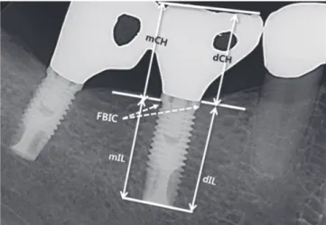
For evaluation of the C/I ratio and change in marginal bone level, periapical radiographs were taken using a long-cone paralleling technique [14]. The images were digitized for mea- surement using specialized software (ImageTool, Ver. 1.28, University of Texas Health Science Center at San Antonio, San Antonio, TX, USA). A computer-assisted calibration was performed for each radiograph by evaluation of the previous known values (e.g., fixture length). Unclear radiographs or implant sites were excluded. The following linear measure- ments between landmarks were taken [15]: 1) crown height (CH) was measured from the top of the restoration to the first bone-to-implant contact (FBIC) on both mesial and distal sides; 2) implant length (IL) was measured from the apex to
the FBIC on both mesial and distal sides. To obtain the mesi- al C/I ratio, the mesial CH was divided by the mesial IL. The distal C/I ratio was also determined, and mean mesiodistal C/I ratio was then obtained for each implant (Fig. 1). The pe- riimplant marginal bone level was measured from the refer- ence point to the FBIC. The reference point was the fixture- abutment interface. The mesial and distal values of the mar- ginal bone loss were averaged to one value. The mean mar- ginal bone loss was calculated as the difference between the initial marginal bone level and the marginal bone level at the follow-up examination. All radiographic measurements were recorded to the nearest 0.1 mm and assessed three times by one examiner.
Statistical analysis
Descriptive statistics were performed, the absolute and rel- ative frequency distribution were calculated for qualitative variables, and the mean±standard deviation was calculated for quantitative variables. Comparative statistics were per- formed, mean marginal bone loss was analyzed for different C/I ratios (divided into two categories: <1 and ≥1), implant diameter (divided into two categories: 3.75 to 4 as regular and
5 mm as wide), implant location (maxilla and mandible), im- plant system (divided into three categories: system I, system II, and system III), prosthesis type (divided into two catego- ries: single and splinted), and GBR procedure (divided into two categories depending on the use or not of GBR). The Stu- dent’s t-test was used for comparison of the means of the two groups. However, for comparison of the means of three or more groups, the one-way parametric analysis of variance test was applied and post hoc examination of group mean dif- ferences was performed using Tukey’s test. The Kruskal-Wal- lis test was used for three or more groups when required.
Pearson’s correlation was applied for determination of asso- ciations between the C/I ratio and periimplant marginal bone loss. A P-value of less than 0.05 was considered statistically significant. A statistical software package SPSS ver. 17.0 (SPSS Inc., Chicago, IL, USA) was used in performance of all statisti- cal analyses.
RESULTS
A total of 259 implants from 175 patients were analyzed in this study. The mean follow-up period amounted to 5.7±2.0 years. The study population consisted of 81 men and 94 wom- en with a mean age of 54.2±15.6 years (range, 21 to 76 years).
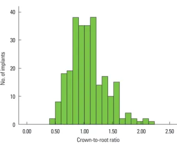
The frequency of the C/I ratios of implants is shown in Fig. 2.
In this study, C/I ratios were sorted into two groups based on 259 measurements: 44.7% of the cases belonged to the <1 group, and 55.3% to the ≥1 group. The mean C/I ratio was 1.06±0.42. The mean periimplant marginal bone loss at the five-year follow-up was 0.93±0.15 mm (range, 0.05 to 1.89 mm).
to-implant (C/I) ratio and site-related factors (n=259).
Implant factor No. (%) MBL (mm), mean±SD
C/I ratio
<1 116 (44.8) 1.18±0.18a)
≥1 143 (55.2) 0.73±0.13
Implant diameter
Regular 124 (47.9) 0.99±0.15
Wide 135 (52.1) 0.88±0.22
Implant location
Maxilla 126 (48.6) 0.96±0.11
Mandible 133 (51.4) 0.91±0.14
Prosthesis type
Splinted 186 (47.1) 0.97±0.13
Single 73 (52.9) 0.90±0.21
GBR procedure
With 78 (30.1) 0.87±0.21
Without 181 (69.9) 0.96±0.13
Implanted system
System I 125 (48.3) 0.81±0.15b)
System II 76 (29.3) 0.65±0.12b)
System III 58 (22.4) 1.57±0.25
MBL: mean periimplant marginal bone loss, SD: standard deviation, GBR: guided bone regeneration.
a)Statistically significant difference compared to C/I ratio≥1 (Student’s t-test;
P<0.05).
b)Statistically significant difference compared to system III (analysis of variance/
Tukey’s test; P<0.001).
Figure 1. Formula for calculation of crown-to-implant (C/I) ratio.
mCH/mIL: mesial C/I ratio, dCH/dIL: distal C/I ratio. An average mesiodistal C/I ratio was obtained per implant restoration. mCH:
mesial clinical crown height, mIL: mesial implant length, dCH: dis- tal clinical crown height, dIL: distal implant length, FBIC: the first bone-to-implant contact.
The effect of C/I ratio on periimplant marginal bone loss af- ter insertion of definitive restoration was evaluated and is shown in Table 1. Implants with a lower C/I ratio showed a statistically significantly greater periimplant marginal bone loss than implants with a higher C/I ratio (P<0.05). Results of Pearson’s correlation analysis revealed a statistically signifi- cant inverse relationship between C/I ratio and periimplant marginal bone loss (r=-0.258, P<0.001).
Except for the implant system, site-related factors did not have an influence on periimplant marginal bone loss (Table 1). Compared with the system I and system II implants, the system III implant showed statistically significantly greater bone loss (P<0.001). In analysis of the combination effect of the C/I ratio and site-related factors (Table 2), the C/I ratio was the more dominant factor influencing the change in pe- riimplant marginal bone level, compared with implant diam- eter, prosthesis type, implant location, and GBR procedure. In terms of implant location, implants with a lower C/I ratio had greater periimplant marginal bone loss than implants with a higher C/I ratio in the maxilla (P<0.05), while the C/I ratio had no influence on periimplant marginal bone loss in the mandible (P>0.05). Meanwhile, periimplant marginal bone loss was more affected by the implant system than the C/I ra- tio in posterior areas.
DISCUSSION
Implant surface, implant location, and surgical procedure as well as loading after prosthetic rehabilitation could affect the change in the periimplant marginal bone level. The C/I ratio has been regarded as one of the geometric loading fac- tors that could increase biomechanical complications [16]. In
mathematical models, the higher the effect of bending mo- ments, the greater the possibility of occurrence of bone loss around implants with long lever arms, such as those with an unfavorable C/I ratio. Nevertheless, the current study found that implants with a higher C/I ratio showed statistically sig- nificantly less periimplant marginal bone loss than implants with a lower C/I ratio. This finding can be explained by the fact that the stress concentration at the bone crest caused by masticatory forces may stimulate bone formation around some implant fixtures. This biologic phenomenon is support- ed by the ‘stress-shielding’ effect described in previous stud- ies [17,18]. According to these authors, clinical scenarios in which bone stress is reduced may cause disuse atrophy and eventual marginal bone loss.
Blanes et al. [19] evaluated both the clinical C/I ratio and an- atomical C/I ratio of nonsubmerged internal implants. The clinical C/I ratio applied in the current study was measured from the top of the crestal bone in contact with the implant to the top of the crown as the CH; thus, the length of the im- plant was measured from the bottom of the fixture to the top of the crestal bone in contact with the implant, whereas, the anatomical C/I ratio was measured from the bottom of the fixture to the implant-abutment connection and then from tion of crown-to-implant (C/I) ratio and site-related factors (n=259).
Implant factor C/I ratio<1 C/I ratio≥1
No. MBL (mm) No. MBL (mm)
Implant diameter
Regular 53 1.29±0.27a) 71 0.77±0.21
Wide 60 1.20±0.24a) 75 0.63±0.13
Implant location
Maxilla 50 1.26±0.30a) 76 0.77±0.20
Mandible 66 1.05±0.21 67 0.78±0.15
Prosthesis type
Splinted 63 1.27±0.28a) 59 0.70±0.19
Single 53 1.12±0.21a) 84 0.59±0.18
GBR procedure
With 25 1.24±0.30a) 53 0.70±0.21
Without 91 1.17±0.21a) 90 0.75±0.20
Implant surface
System I 56 0.86±0.27b) 69 0.77±0.21c)
System II 21 0.71±0.14b) 55 0.63±0.20c)
System III 39 1.65±0.33 19 1.41±0.36
Values are presented as mean±standard deviation.
MBL: mean periimplant marginal bone loss, GBR: guided bone regeneration.
a)Statistically significant difference compared to C/I ratio≥1 (Student’s t-test;
P<0.05).
b,c)Statistically significant difference compared to system III (analysis of variance/
Tukey’s test; P<0.001).
30
20
10
0 0.00 0.50 1.00 1.50 2.00 2.50
Crown-to-root ratio
No. of implants
Figure 2. Distribution of the implants according to their crown-to- implant ratio.
ference due to the fact that an implant of conventional length may exhibit a high C/I ratio if the marginal bone has remod- eled to a level far below the implant-abutment connection.
Rokni et al. [18] found no association between the C/I ratio and FBIC levels, but did find an association between decreas- ing FBIC levels and increasing IL, as well as splinting of res- torations. However, Blanes et al. [19] reported that higher clin- ical C/I ratios showed lower average bone loss when com- pared with lower C/I ratios. This observation is in agreement with the results of our study. The anatomical C/I ratio offers a more favorable biomechanical scenario, as the lever arm is shorter than that in the clinical C/I ratio. Nevertheless, be- cause the stiffness of components connected to the implant is greater than that of the cortical bone, the clinical C/I ratio measured in our study appears to offer a more realistic clini- cal scenario for evaluation of the effect of the C/I ratio on com- plications of implant restoration [20]. Therefore, the most coronal bone-to-implant contact is the location where the applied forces and strains they create are resisted by bone.
Ultimately, for determination of change in marginal bone loss related to the C/I ratio, the clinical C/I ratio is a more ap- propriate parameter than the anatomical C/I ratio.
In this study, the implant location had an influence on peri- implant marginal bone loss related to the C/I ratio. Implants with a lower C/I ratio had more periimplant marginal bone loss than implants with a higher C/I ratio in the maxilla, while the C/I ratio had no influence on periimplant marginal bone loss in the mandible. Bone density is directly related to the elastic modulus of bone [21]. Due to its lower elastic modulus, the cancellous bone exhibited a lower stress concentration and less variation than the cortical bone [22]. The explanation could be that the difference of bone density induces this re- sult. In the maxilla posterior area, the C/I ratio could act as a more dominant loading factor, which may increase the peri- implant marginal bone loss, compared with the bone density.
Interestingly, in our study, a statistically significant differ- ence in periimplant marginal bone loss was observed accord- ing to the implant system. It appears that, among the implant systems, differences in surface texture of the implant result in significant differences in the magnitude of the marginal bone resorption. Radiographic evaluation of marginal bone levels around different implant systems showed that implants with a hybrid surface were associated with greater change in crestal bone levels than implants with an anodized or RBM surface (P<0.001). In analysis of the combined effect of the C/
I ratio and implant system on periimplant marginal bone loss, the implant system was a more dominant factor influ- encing the change in periimplant marginal bone level than the C/I ratio. However, our results differ from those of previ-
teristics on periimplant marginal bone loss. A recent clinical study found that among TiUnite, Osseotite, and machined dental implants, no statistically significant differences in bone loss were observed at two years postloading [23]. Until now, no specific implant surface characteristic has been found to be superior in marginal bone preservation [24]. As this study included a relatively small sample size, and, other than the configuration of the surface, the implants studied had no other differences that may have influenced the results, con- ducting further studies on other fixture designs is necessary in order to clarify the mechanism and the relationship be- tween the design and marginal bone loss in dental implants.
Although the results of the current study showed an inverse relationship between the C/I ratio and marginal bone loss, due to the following limitations, our result should be inter- preted with caution. A recent study found no correlation be- tween the C/I ratio and periimplant marginal bone loss [25].
The reports established different categories of C/I ratio from ours. The C/I ratio was sorted into six groups based on 234 measurements (<1, 1–1.2, 1.21–1.4, 1.41–1.6, 1.61–2, and >2).
However, because the number of implant-supported restora- tions with a C/I ratio greater than 2 was very low, we only classified our cases into two groups based on the C/I ratio of 1. Furthermore, our results came from a retrospective study.
Due to issues of bias and confounding factors, this type of study has less validity than a randomized, prospective study.
Within the limitations of this study, it is suggested that the C/I ratio and implant system were factors that should be consid- ered as affecting marginal bone loss around the implant in the posterior region more than implant diameter, prosthesis type, implant location, or GBR procedure, especially in the maxilla.
CONFLICT OF INTEREST
No potential conflict of interest relevant to this article was reported.
ACKNOWLEDGEMENTS
This research was supported by Kyungpook National Uni- versity Research Fund 2012.
REFERENCES
1. Grossmann Y, Sadan A. The prosthodontic concept of crown-to-root ratio: a review of the literature. J Prosthet Dent 2005;93:559-62.
2. Dykema RW. Diagnosis and Treatment Planning. In: John- ston JF, Dykema LW, Goodacre CJ, Phillips RW. Johnston’s
phia: WB Saunders; 1986. p.8-21.
3. Shillingburg HT Jr. Treatment Planning for the Replace- ment of Missing Teeth. In: Shillingburg HT Jr, Hobo S, Whitsett LD, Jacobi R, Brackett SE. Fundamentals of fixed prosthodontics. 3rd ed. Chicago: Quintessence Publish- ing; 1997. p.89-90.
4. Nyman S, Ericsson I. The capacity of reduced periodontal tissues to support fixed bridgework. J Clin Periodontol 1982;9:409-14.
5. Laurell L, Lundgren D, Falk H, Hugoson A. Long-term prognosis of extensive polyunit cantilevered fixed partial dentures. J Prosthet Dent 1991;66:545-52.
6. Yi SW, Ericsson I, Carlsson GE, Wennström JL. Long-term follow-up of cross-arch fixed partial dentures in patients with advanced periodontal destruction. Evaluation of the supporting tissues. Acta Odontol Scand 1995;53:242-8.
7. Bidez MW, Misch CE. Clinical Biomechanics in Implant Dentistry. In: Misch CE. Contemporary implant dentistry.
St. Louis: Mosby Elsevier; 2008. p.543-55.
8. Brose MO, Avers RJ, Rieger MR, Duckworth JE. Submerged alumina dental root implants in humans: five-year evalu- ation. J Prosthet Dent 1989;61:594-601.
9. Nasr HF, Meffert RM. A proposed radiographic index for assessment of the current status of osseointegration. Int J Oral Maxillofac Implants 1993;8:323-8.
10. Haas R, Mensdorff-Pouilly N, Mailath G, Watzek G. Bråne- mark single tooth implants: a preliminary report of 76 im- plants. J Prosthet Dent 1995;73:274-9.
11. Glantz PO, Nilner K. Biomechanical aspects of prosthetic implant-borne reconstructions. Periodontol 2000 1998;
17:119-24.
12. Schulte J, Flores AM, Weed M. Crown-to-implant ratios of single tooth implant-supported restorations. J Prosthet Dent 2007;98:1-5.
13. Verhoeven JW, Cune MS, de Putter C. Reliability of some clinical parameters of evaluation in implant dentistry. J Oral Rehabil 2000;27:211-6.
14. Updegrave WJ. Right-angle dental radiography. Dent Clin North Am 1968;12:571-9.
15. Urdaneta RA, Rodriguez S, McNeil DC, Weed M, Chuang SK. The effect of increased crown-to-implant ratio on sin-
plants 2010;25:729-43.
16. Rangert BR, Sullivan RM, Jemt TM. Load factor control for implants in the posterior partially edentulous segment.
Int J Oral Maxillofac Implants 1997;12:360-70.
17. Pilliar RM, Cameron HU, Binnington AG, Szivek J, Macnab I. Bone ingrowth and stress shielding with a porous sur- face coated fracture fixation plate. J Biomed Mater Res 1979;13:799-810.
18. Rokni S, Todescan R, Watson P, Pharoah M, Adegbembo AO, Deporter D. An assessment of crown-to-root ratios with short sintered porous-surfaced implants supporting prostheses in partially edentulous patients. Int J Oral Max- illofac Implants 2005;20:69-76.
19. Blanes RJ, Bernard JP, Blanes ZM, Belser UC. A 10-year pro- spective study of ITI dental implants placed in the poste- rior region. II: Influence of the crown-to-implant ratio and different prosthetic treatment modalities on crestal bone loss. Clin Oral Implants Res 2007;18:707-14.
20. Sekine H, Komiyama Y, Hotta H, Yoshida T. Mobility Char- acteristics and Tactile Sensitivity of Osseointegrated Im- plant-Supporting Systems. In: Van Steenberghe D. Tissue integration in oral and maxillofacial reconstruction. Am- sterdam: Excerpta Medica; 1986. p.326-39.
21. Misch CE, Qu Z, Bidez MW. Mechanical properties of tra- becular bone in the human mandible: implications for dental implant treatment planning and surgical placement.
J Oral Maxillofac Surg 1999;57:700-6.
22. Akca K, Cehreli MC. Biomechanical consequences of pro- gressive marginal bone loss around oral implants: a finite element stress analysis. Med Biol Eng Comput 2006;44:
527-35.
23. Aalam AA, Nowzari H. Clinical evaluation of dental im- plants with surfaces roughened by anodic oxidation, dual acid-etched implants, and machined implants. Int J Oral Maxillofac Implants 2005;20:793-8.
24. Abrahamsson I, Berglundh T. Effects of different implant surfaces and designs on marginal bone-level alterations:
a review. Clin Oral Implants Res 2009;20 Suppl 4:207-15.
25. Tawil G, Aboujaoude N, Younan R. Influence of prosthetic parameters on the survival and complication rates of short implants. Int J Oral Maxillofac Implants 2006;21:275-82.