CASE REPORT
심한 협착이 동반된 크론병 환자에서 인플리시맵 치료 후 발생한 소장 천공 1예
임창섭1, 문 원1,4, 박선자1, 박무인1, 김형훈1, 김종빈1, 최정문1, 장희경2, 이승현3
고신대학교 의과대학 내과학교실1, 병리학교실2, 외과학교실3, 의과학연구소4
A Rare Case of Free Bowel Perforation Associated with Infliximab Treatment for Stricturing Crohn’s Disease
Chang Sup Lim1, Won Moon1,4, Seun Ja Park1, Moo In Park1, Hyung Hun Kim1, Jong Bin Kim1, Jeong Moon Choi1, Hee Kyung Chang2 and Seung Hyun Lee3
Departments of Internal Medicine1, Pathology2, and Surgery3, Institute for Medical Science4, Kosin University College of Medicine, Busan, Korea
Crohn’s disease is characterized by chronic transmural inflammation of the bowel and is associated with serious complications, such as bowel strictures, abscesses, fistula formation, and perforation. As neither medical nor surgical therapy provides a cure for Crohn’s disease, the primary goals of therapy are to induce and maintain remission and prevent complications. As a biologic agent, infliximab, a monoclonal antibody to tumor necrosis factor, is indicated for refractory luminal and fistulizing Crohn’s disease that does not respond to other medical therapies or surgery. Infliximab has proven to be very effective for inducing and maintaining remission in Crohn’s disease; however, infliximab treatment has several potential complications.
Here, we report a case of free perforation following a therapeutic response after an initial dose of infliximab for Crohn’s disease. This is the first case report describing a free perforation in a Crohn’s disease patient after an initial dose of infliximab.
(Korean J Gastroenterol 2013;62:169-173)
Key Words: Crohn’s disease; Stricture; Infliximab; Perforation
Received November 8, 2012. Revised February 25, 2013. Accepted February 26, 2013.
CC This is an open access article distributed under the terms of the Creative Commons Attribution Non-Commercial License (http://creativecommons.org/licenses/
by-nc/3.0) which permits unrestricted non-commercial use, distribution, and reproduction in any medium, provided the original work is properly cited.
교신저자: 문 원, 602-702, 부산시 서구 감천로 262, 고신대학교 의과대학 내과학교실, 의과학연구소
Correspondence to: Won Moon, Department of Internal Medicine and Institute for Medical Science, Kosin University College of Medicine, 262 Gamcheon-ro, Seo-gu, Busan 602-702, Korea. Tel: +82-51-990-5205, Fax: +82-51-990-3005, E-mail: [email protected]
Financial support: None. Conflict of interest: None.
INTRODUCTION
Crohn’s disease is characterized by chronic transmural in- flammation of the bowel. It can involve any portion of the gas- trointestinal tract and is associated with serious complica- tions, including bowel strictures, abscesses, fistula for- mation, and perforation. As neither medical nor surgical ther- apy provides a cure for Crohn’s disease, the primary goals of therapy are to induce and maintain remission and prevent complications.
Treatment for Crohn’s disease usually involves drug ther- apy or, in certain cases, surgery. Medical therapies include aminosalicylates, corticosteroids, immunosuppressants, and biologic agents.1 As a biologic agent, infliximab, a mono- clonal antibody to tumor necrosis factor (TNF), is indicated for refractory luminal and fistulizing Crohn’s disease that does not respond to other medical therapies or surgery.2,3 Additio- nally, infliximab therapy increases mucosal healing and de- creases the need for hospitalization and surgery. However, after one decade of experience with this potent anti-TNF ther-
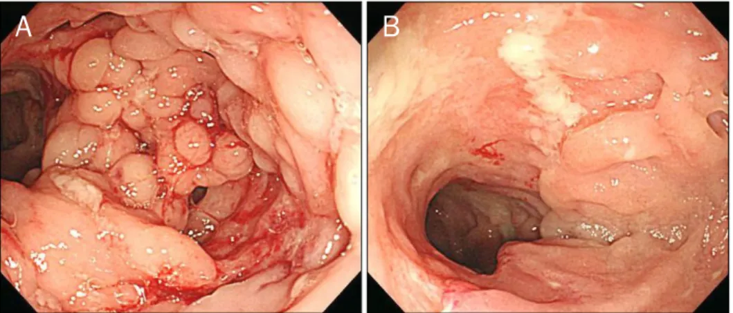
Fig. 1. Colonoscopic findings. (A) Co- bble-stone appearance in the ascen- ding colon consistent with Crohn’s disease. (B) Longitudinal ulcers and luminal narrowing in the terminal ileum consistent with Crohn’s disease.
Fig. 2. A small bowel follow-through at diagnosis demonstrating multiple strictures and pseudosacculations (arrowhead, stricture;
arrow, pseudosacculation).
apy, it seems appropriate to evaluate the associated safety issues.4,5
A retrospective observational study reported that in- fliximab may be a risk factor for free perforation in Crohn’s disease;6 however, there was no specific case of this in the literature at that time. Here, we report a case of free perfo- ration in Crohn’s disease with strictures following infliximab treatment, despite effectiveness of the therapy. To our knowl- edge, there are no case reports describing free perforation in Crohn’s disease following an initial dose of infliximab.
CASE REPORT
A 17-year-old Korean woman presented to the emergency room with an abrupt onset of severe abdominal pain of one hour’s duration. She had been diagnosed with Crohn’s dis- ease (A2L3B2 Montreal Classification for Crohn’s disease 2005: ileocolonic type and stricturing behavior) eight months prior. She had suffered from longstanding abdomi- nal pain, diarrhea, and poor oral intake. Initial colonoscopy findings were compatible with Crohn’s disease, including a cobblestone appearance and longitudinal ulcers (Fig. 1). A small bowel follow-through demonstrated multiple strictures with pseudosacculations in the distal ileum (Fig. 2). Her Crohn’s Disease Activity Index (CDAI) was 198 points at diagnosis. She had been treated with 5-aminosalicylic acid and azathioprine (1.2 mg/kg/day). During treatment, her CDAI improved to 109 points and her hemoglobin, albumin, and BMI increased. Despite this improvement, she could not tolerate a full dose of azathioprine due to severe anorexia and generalized fatigue. She was admitted to the hospital twice due to poor oral intake. Her anorexia and poor oral intake
were attributed to azathioprine, with a contribution from the ileal strictures. Surgery was recommended, but her parents refused. Accordingly, intravenous infliximab (5 mg/kg) ther- apy was added to her previous azathioprine regimen, follow- ing a normal chest X-ray, negative Mantoux testing, Quanti- FERONⓇ-TB Gold (Cellestis Ltd., Victoria, Australia), and hep- atitis B surface antigen. There were no immediate complica- tions following the infliximab infusion. Seven days after the first infliximab infusion, her general condition and appetite rapidly and markedly improved. She was able to tolerate fre- quent high-fiber meals without difficulty. Before infliximab treatment, her height was 151 cm, she weighted 42 kg, and her BMI was 18.42 kg/m2. Following treatment, her weight increased to 45 kg and her BMI increased to 19.74 kg/m2.
Fig. 3. Abdominal CT at the time of the emergency room presentation demonstrating a focal, asymmetric wall defect in the distal ileum with extraluminal free fecal density in the peritoneal space. A stricture is noted in the distal part of the perforation lesion. The wall is relatively thin at the level of the distal ileum perforation (arrow, wall defect of the distal ileum; arrowheads, extraluminal free fecal density in the peritoneal space; open arrows, stricture).
Fig. 5. Microscopic findings of the surgical specimen (H&E). (A) At the level of the perforation, only mild infiltration of inflammatory cells and fibrosis and a consequently thinned intestinal wall is noted (×40; arrows, perforated margin). (B) Non-caseating epithelioid granulomas are noted (×200; arrows, non-caseating epithelioid granulomas).
Fig. 4. Gross findings of the surgical specimen revealing a perforated lesion with a relatively thin ileal wall (arrows).
However, thirteen days after the first infliximab treatment, she experienced a sudden onset of abdominal pain. On phys- ical examination, blood pressure was 100/70 mmHg, pulse rate was 104/minute, and body temperature was 37.4oC.
She was acutely ill-appearing and her abdomen displayed signs of generalized peritonitis. Initial blood work revealed a hemoglobin level of 9.4 g/dL and a total white blood cell count of 15,700/μL; her high-sensitive CRP (HS-CRP) was 0.06 mg/dL; all results were within the normal range (shortly before infliximab treatment, blood work revealed a total
white blood cell count of 7,900/μL and a HS-CRP level of 1.431 mg/dL). Chest and abdominal X-rays did not convinc- ingly demonstrate the presence of free air. After 10 hours, fol- low-up blood work revealed a total white blood cell count of 16,900/μL and an elevated HS-CRP of 4.9 mg/dL. Computed tomography demonstrated a focal asymmetric wall defect in the distal ileum with extraluminal free fecal density in the per- itoneal space (Fig. 3). Emergency small bowel segmental re- section and ileostomy were performed. During the operation, a large perforation of the distal ileum was discovered and it was accompanied by the leakage of a large amount of pine- apple-like, high-fiber, undigested food into the peritoneal cav- ity through the perforation. Surgical pathology demonstrated that a perforation was present in the distal ileum, at the prox-
imal site of the stricture, without adhesions (Fig. 4). Microsco- pic findings at the level of the perforation demonstrated only mild infiltration of inflammatory cells and fibrosis with a re- sulting thinned intestinal wall (Fig. 5A) and several discrete non-caseating epithelioid granulomas (Fig. 5B). The post- operative course was uneventful and subsequent medical treatment of the underlying Crohn’s disease proved success- ful, with clinical and biological parameters of inflammation returning to normal within several days. After surgery, she tol- erated azathioprine treatment and is currently being treated with azathioprine 2.3 mg/kg/day without infliximab.
DISCUSSION
Burrill Bernard Crohn stated in a 1957 paper that “free per- foration of ileitis into the peritoneal cavity never occurs or at least I have not seen it.”7 In 1965 however, he reported seven cases of free perforation.8 Because Crohn’s disease is a transmural inflammatory process, serosal adhesions devel- op that provide direct pathways for fistula formation and re- duce the incidence of free perforation. Due to this, the etiol- ogy of free perforations is unknown. It is generally accepted that 1-3% of patients with Crohn’s disease will present with a free perforation, either initially or later in their disease course.9-11
Infliximab’s mechanism of action is targeting TNF, which is responsible for the majority of the cases of inflammation associated with Crohn’s disease. With the introduction of TNF alpha blockers, treatment options for Crohn’s disease have improved significantly. Infliximab is very effective for the treatment of Crohn’s disease, and is used to induce and maintain remission.12 Unfortunately, infliximab treatment al- so has potential adverse events, including acute infusion re- actions, severe serum sickness, and increased risk of in- fections, particularly reactivation of latent tuberculosis.
Rarely, infliximab has been associated with optic neuritis, seizures, new onset or exacerbation of clinical symptoms, and radiographic evidence of central nervous system demye- linating disorders, including multiple sclerosis. It may also ex- acerbate symptoms in patients with New York Heart Association functional class III/IV heart failure.13,14
Recently, Eshuis et al.6 reported that anti-TNF treatment for Crohn’s disease was associated with a significantly higher rate of free perforation requiring surgery. Although there was
an attempt to equate the disease activity between cases and controls, it was quite clear that the disease activity compar- ison was poorly quantified. Therefore, the study demon- strated only a significant association between the occur- rence of free perforation in Crohn’s disease patients and the use of anti-TNF therapy, but did not prove a cause-and-effect relationship. However, the current report is the first case pre- sentation describing free perforation in Crohn’s disease pa- tients after infliximab treatment and strongly suggests that infliximab may be a risk factor for free perforation in Crohn’s disease, especially in cases with severe strictures.
Greenstein et al.10 suggested that increased intraluminal pressure proximal to the stricture and ischemia due to in- flammation and abscesses caused by Crohn’s disease may be potential causes of perforation. But, free perforations in Crohn’s disease are relatively rare because adjacent organs and the omentum usually encapsulate the imminent perfo- ration site by creating an inflammatory mass. In patients un- dergoing infliximab therapy, anti-TNF may block develop- ment of this protective inflammatory mass.6
In the current case, free perforation was likely caused by rapid and markedly increased intraluminal pressure prox- imal to the severe strictures, secondary to a large amount of food intake over a short time due to the therapeutic effective- ness of infliximab and relief of inflammation of the intestinal wall due to the effects of the drug. Previous studies have re- ported that cytokines, such as TNF-α, can influence human behavior during inflammatory conditions. Changes in behav- ior include decreased sexual drive, increased sleepiness, de- creased appetite, and fatigue.15,16 Infliximab decreases the amount of circulating TNF-α, which is increased in Crohn’s disease and, as a result, may influence behavior in Crohn’s disease.17 Therefore, infliximab might contribute to overea- ting. In general, disease-associated fibrous reactions and ad- hesions to adjacent organs appear to be protective against free perforation. If a minimal perforation does occur, then ei- ther an abscess will develop within the inflammatory mass or a fistula to an adjacent organ will develop. But in the current case, infliximab blocked the inflammatory response as a
“treatment” effect, and contributed to the occurrence of free perforation by preventing the formation of an inflammatory mass to seal off the imminent perforation.
This case report and discussion emphasize that during or after infliximab treatment for Crohn’s disease accompanied
by severe strictures, a large food intake should be avoided.
Although food tolerance is an important signal of treatment effectiveness, slow and incremental increases in intake are advised.
REFERENCES
1. Hanauer SB, Sandborn W; Practice Parameters Committee of the American College of Gastroenterology. Management of Crohn's disease in adults. Am J Gastroenterol 2001;96:635- 643.
2. Present DH, Rutgeerts P, Targan S, et al. Infliximab for the treat- ment of fistulas in patients with Crohn's disease. N Engl J Med 1999;340:1398-1405.
3. Rutgeerts P, Van Assche G, Vermeire S. Optimizing anti-TNF trea- tment in inflammatory bowel disease. Gastroenterology 2004;
126:1593-1610.
4. de Vries HS, van Oijen MG, de Jong DJ. Serious events with in- fliximab in patients with inflammatory bowel disease: a 9-year cohort study in the Netherlands. Drug Saf 2008;31:1135-1144.
5. Fidder H, Schnitzler F, Ferrante M, et al. Long-term safety of in- fliximab for the treatment of inflammatory bowel disease: a sin- gle-centre cohort study. Gut 2009;58:501-508.
6. Eshuis EJ, Griffioen GH, Stokkers PC, Ubbink DT, Bemelman WA.
Anti tumour necrosis factor as risk factor for free perforations in Crohn's disease? A case-control study. Colorectal Dis 2012;14:
578-584.
7. Crohn BB. Indications for surgical intervention in regional ileitis.
AMA Arch Surg 1957;74:305-311.
8. Crohn BB. Acute regional ileitis; clinical aspects and follow-up studies. N Y State J Med 1965;65:641-644.
9. Greenstein AJ, Mann D, Sachar DB, Aufses AH Jr. Free perfo- ration in Crohn's disease: I. A survey of 99 cases. Am J Gastroen- terol 1985;80:682-689.
10. Greenstein AJ, Sachar DB, Mann D, Lachman P, Heimann T, Aufses AH Jr. Spontaneous free perforation and perforated ab- scess in 30 patients with Crohn's disease. Ann Surg 1987;205:
72-76.
11. Katz S, Schulman N, Levin L. Free perforation in Crohn's disease:
a report of 33 cases and review of literature. Am J Gastroenterol 1986;81:38-43.
12. Hanauer SB, Feagan BG, Lichtenstein GR, et al; ACCENT I Study Group. Maintenance infliximab for Crohn's disease: the ACCENT I randomised trial. Lancet 2002;359:1541-1549.
13. Sleisenger MH, Feldman M, Friedman LS, Brandt LJ. Sleisenger and Fordtran's gastrointestinal and liver disease. 9th ed. Phila- delphia: Saunders, 2010:1967-1969.
14. Colombel JF, Loftus EV Jr, Tremaine WJ, et al. The safety profile of infliximab in patients with Crohn's disease: the Mayo clinic ex- perience in 500 patients. Gastroenterology 2004;126:19-31.
15. Kronfol Z, Remick DG. Cytokines and the brain: implications for clinical psychiatry. Am J Psychiatry 2000;157:683-694.
16. Reichenberg A, Yirmiya R, Schuld A, et al. Cytokine-associated emotional and cognitive disturbances in humans. Arch Gen Psychiatry 2001;58:445-452.
17. van Balkom BP, Schoon EJ, Stockbrügger RW, et al. Effects of an- ti-tumour necrosis factor-alpha therapy on the quality of life in Crohn's disease. Aliment Pharmacol Ther 2002;16:1101-1107.