Hip Pelvis 33(3): 154-161, 2021 https://doi.org/10.5371/hp.2021.33.3.154
Factors Associated with Mechanical
Complications in Intertrochanteric Fracture Treated with Proximal Femoral Nail Antirotation
Oog-Jin Shon, PhD, Chang Hyun Choi, MD, Chan Ho Park, PhD
Department of Orthopedic Surgery, Yeungnam University Medical Center, Daegu, Korea
Purpose: Although proximal femoral nail antirotation (PFNA; Synthes, Switzerland) has demonstrated satisfac- tory results when used for the treatment of intertrochanteric fractures, mechanical complications may occur. To better quantify the risk of mechanical complications when proximal femoral nail antirotation is used to treat intertrochanteric fractures, this study aimed to: (1) characterize the frequency of mechanical complications and extent of blade sliding and their correlation with reduction quality and (2) identify factors associated with mechanical complications.
Materials and Methods: A review of medical records from 93 patients treated for intertrochanteric fractures with a minimum of 6-months of follow-up between February 2014 and February 2019 was conducted. Blade position was evaluated using Tip-apex distance (TAD) and Cleveland index. The extent of blade sliding was evaluated using the adjusted Doppelt’s method for intramedullary nailing. Individuals were classified as having or not having mechanical complications, and reduction quality and radiologic outcomes were compared between the two groups.
Results: Mechanical complications occurred in 12 of 94 hips (12.8%), with 11 out of 12 being from the intramedullary reduction group. There was no significant difference in TAD between groups; however, there were significant differences were noted in Cleveland index, AO/OTA classification, reduction quality and extent of blade sliding. The mean blade sliding distance was 1.17 mm (anatomical group), 3.28 mm (extramedullary group), and 6.11 mm (intramedullary group), respectively (P<0.001). Data revealed that blade sliding was an associated factor for mechanical complications (odds ratio 1.25, 95% confidence interval 1.03-1.51).
Conclusion: The extent of blade sliding determined using the adjusted Doppelt’s method was significantly asso- ciated with mechanical complications suggesting that prevention of excessive sliding through proper intraopera- tive reduction is important to help achieve satisfactory treatment outcomes.
Key Words: Blade sliding, Femur, Hip fractures, Intramedullary reduction
Submitted:September 21, 2020 1st revision:January 11, 2021 Final acceptance:January 19, 2021
Address reprint request to Chan Ho Park, PhD
(https://orcid.org/0000-0003-0409-8132)
Department of Orthopedic Surgery, Yeungnam University Medical Center, 170 Hyeonchung-ro, Nam-gu, Daegu 42415, Korea TEL:+82-53-620-3642 FAX:+82-53-628-4020
E-mail:[email protected]
This is an Open Access article distributed under the terms of the Creative Commons Attribution Non-Commercial License (http://creativecommons.
org/licenses/by-nc/4.0) which permits unrestricted non-commercial use, dis- tribution, and reproduction in any medium, provided the original work is properly cited.
INTRODUCTION
The number of osteoporotic hip fracture increases in the elderly population as life expectancy increases. Additionally, the incidence of osteoporotic hip fractures has also increased over the years1,2). The 1-year mortality rate following hip frac- tures in previous studies ranges from 8.4% to 36%3). The goal of treatment for hip fractures is to lower mortality rate through early recovery of ambulatory function4,5).
Patients with intertrochanteric fractures tend to be older and have more severe osteoporosis compared with patients with femoral neck fractures6,7). Although some studies report that arthroplasty is better for early rehabilitation, internal fixation using various devices is currently the treatment of choice for intertrochanteric fractures8). Cephalomedullary nailing has several advantages (e.g., shorter operating time, biomechanical stability) for the treatment of intertrochanteric fractures9,10). Among several types of cephalomedullary nails, proximal femoral nail antirotation (PFNA; Synthes, Solothurn, Switzerland) is characterized by the anti-rotation helical blade which is more resistant to rotational deformity compared with lag screws11,12).
Although the PFNA system has demonstrated satisfactory results, the rate of mechanical failure including nonunion, cut-through or cut-out, excessive migration of blade, peri- implant fracture and implant breakage ranges from 2.6% to
13%13,14). For the prevention of mechanical complications,
appropriate fracture reduction and blade position are essen-
tial15,16). Anatomical reduction or extramedullary reduction
with medial cortical overlap known as the Wayne-County technique are associated with biomechanical stability com- pared with intramedullary reduction17). Intramedullary reduc- tion in comminuted intertrochanteric fractures without pos- teromedial cortical support is prone to excessive sliding and varus malposition of proximal fragment leading to mechan- ical failures. We hypothesized that the extent of blade slid- ing is different according to the reduction quality. In addi- tion, we consider that excessive blade migration is a factor associated with mechanical complications.
Therefore, the purpose of this study was to: (1) determine the proportion of mechanical complications according to reduction quality and (2) to identify factors associated with mechanical complications in patients with intertrochanteric fracture treated by PFNA.
MATERIALS AND METHODS
This study was approved by the Institutional Review Board (IRB) of Yeungnam University Medical Center, and the informed consent was waived by the IRB (YUMC 2020-03-012). A retrospective study was conducted at a tertiary referral hospital. Medical records and radiographs of patients who were surgically treated with PFNA for intertrochanteric fractures between February 2014 and February 2019 were evaluated. During the study period, all patients with intertrochanteric fractures who visited our institution underwent osteosynthesis surgery. Only patients treated with implants using a helical blade were included in this study. The inclusion criteria were: (1) over 55 years of age with osteoporotic intertrochanteric fracture due to low energy trauma like simple fall18)and (2) minimum of 6- month post-surgical follow-up. Patients with high energy trauma, reoperation, subtrochanteric or atypical femoral frac- tures were excluded. During the study period, 491 patients (492 hips) underwent intramedullary nailing at our institu- tion for treatment of intertrochanteric fractures; all surg- eries were conducted by a single surgeon. Among these patients, 62 were excluded (62 hips) because they were not treated by implants with a helical blade. Of the remaining 429 patients (430 hips) who underwent surgery using PFNA, only 93 patients (94 hips) had at least 6 months of post-surgical follow-up and were thus included in the final analysis (Fig. 1). Of the 93 patients included, 22 and 71 were male and female, respectively; mean age at time of surgery was 77.6±7.8 years (range, 55.0-95.0 years). Mean body mass index was 22.2±4.0 kg/m2(range, 13.6-32.4 kg/m2) and the mean duration from admission to opera- tion was 1.11±1.5 days (range, 0-6 days). Operations were not delayed beyond 48 hours as except in the rare case that patients were not able to undergo surgery because of poor general condition. The medical status of patients was eval- uated according to the American Society of Anesthesiologists (ASA) classification and Charlson comorbidity index (CCI).
The median preoperative ASA classification and CCI were 2.5 (range, 2-4) and 4.5 (range, 2-8), respectively.
All fractures were classified according to AO/OTA guide- lines based on preoperative computed tomography scans;
31A2.2 was the most common classification (n=34 hips).
The percentages of stable and unstable fractures were 40.4%
and 59.6%, respectively according to the AO classification (31A1=stable; 31A2=unstable).
The mean postoperative follow-up period was 18.1 months (range, 6-56 months). PFNA nails with a centrum-collum-
diaphyseal (CCD) angle of 125。or 130。were selected based on the CCD angle of the contralateral side. Nails with 125。
and 130。CCD angles were used in 81 hips (86.2%) and 13 hips (13.8%), respectively.
Using radiographs gathered immediately after surgery, two independent orthopedic surgeons evaluated tip-apex distance (TAD)16), blade position in the femoral head (using the Cleveland index)19)and classified fracture reduction qual- ity into one of three categories (anatomical, extramedullary, and intramedullary) as described by Ito et al.15). Intramedullary reduction classification required observation in at least one
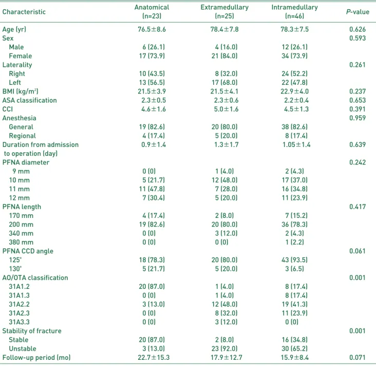
of the anteroposterior and lateral radiographs. If the radi- ologic outcome was not in agreement, results were confirmed after discussion. The quality of reduction was anatomical reduction (n=23 hips), extramedullary reduction (n=25 hips), and intramedullary reduction (n=46 hips). Blade sliding was evaluated using the Doppelt’s method adjusted suitably for intramedullary nailing comparing initial postoperative radi- ograph with the last follow-up (Fig. 2)20). To minimize mea- surement error, femur rotation was confirmed by comparing the size of the lesser trochanter immediately after surgery to the radiograph captured at final follow-up. Mechanical com- plications included non-union, cut-out or cut-through, exces- sive migration of blade, peri-implant fracture, and implant breakage14). Demographic data of patients according to reduc- tion quality are summarized in Table 1.
Statistical analyses were performed with univariate com- parisons using independent t-test or ANOVA test for con- tinuous variables and chi-square test for categorized data.
Then, multivariable logistic regression analyses were per- formed to identify potential factors associated with mechan- ical complications. Differences were considered significant if P-values were <0.05. All analyses were performed using IBM SPSS Statistics for Windows (ver. 20.0; IBM, Armonk, NY, USA).
RESULTS
During the follow-up period, mechanical complications occurred in 12 of 94 hips (12.8%). Although TAD was mea- sured as the mean 20.1±5.07 mm (range, 11-34 mm) in all patients, TAD exceeded 25 mm in 22 hips (23.4%). There was no significant difference in TAD between those with F
Fiigg.. 11.. Study flow chart.
F
Fiigg.. 22.. Adjusted Doppelt’s method for intramedullary nail- ing (AA) immediate postoperative radiograph (BB) subsequent radiograph after sliding. Correction factor=b/b’, the extent of blade sliding=a-(a’××b/b’).
A B
and without mechanical complications. Blades were insert- ed into a safe zone (Cleveland zones 5, 6, 8, and 9) in 82 hips (87.2%). The proportion of AO/OTA classification and Cleveland index was significantly different between those with and without mechanical complications (Table 2).
Among the 12 hips with mechanical complications, bony union was achieved in 4 hips through a revision osteosyn- thesis operation within 6 months following the initial oper-
ation; treatment failure occurred in 8 hips (8.5%). Conversion to hip arthroplasty was performed in 5 patients due to cut- out or cut through and osteonecrosis of femoral head. An additional 3 patients refused to undergo additional oper- ations. All patients achieved bony union at a mean of 7.2 months post operation except for those patients with treat- ment failure.
The mean distance of blade sliding was 1.17 mm, 3.28 mm,
Table 1. Patients Demographics
Characteristic Anatomical Extramedullary Intramedullary
P-value
(n=23) (n=25) (n=46)
Age (yr) 76.5±±8.6 78.4±±7.8 78.3±±7.5 0.626
Sex 0.593
Male 06 (26.1) 04 (16.0) 12 (26.1)
Female 17 (73.9) 21 (84.0) 34 (73.9)
Laterality 0.261
Right 10 (43.5) 08 (32.0) 24 (52.2)
Left 13 (56.5) 17 (68.0) 22 (47.8)
BMI (kg/m2) 21.5±±3.9 21.5±±4.1 22.9±±4.0 0.237
ASA classification 02.3±±0.5 02.3±±0.6 02.2±±0.4 0.653
CCI 04.6±±1.6 05.0±±1.6 04.5±±1.3 0.391
Anesthesia 0.959
General 19 (82.6) 20 (80.0) 38 (82.6)
Regional 04 (17.4) 05 (20.0) 08 (17.4)
Duration from admission 00.9±±1.4 01.3±±1.7 01.05±±1.4 0.639
to operation (day)
PFNA diameter 0.242
09 mm 0 (0)0. 1 (4.0) 2 (4.3)
10 mm 05 (21.7) 12 (48.0) 17 (37.0)
11 mm 11 (47.8) 07 (28.0) 16 (34.8)
12 mm 07 (30.4) 05 (20.0) 11 (23.9)
PFNA length 0.417
170 mm 04 (17.4) 2 (8.0) 07 (15.2)
200 mm 19 (82.6) 20 (80.0) 36 (78.3)
340 mm 0 (0)0. 03 (12.0) 2 (4.3)
380 mm 0 (0)0. 0 (0)0. 1 (2.2)
PFNA CCD angle 0.061
125。 18 (78.3) 20 (80.0) 43 (93.5)
130。 05 (21.7) 05 (20.0) 3 (6.5)
AO/OTA classification 0.001
31A1.2 20 (87.0) 1 (4.0) 08 (17.4)
31A1.3 0 (0)0. 1 (4.0) 08 (17.4)
31A2.2 03 (13.0) 12 (48.0) 19 (41.3)
31A2.3 0 (0)0. 08 (32.0) 11 (23.9)
31A3.3 0 (0)0. 03 (12.0) 0 (0)0.
Stability of fracture 0.001
Stable 20 (87.0) 2 (8.0) 16 (34.8)
Unstable 03 (13.0) 23 (92.0) 30 (65.2)
Follow-up period (mo) 022.7±±15.3 017.9±±12.7 15.9±±8.4 0.071
Values are presented as mean±±standard deviation or number (%).
BMI: body mass index, ASA classification: American Society of Anesthesiologists classification, CCI: Charlson comorbidity index, PFNA: proximal femoral nail antirotation, CCD: centrum-collum-diaphyseal.
and 6.11 mm in the anatomical reduction, extramedullary reduction, and intramedullary reduction groups, respective- ly (P<0.001) (Fig. 3).
There were no cases of mechanical complications in the anatomical reduction group. Although excessive blade slid- ing (>5 mm) occurred in 1 hip in the extramedullary reduc- tion group, bony union was achieved after blade exchange to a shorter one was performed. Most mechanical compli- cations occurred in the 11 hips in the intramedullary reduc- tion group.
When patients were classified into two groups (i.e., those with and those without mechanical complications), a uni- variable analysis revealed significant differences in among the groups. Multivariable logistic regression analysis includ- ing these three factors (i.e., reduction quality, AO/OTA clas- sification, extent of blade sliding), only the extent of blade sliding was associated with a mechanical complication after adjustment (odds ratio 1.25, 95% confidence interval 1.03- 1.51) (Fig. 4).
DISCUSSION
Internal fixation remains the treatment of choice for
intertrochanteric fractures, however some arthroplasty stud- ies have demonstrated satisfactory results (e.g., mortality, risk of complications)21). Reduction quality is important dur- ing internal fixation, intramedullary reduction in commin- Table 2. Radiologic Outcomes between Groups with or without Mechanical Complications
Characteristic Normal Mechanical complications
P-value
(n=82) (n=12)
TAD: tip-apex distance (mm) 20.9±±4.9 21.6±±6.5 0.708
Reduction quality 0.003
Anatomical 23 (28.0) 0 (0)0.
Extramedullary 24 (29.3) 1 (8.3)
Intramedullary 35 (42.7) 11 (91.7)
Cleveland index 0.004
4 7 (8.5) 02 (16.7)
5 43 (52.4) 1 (8.3)
6 5 (6.1) 03 (25.0)
7 1 (1.2) 02 (16.7)
8 19 (23.2) 03 (25.0)
9 7 (8.5) 1 (8.3)
AO/OTA classification 0.001
31A1.2 23 (28.0) 0 (0)0.
31A1.3 12 (14.6) 03 (25.0)
31A2.2 31 (37.8) 03 (25.0)
31A2.3 13 (15.9) 06 (50.0)
31A3.3 3 (3.7) 0 (0)0.
Stability of fracture 0.395
Stable 35 (42.7) 03 (25.0)
Unstable 47 (57.3) 09 (75.0)
The extent of blade sliding (mm) 03.5±±2.9 08.1±±5.7 0.018
Values are presented as mean±±standard deviation or number (%).
TAD: tip-apex index.
F
Fiigg.. 33.. The extent of blade sliding according to reduction qual- ity.
** P<0.001.
uted intertrochanteric fracture without posteromedial corti- cal support may lead to varus malpostion of the proximal fragment. Excessive blade sliding–defined as sliding more than 5 mm–may eventually cause treatment failure22). In the present study, the extent of blade sliding occurred more fre- quently in the intramedullary reduction group compared with the two other groups. In addition, blade slides was identified as a factor associated with mechanical complications. To prevent excessive blade sliding, reduction quality is essen- tial. In elderly patients with severe osteoporosis specifical- ly, it is exceedingly difficult to achieve appropriate reduc- tion of comminuted fragments in posteromedial cortex.
Therefore, if anteromedial cortex is not reduced properly, treatment failure is likely23,24). Yoon et al.22)emphasized the importance of achieving continuity of the medial and anteri- or cortical line in anteroposterior and axial images intraop- eratively (Fig. 5).
Although the number of conversion arthroplasties includ- ed in this study was too small to identify statistical signifi- cance, all conversion arthroplasty occurred in the intramedullary group because cut-out eventually occurred due to excessive sliding in this group. On the other hand, blade exchange alone did not cause further sliding and bone union was achieved in the anatomical and extramedullary reduction groups; if blade sliding did occur in these groups, the blade was not able to slide excessively after the cortical apposi-
tion. However, blade sliding was not blocked by cortical apposition in the intramedullary reduction group, and after that, cut-out occurred following varus malposition and rota- tion of proximal fragment. A previous biomechanical study demonstrated results similar to those in the extamedullary reduction group which had better resistance against axial load- ing compared with the intramedullary reduction group17).
Implant design has been developing to prevent excessive sliding–namely sliding that adversely affects clinical results.
Although this study only determined the results associated with blade-type cephalomedullary nails, Gamma nail (Stryker Trauma, Schoenkirchen, Germany), which is the lag-type screw cephalomedullary nail, early versions were associ- ated with higher rates of cut-out compared with dynamic hip screw25). Gamma-3 nails, a third-generation version, employ a U-blade to withstand varus and rotational deforming force.
Comparing the Gamma 3 nail without U-blade, U-blade significantly prevented lag screw sliding26). After all, sur- geons must try to reduce sliding through appropriate reduc- tion and implant design because the extent of blade or lag screw sliding is a risk factor for treatment failure in this study.
The present study had several limitations. First, the num- ber of cases was small because the rate of follow-up loss and mortality in elderly patients with hip fractures were rel- atively high. Second, there might be selection bias because only patients followed-up for a minimum of 6 months were F
Fiigg.. 44.. Odds ratio (OR) plot for mechanical complications.
CI: confidence interval.
* P<0.05.
included. For this reason, the rate of treatment failure and mechanical complication was overestimated and higher than previous studies. Third, the reliability of radiologic and clin- ical outcomes as related to blade sliding (e.g., trochanter pain, limping) were not evaluated.
Nevertheless, the extent of blade sliding was more accu- rately assessed using the adjusted Doppelt’s method for intramedullary nailing. In addition, the extent of sliding was deemed an important factor impacting treatment outcomes.
CONCLUSION
The extent of sliding was significantly different depend- ing on reduction quality, a factor associated with mechan- ical complications. Preventing excessive sliding through proper intraoperative reduction is important to achieve sat- isfactory treatment outcomes.
CONFLICT OF INTEREST
The authors declare that there is no potential conflict
of interest relevant to this article.
REFERENCES
01. Kanis JA, Odén A, McCloskey EV, Johansson H, Wahl DA, Cooper C. A systematic review of hip fracture incidence and probability of fracture worldwide. Osteoporos Int. 2012;23:
2239-56.
02. Park C, Ha YC, Jang S, Jang S, Yoon HK, Lee YK. The incidence and residual lifetime risk of osteoporosis-related fractures in Korea. J Bone Miner Metab. 2011;29:744-51.
03. Abrahamsen B, van Staa T, Ariely R, Olson M, Cooper C.
Excess mortality following hip fracture: a systematic epi- demiological review. Osteoporos Int. 2009;20:1633-50.
04. Halbert J, Crotty M, Whitehead C, et al. Multi-disciplinary rehabilitation after hip fracture is associated with improved outcome: a systematic review. J Rehabil Med. 2007;39:507- 12.
05. Shyu YI, Liang J, Tseng MY, et al. Comprehensive care improves health outcomes among elderly Taiwanese patients with hip fracture. J Gerontol A Biol Sci Med Sci. 2013;68:
188-97.
06. Cho Y, Lee I, Ha SH, Park JH, Park JH. Comparison of hip subregion bone mineral density to the type of proximal femur fracture. Arch Osteoporos. 2020;15:122.
F
Fiigg.. 55.. (AA) A 75-year-old women sustained a 31A2.2 intertrochanteric fracture. (BB) Intramedullary reduction visual in an intra- operative anteroposterior radiograph. (CC) Anterior cortical line was well reduced as noted in an intraoperative axial radi- ograph. (DD, EE) Bone union was achieved after 6.75 mm sliding of blade as noted by a comparison of a radiograph captured immediately after surgery to a radiograph collected 1-year postoperatively.
A B C
D E
07. Hey HW, Sng WJ, Lim JL, et al. Interpretation of hip frac- ture patterns using areal bone mineral density in the proxi- mal femur. Arch Orthop Trauma Surg. 2015;135:1647-53.
08. Yoo JI, Ha YC, Lim JY, Kang H, Yoon BH, Kim H. Early rehabilitation in elderly after arthroplasty versus internal fix- ation for unstable intertrochanteric fractures of femur: sys- tematic review and meta-analysis. J Korean Med Sci. 2017;
32:858-67.
09. Kim BS, Lim JY, Ha YC. Recent epidemiology of hip frac- tures in South Korea. Hip Pelvis. 2020;32:119-24.
10. Strauss E, Frank J, Lee J, Kummer FJ, Tejwani N. Helical blade versus sliding hip screw for treatment of unstable intertrochanteric hip fractures: a biomechanical evalua- tion. Injury. 2006;37:984-9.
11. Gardenbroek TJ, Segers MJ, Simmermacher RK, Hammacher ER. The proximal femur nail antirotation: an identifiable improvement in the treatment of unstable pertrochanteric fractures? J Trauma. 2011;71:169-74.
12. Al-Munajjed AA, Hammer J, Mayr E, Nerlich M, Lenich A. Biomechanical characterisation of osteosyntheses for proximal femur fractures: helical blade versus screw. Stud Health Technol Inform. 2008;133:1-10.
13. Kashigar A, Vincent A, Gunton MJ, Backstein D, Safir O, Kuzyk PR. Predictors of failure for cephalomedullary nail- ing of proximal femoral fractures. Bone Joint J. 2014;96- B:1029-34.
14. Zhang WQ, Sun J, Liu CY, Zhao HY, Sun YF. Comparing the intramedullary nail and extramedullary fixation in treatment of unstable intertrochanteric fractures. Sci Rep.
2018;8:2321.
15. Ito J, Takakubo Y, Sasaki K, Sasaki J, Owashi K, Takagi M.
Prevention of excessive postoperative sliding of the short femoral nail in femoral trochanteric fractures. Arch Orthop Trauma Surg. 2015;135:651-7.
16. Baumgaertner MR, Curtin SL, Lindskog DM, Keggi JM. The value of the tip-apex distance in predicting failure of fixa-
tion of peritrochanteric fractures of the hip. J Bone Joint Surg Am. 1995;77:1058-64.
17. Park YC, Yoon SP, Yang KH. The effects of extramedullary reduction in unstable intertrochanteric fracture: a biome- chanical study using cadaver bone. J Korean Fract Soc.
2018;31:79-86.
18. Yoo JH, Moon SH, Ha YC, et al. Osteoporotic fracture: 2015 position statement of the Korean Society for Bone and Mineral Research. J Bone Metab. 2015;22:175-81.
19. Cleveland M, Bosworth DM, Thompson FR, Wilson HJ Jr, Ishizuka T. A ten-year analysis of intertrochanteric fractures of the femur. J Bone Joint Surg Am. 1959;41-A:1399-408.
20. Doppelt SH. The sliding compression screw--today’s best answer for stabilization of intertrochanteric hip fractures.
Orthop Clin North Am. 1980;11:507-23.
21. Yoo JI, Cha YH, Kim KJ, Kim HY, Choy WS, Hwang SC.
Comparison between cementless and cemented bipolar hemi- arthroplasty for treatment of unstable intertrochanteric fractures: systematic review and meta-analysis. Hip Pelvis.
2018;30:241-53.
22. Yoon YC, Oh CW, Sim JA, Oh JK. Intraoperative assessment of reduction quality during nail fixation of intertrochanteric fractures. Injury. 2020;51:400-6.
23. Sharma G, Gn KK, Khatri K, Singh R, Gamanagatti S, Sharma V. Morphology of the posteromedial fragment in pertrochanteric fractures: a three-dimensional computed tomography analysis.
Injury. 2017;48:419-31.
24. Pesce V, Speciale D, Sammarco G, Patella S, Spinarelli A, Patella V. Surgical approach to bone healing in osteoporosis.
Clin Cases Miner Bone Metab. 2009;6:131-5.
25. Ahrengart L, Törnkvist H, Fornander P, et al. A randomized study of the compression hip screw and Gamma nail in 426 fractures. Clin Orthop Relat Res. 2002;(401):209-22.
26. Ryu HG, Choi YT, Kim SM, Seo JS. A comparison of U- blade Gamma3 and Gamma3 nails used for the treatment of intertrochanteric fractures. Hip Pelvis. 2020;32:50-7.