CASE REPORT
내시경 치료 후 폐쇄성 황달을 동반한 십이지장의 장벽내 혈종 1예
김학수1, 김희경2, 김원희3, 홍성표3, 조주영3
인천사랑병원 소화기내과1, 청구성심병원 소화기내과2, 차의과학대학교 분당차병원 소화기센터3
Huge Intramural Duodenal Hematoma Complicated with Obstructive Jaundice following Endoscopic Hemostasis
Hak Su Kim1, Hee Kyoung Kim2, Won Hee Kim3, Sung Pyo Hong3 and Joo Young Cho3
Department of Gastroenterology and Hepatology, Incheon Sarang Hospital1, Incheon; Department of Gastroenterology and Hepatology, Cheonggu Sungsim Hospital2, Seoul; Digestive Disease Center, CHA Bundang Medical Center, CHA University3, Seongnam, Korea
Intramural hematoma of the duodenum is a relatively unusual complication associated with the endoscopic treatment of bleeding peptic ulcers. Intramural hematomas are typically resolved spontaneously with conservative treatment alone. We report a case of an intramural duodenal hematoma following endoscopic hemostasis with epinephrine injection therapy, which was associated with transient obstructive jaundice in a patient undergoing hemodialysis. The patient developed biliary sepsis due to obstruction of the common bile duct secondary to the huge hematoma. He was treated with fluoroscopy-guided drainage catheter insertion, which spon- taneously resolved the biliary sepsis through conservative treatment in 6 weeks. Fluoroscopy-guided drainage may impact the treat- ment of intramural hematomas that involve life-threatening complications. (Korean J Gastroenterol 2019;73:39-44)
Key Words: Duodenal ulcer; Hematoma; Cholestasis; Drainage
Received June 1, 2018. Revised June 21, 2018. Accepted June 29, 2018.
CC This is an open access article distributed under the terms of the Creative Commons Attribution Non-Commercial License (http://creativecommons.org/licenses/
by-nc/4.0) which permits unrestricted non-commercial use, distribution, and reproduction in any medium, provided the original work is properly cited.
Copyright © 2019. Korean Society of Gastroenterology.
교신저자: 김원희, 13496, 성남시 분당구 야탑로 59, 차의과학대학교 분당차병원 소화기센터
Correspondence to: Won Hee Kim, Digestive Disease Center, CHA Bundang Medical Center, CHA University, 59 Yatap-ro, Bundang-gu, Seongnam 13496, Korea. Tel:
+82-31-780-5212, Fax: +82-31-780-5219, E-mail: [email protected], ORCID: https://orcid.org/0000-0003-0013-0211 Financial support: None. Conflict of interest: None.
INTRODUCTION
Intramural hematoma of the gastrointestinal tract is a rela- tively uncommon condition. It is mainly caused by blunt trauma, bleeding disorders, or anticoagulation therapy.1 Recently, re- ports of intramural hematoma have increased with the use of therapeutic endoscopy for gastro-duodenal peptic ulcer bleeding.1,2 The risk of complications associated with endo- scopic intervention is very low and mainly includes aspiration pneumonia and gut perforation.3 Intramural duodenal hema- toma is a rare complication after an endoscopic intervention, although patients susceptible to bleeding such as those with
end-stage renal disease (ESRD), liver cirrhosis, or those receiv- ing anticoagulant therapy are more susceptible.4-6 We report a case of an intramural hematoma occurring after the endo- scopic treatment of a duodenal ulcer in a patient undergoing hemodialysis (HD), followed by a secondary hematoma-in- duced common bile duct obstruction.
CASE REPORT
A 45-year-old man presented to emergency room of CHA Bundang Medical Center, complaining of multiple episodes of melena in 1 day. The patient had been on maintenance
Fig. 1. Endoscopic findings. (A) The esophago-gastro-duodenoscopy on admission revealed a deep ulcer and an exposed vessel on the base was noted at the duodenal bulb. (B) An epinephrine injection (2 mL, 1 mL) was administered to control the blood oozing from the vessel. Complete hemostasis was acquired with four hemoclippings and 2 mL of fibrin glue injection.
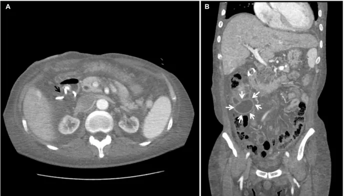
Fig. 2. On the 2nd hospital day, the contrast-enhanced abdominal computed tomography scan showed a huge intramural hematoma in the 2nd to 4th portions of the duodenum with compression of the common bile duct (black arrows, intramural hematoma; white arrows, common bile duct). (A) Axial image. (B) Coronal image.
HD three times per week 6 years ago. He had a history of undergoing the Hartmann operation to treat left colic artery bleeding and colonic perforation a year ago. He had not tak- en non-steroidal anti-inflammatory drugs, antiplatelet drugs, or anticoagulants before hospitalization. At presentation, his vital signs were normal, and a physical examination re- vealed anemic conjunctiva without icteric sclera. The rele- vant laboratory investigations were as follows; hemoglobin 8.1 g/dL, platelet count 242,000/mm3, white blood cell count 10,810/mm3, INR 1.04, serum total bilirubin 0.35
mg/dL, serum AST 10 IU/L, serum ALT 16 IU/L, ALP 134 IU/L, CRP 5.98 mg/dL, BUN 116.1 mg/dL, and serum cre- atinine 7.8 mg/dL.
An emergency esophago-gastro-duodenoscopy (EGD) was performed due to suspected active gastrointestinal bleeding.
The endoscopy revealed a deep ulcer with exposed vessel at the duodenal bulb (Fig. 1A). A total of 3 mL of 0.2% epi- nephrine injection, with subsequent hemoclipping, was ad- ministered to control the continuous oozing of blood from the exposed vessel. Moreover, 2 mL of fibrin glue injection was administered to treat the active bleeding from the duodenal ulcer (Fig. 1B). He was conservatively treated with an intra- venous proton pump inhibitor.
On the second day after the endoscopic treatment, he com- plained of abdominal pain, a palpable mass and jaundice.
The mass was palpated about 15 cm from the right upper quadrant site. The serum total bilirubin level had increased from 0.35 mg/dL, on admission, to 12.54 mg/dL, with ele- vations of the serum AST to 45 IU/L and ALT to 95 IU/L.
An emergency abdominal CT scan revealed a common bile duct obstruction by a large hematoma of up to 20 cm in the submucosal area in the second to fourth portions of the duo- denum (Fig. 2). He was conservatively managed with an intra-
A B
A B
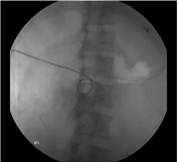
Fig. 3. Fluoroscopic finding. An 8-Fr pigtail catheter was inserted through the right transhepatic access to the duodenal submucosal hematoma.
Fig. 4. Six days after catheter insertion, the follow-up abdominal computed tomography showed partial regression of the intramural hematoma and mild improvement of the common bile duct obstruction. A marked increase in loculated fluid collection was shown in the right abdomen with fistula formation leading to the retroperitoneum from the duodenal submucosal hematoma (black arrows, hematoma fluid loculation;
arrowheads, fistula; white arrows, common bile duct). (A) Axial image. (B) Coronal image.
venous proton pump inhibitor, continuous nasogastric suction, and total parenteral nutrition. For ESRD, the patient was ad- ministered heparin-free HD thrice weekly.
The next day, the patient developed fever (38.5℃). His pulse
rate, respiratory rate, and blood pressure were 114/min, 18/min, and 90/60 mmHg, respectively. His white blood cell count increased to 16,720/mm3, and his CRP level was ele- vated to 19.87 mg/dL. Serum total bilirubin and serum amy- lase levels increased to 13.85 mg/dL and 511 IU/L, respectively. Sepsis was diagnosed due to the systemic in- flammatory response syndrome and elevated infection markers. Empirical treatment with intravenous cefoper- azone-sulbactam and metronidazole was started after two blood cultures. After fluid resuscitation, we attempted fluoro- scopy-guided drainage catheter insertion because the patient was in biliary sepsis. An 8-Fr pigtail catheter was inserted through the right transhepatic access under a fluoroscopy guide and 170 mL of old blood was drained (Fig. 3). Blood culture and aspirate culture showed no growth. Six days after catheter insertion, follow-up abdominal CT scans revealed a decrease in the size of the intramural hematoma with an im- provement of the common bile duct obstruction. However, ocu- lated fluid collection was markedly increased in the right abdo- men due to fistula formation leading to the retroperitoneum from the duodenal submucosal hematoma (Fig. 4).
Total parenteral nutrition was discontinued on the 24th day
A B
Fig. 6. Esophago-gastro-duodenoscopy at 8 weeks revealed the healed duodenal ulcer without any evidence of hematoma.
Fig. 5. After 4 weeks of catheter drainage, the follow-up abdominal computed tomography showed near resorption of the hematoma in the submucosal area of the duodenum and decreased loculated fluid collection in the right abdomen (black arrow, resolved intramural hematoma;
white arrows, decreased loculated fluid). (A) Axial image. (B) Coronal image.
and the patient was slowly and carefully given liquids and then a soft meal. After 4 weeks of catheter drainage, follow-up abdominal CT findings showed progressive resolution of the hematoma (Fig. 5). When the amount of daily drainage was less than 5 mL and serum total bilirubin level was decreased to 4.76 mg/dL, the drainage catheter was removed. On the follow-up at 8 weeks, a repeat EGD revealed a healed duode- nal ulcer with no evidence of hematoma (Fig. 6).
DISCUSSION
Intramural hematoma of the duodenum is a rare complica- tion of endoscopic treatment. It is typically known to occur secondary to blunt abdominal trauma in children and young adults.1,2 However, this condition can occur in adults with un- derlying diseases that are susceptible to bleeding (e.g., liver cirrhosis, ESRD, leukemia, and any disease requiring anti-co- agulation treatment), even with minimal trauma such as an endoscopic procedure.2-5 Intramural hematomas are also re- ported in healthy patients, without risk factors, after endo- scopic biopsies.7 In our case, the coagulation parameters and the platelet count were within the reference range. However,
he had been on HD for a long time and the occurrence of hematoma in ESRD patients is well known.
In previously reported cases of intramural hematoma, most patients showed hemoperitoneum, pancreatitis, and partial or complete bowel obstruction with hematoma.1,2,4-8 Our case is rarer because the common bile duct obstruction occurred secondary to the large intramural duodenal hematoma. The development of fever, leukocytosis, and hyperbilirubinemia af-
A B
ter repeated endoscopic procedures suggested the presence of biliary sepsis due to complications associated with the intra- mural hematoma. However, the patient had been diagnosed with ESRD and was receiving HD. And he had undergone a surgery for a large bowel perforation a year ago. It was there- fore difficult to suggest surgical intervention for intramural hematoma removal. In addition, this patient had biliary sepsis caused by the common bile duct obstruction with jaundice, which also increased the risks of surgical intervention.
Therefore, we tried hematoma decompression through fluoro- scopy-guided drainage catheter insertion despite the risk of catheter bleeding. The common bile duct obstruction was re- solved as the intramural hematoma reduced in size.
The mechanism of hematoma formation due to endoscopic intervention is not yet clear. However, various factors involving iatrogenic trauma (e.g., needle, scope, instrument), as well as underlying conditions, may contribute to its development.
First, rupture of the arterial blood vessel by needle injection may result in the subsequent formation of a hematoma in predisposed individuals because the gastro-duodenal wall is fixed by the retroperitoneal location and the rich submucosal vascular supply.8 Second, it is possible that the mucosa of the gastrointestinal tract wall may have been excessively pulled up by forceps during the endoscopic treatment proce- dure, which might damage the submucosal blood vessels.
Zinelis et al.7 suggested that a significant amount of mucosal tissue can be pulled out and away from the fixed wall of the mucosa underneath, with more than 3 cm from the endo- scope tip during the biopsy forceps procedure. In our case, we did not use biopsy forceps; however, we presume that the hematoma occurred due to injection during the endo- scopic hemostasis.
The diagnosis of intramural duodenal hematoma is based on symptoms of bowel obstruction and a decrease in hemo- globin level without overt signs of bleeding. An enhanced ab- dominal CT serves as the best imaging tool for the diagnosis.9 In principle, the management of this condition is typically con- servative treatments. Because of the rich blood supply of the duodenal wall, the hematoma is anticipated to be absorbed with time. Conservative treatments include total parenteral nutrition and electrolyte replacement, nasogastric tube de- compression and careful observation.
On the other hand, surgical treatment may be considered when there is a septic condition, with obstruction secondary
to the hematoma, such as the current case. In addition, ag- gressive treatment should be considered when there is no evidence of partial resolution after conservative treatment, or in cases of perforation or peritonitis, which increases the size of the hematoma.7,8 Recently, several new therapeutic strategies have been reported. Kwon et al.6 reported a case of intramural hematoma of the duodenum, which was treated with percutaneous drainage and embolization of the bleeding focus, similar to our case. They also recommended endo- scopic incision and drainage for treatment.8 Aizawa et al.10 reported that a patient with a duodenal hematoma was treat- ed with ultrasound-guided drainage and balloon dilatation.
In conclusion, an intramural duodenal hematoma can oc- cur following a common endoscopic treatment in patients sus- ceptible to bleeding. Therefore, the possibility of an intramural hematoma should be considered in any patient with symp- toms such as gastrointestinal obstruction or pancreatitis, ab- dominal pain, nausea, vomiting, and jaundice following an en- doscopic treatment. Noninvasive diagnostic tools such as EGD or abdominal CT scans may help in early diagnosis of intra- mural hematomas. We suggest that fluoroscopy-guided drain- age may also play a role in the treatment of intramural hema- tomas, if conservative treatment is limited.
REFERENCES
1. Rohrer B, Schreiner J, Lehnert P, Waldner H, Heldwein W.
Gastrointestinal intramural hematoma, a complication of endo- scopic injection methods for bleeding peptic ulcers: a case series. Endoscopy 1994;26:617-621.
2. Dhawan V, Mohamed A, Fedorak RN. Gastric intramural hema- toma: a case report and literature review. Can J Gastroenterol 2009;23:19-22.
3. Laine L, McQuaid KR. Endoscopic therapy for bleeding ulcers: an evidence-based approach based on meta-analyses of random- ized controlled trials. Clin Gastroenterol Hepatol 2009;7:33-47;
quiz 1-2.
4. Chung S, Park CW, Chung HW, Shin SJ, Chang YS. Intramural duo- denal hematoma and hemoperitoneum after endoscopic treat- ment in a patient with chronic renal failure on hemodialysis: a case report. Cases J 2009;2:9083.
5. Sugai K, Kajiwara E, Mochizuki Y, et al. Intramural duodenal hem- atoma after endoscopic therapy for a bleeding duodenal ulcer in a patient with liver cirrhosis. Intern Med 2005;44:954-957.
6. Kwon CI, Choi KH, Ko EH, et al. A case of duodenal intramural hematoma treated by percutaneous external drainage. Korean J Gastroenterol 2007;49:45-49.
7. Zinelis SA, Hershenson LM, Ennis MF, Boller M, Ismail-Beigi F.
Intramural duodenal hematoma following upper gastro- intestinal endoscopic biopsy. Dig Dis Sci 1989;34:289-291.
8. Kwon CI, Ko KH, Kim HY, et al. Bowel obstruction caused by an intramural duodenal hematoma: a case report of endoscopic in- cision and drainage. J Korean Med Sci 2009;24:179-183.
9. Plojoux O, Hauser H, Wettstein P. Computed tomography of intra- mural hematoma of the small intestine: a report of 3 cases.
Radiology 1982;144:559-561.
10. Aizawa K, Tokuyama H, Yonezawa T, et al. A case of traumatic in- tramural hematoma of the duodenum effectively treated with ul- trasonically guided aspiration drainage and endoscopic balloon catheter dilation. Gastroenterol Jpn 1991;26:218-223.