INTRODUCTION
Phlebosclerotic colitis, a rare disease entity of ischemic coli- tis, is known to be characterized by the thickening of the colonic wall due to fibrotic sclerosis of the colono-mesenter- ic vein and fibrous degeneration of the submucosal layer of involved colon, resulting in the disturbed venous return from the colon (1, 2). The pathophysiologic mechanism of this dis- ease remains unclear although several reports have suggest- ed that it might be related to portal hypertension secondary to liver cirrhosis or other chronic liver diseases (2, 3). The diag- nosis is usually achieved to be based on a combination of radi- ologic, endoscopic, and histologic findings along with clini- cal manifestations. There are several cases about this disease entity those have been reported in authorized medical jour- nals around the world but rare in Korea (4-11). Here we report the first case of phlebosclerotic colitis confirmed surgically in a 61-yr-old cirrhotic patient with portal hypertension in Korea.
CASE REPORT
A 61-yr-old man was admitted to hospital because of right lower quadrant abdominal pain and bloody stool for one month.
He had a medical history of regular follow-up for alcoholic liver cirrhosis since four years ago. He had experienced the
endoscopic histoacryl injection sclerotherapy for gastric varix with bleeding eight months ago. At the time of admission, physical examination revealed tenderness of the right lower abdomen but no rebound tenderness. Laboratory tests showed a white blood cell count of 2,990/μL, hemoglobin of 11.6 g/
dL, platelet count of 119,000/μL, but there were no other ab- normal findings in the blood chemistry with liver tests, elec- trolytes, and prothrombin time. Tumor markers such as alpha- fetoprotein, carcinoembryogenic antigen (CEA), and CA19- 9 were within normal limit.
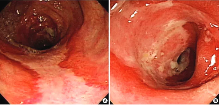
A colonoscopy revealed multiple, geographic, and circum- ferential ulcerations in the distal ascending and transverse colon and could not get through further proximal portion of ascend- ing colon due to luminal narrowing with fibrotic stenosis (Fig. 1A). Endoscopic biopsy showed ulcerative inflamma- tion without malignant cells. The tests for cytomegalovirus and tuberculosis in biopsy specimens were negative. A fol- low-up colonoscopy performed one month later showed still remained multiple ulcerations in the transverse colon. The ascending colon was not observed because of no more further advance of endoscopy due to marked stenosis (Fig. 1B).
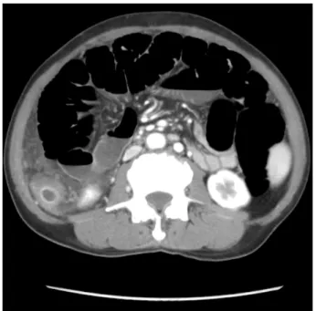
Abdominal computed tomography (CT) demonstrated cir- rhotic liver with splenomegaly and multiple porto-systemic venous collaterals. The proximal to mid ascending colon were thick-walled with increased density in the surrounding fatty tissue and diffuse vascular engorgement (Fig. 2A). No defi- nite calcification in the thickened wall and vessels was iden-
1195
Division of Hepatology, Department of Internal Medicine, Dankook University College of Medicine, Cheonan, Korea
Address for correspondence Il Han Song, M.D.
Division of Hepatology, Department of Internal Medicine, Dankook University College of Medicine, 16-5 Anseo-dong, Cheonan 330-715, Korea Tel : +82.41-550-3924, Fax : +82.41-556-3256 E-mail : [email protected]
disturbance of venous return from the colon. The pathogenic mechanism of this entity remains unknown but chronic liver disease with portal hypertension is maybe thought to be one of the speculated mechanisms. Here we first report the case of surgically confirmed phlebosclerotic colitis, that was in the early stage but showed the aggressive nature, in a 61-yr-old cirrhotic patients with portal hypertension in Korea.
Key Words : Colitis, Ischemic; Liver Cirrhosis; Hypertension, Portal; Sclerosis
Received : 30 January 2009 Accepted : 31 March 2009
tified. Virtual CT colonography showed marked luminal nar- rowing with shortening of ascending colon (Fig. 2B).
Superior mesenteric angiography revealed no occlusion of main branch at arterial phase, but indicated venous pooling along the ascending colon at the delayed phase (Fig. 3).
The right lower abdominal pain of the patient was progres- sively worsening even though the conservative treatments
with total parenteral nutrition for 10 days after admission.
His abdomen was markedly distended and showed distinct tenderness with rebound tenderness. Finally, the marked dila- tation of small bowel caused by colonic obstruction was ob- served on CT scan (Fig. 4) and the passage of stool was not identified.
A right hemicolectomy was performed because of the colonic
Fig. 1. A colonoscopy revealed multiple circumferential ulcerations in the distal ascending and transverse colon (A), and the follow-up colonoscopy performed one month later showed still remained multiple ulcerations in the transverse colon and the scope could not get through the further proximal portion of ascending colon due to stenosis and fibrosis (B).
A B
Fig. 2. Abdominal CT disclosed thickening of the wall of the ascending colon with surrounding fatty infiltration and diffuse vascular engorge- ment (A). Virtual CT colonography showed marked luminal narrowing with shortening of ascending colon (B).
A B
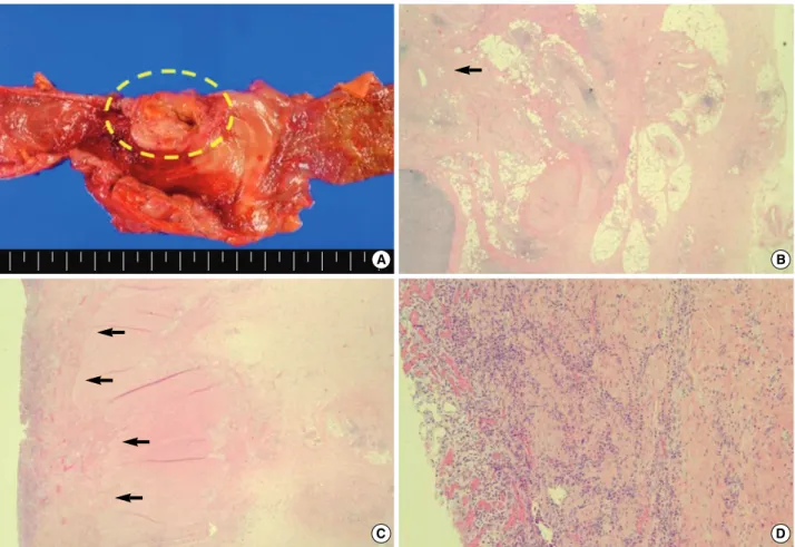
obstruction. Gross findings of operation specimen showed approximately 7×5 cm sized ulcerating mass-like wall thick- ening of the cecum and the proximal ascending colon (Fig.
5A). Microscopic examination showed chronic ulcerative in- flammation, fibrous degeneration of the submucosal layer, and thickening of the venous wall in the mesentery with no evidence of malignancy (Fig. 5B-D).
On the basis of these radiologic, endoscopic, and histologic
findings mentioned above, we diagnosed as a phlebosclerotic colitis occurred during regular follow-up of cirrhotic patients with portal hypertension. Until now, eight months after right hemicolectomy, the patient is being followed in the status of compensated liver cirrhosis without any gastrointestinal symptoms.
DISCUSSION
Phlebosclerotic colitis, first coined by Yao et al. in 2000 (6), is a rare type of ischemic colitis and has different clinicopatho- logic presentations from the classic type of ischemic colitis.
While ischemic colitis is usually caused by insufficient per- fusion of mesenteric arterial blood flow and involves left side colon, especially hepatic flexure or sigmoid regions where are known to be watershed area within the colonic blood supply, phlebosclerotic colitis occurs when sclerotic obstruction of veins in the colonic wall and adjacent mesentery produces chronic congestion of affected colon, and involves the right hemicolon preferentially (5-8). The present case also shows the typical manifestations of phlebosclerotic colitis such as fibrous thickening of affected colono-mesenteric vein and wall thickening of right hemicolon. The pathogenesis of this dis- ease still remains unknown, but there are several clinical con- ditions, diabetes mellitus, hyperlipidemia, cardiac disease, angiitis, CREST syndrome, Churg-Strauss syndrome, lym- phocytic phlebitis and portal hypertension, those might be related to phlebosclerotic colitis (6, 12-15). In the presented case, underlying disease of the patient was alcoholic liver cir- rhosis with obvious portal hypertension. In Korea, to our
Fig. 3. Superior mesenteric angiography revealed no occlusion of main branch at arterial phase (A), and indicated venous pooling (arrows) along the ascending colon at the delayed phase (B).
A B
Fig. 4. Abdominal CT showed markedly dilated small bowel and a portion of colonic wall thickening.
knowledge, a similar case has been reported previously as a term of idiopathic mesenteric phlebosclerosis (11), first des- cribed by Iwashita et al. in 2003 (12), known to be a synonym of phlebosclerotic colitis.
Clinical symptoms include abdominal pain and bloody diarrhea, as our case presented, nausea, vomiting, constipa- tion and ileus, and depend on the severity of the disease (16).
Although this disease usually has long duration of illness in- dicating chronic nature of the disease, progression to mechan- ical ileus resulting from intestinal obstruction is not common.
Surgical intervention, as we did in our case, is not usually necessary unless intestinal obstruction, perforation or mas- sive bleeding occur as postischemic complications (8, 12).
Colonoscopy usually reveals dark purple and edematous mucosa, erosion, ulceration and luminal narrowing. Saito et al. (4) endoscopically observed a patient of phlebosclerotic colitis for three years and considered deep circumferential ulceration on colonoscopy as the aggressive nature of this dis- ease. In our case, the first colonoscopy showed multiple cir- cumferential ulcerations and luminal narrowing, and could not get through the lesion due to stenosis and fibrosis, which
have been worsening in the follow-up colonoscopic finding one month later. We believe these findings suggest as the aggressive form of phlebosclerotic colitis.
The imaging findings of phlebosclerotic colitis are as fol- low (1, 4-6): multiple thread-like or linear calcifications main- ly along the right hemicolon on plain radiographs of abdomen;
thickening of the affected colonic wall with calcification of intramural and mesenteric veins on CT scans; haustral dis- appearance, luminal irregularities, rigidity, narrowing, and thumb-printing on barium enema; and venous pooling and obstruction of the affected colonic wall with dilated collater- al flow on angiography (1, 5). In the present case, contrast- enhanced dynamic CT, virtual CT colonography and superi- or mesenteric angiography also showed typical radiologic fea- tures of phlebosclerotic colitis those were described above, except calcification of involved colonic and adjacent mesen- teric veins.
Histopathologic characteristics include, macroscopically, dark brown appearance of the right hemicolon and thicken- ing of the colonic wall, and microscopically, thickening of the venous walls and tortuous veins, fibrotic degeneration of
Fig. 5. Macroscopic examination showed a huge ulcerating mass-like lesion (dotted circle) at the cecum and the proximal ascending colon measuring about 7×5 cm with colonic wall thickening (A). Microscopic examination showed fibrous thickening (arrow) of the mesenteric vein (B) (H&E, ×200), (C) prominent neurovascular bundles and eosinophilic inflammation in submucosa (arrows) (H&E, ×40), and (D) chronic ulcerative inflammation (H&E, ×100) with no evidence of malignancy.
B
C D
A
bosclerotic colitis, that was in the early stage but showed the aggressive nature, in cirrhotic patients with portal hyperten- sion in Korea.
REFERENCES
1. Tamamoto F, Ishizaki H, Maehara T. Phlebosclerotic colitis. Radiat Med 2008; 26: 164-7.
2. Kusanagi M, Matsui O, Kawashima H, Gabata T, Ida M, Abo H, Isse K. Phlebosclerotic colitis: imaging-pathologic correlation. AJR Am J Roentgenol 2005; 185: 441-7.
3. Kimura Y, Kashima K, Daa T, Tou Y, Hanzawa K, Nakayama I, Yokoyama S. Phlebosclerotic colitis coincident with carcinoma in adenoma. Pathol Int 2003; 53: 721-5.
4. Saito Y, Taniguchi M, Tagawa K, Ibukuro K, Mori M, Emura F.
Phlebosclerotic colitis with deep circumferential ulceration: three- year endoscopic follow-up. Report of a case. Dis Colon Rectum 2005;
48: 2347-51.
5. Oshitani N, Matsumura Y, Kono M, Tamori A, Higuchi K, Matsumo- to T, Seki S, Arakawa T. Asymptomatic chronic intestinal ischemia caused by idiopathic phlebosclerosis of mesenteric vein. Dig Dis Sci 2002; 47: 2711-4.
Education and imaging. Gastrointestinal: phlebosclerotic colitis. J Gastroenterol Hepatol 2008; 23: 670.
11. Jung HG, Koh JW, Lee MY. A case of idiopathic mesenteric phle- bosclerosis. Korean J Gastroentrol 2008; 52: 261-4.
12. Iwashita A, Yao T, Schlemper RJ, Kuwano Y, Yao T, Iida M, Mat- sumoto T, Kikuchi M. Mesenteric phlebosclerosis: a new disease entity causing ischemic colitis. Dis Colon Rectum 2003; 46: 209-20.
13. Ikehata A, Hiwatashi N, Kawarada H, Yamazaki H, Ito K, Kinouchi Y, Kimura M, Noguchi M, Maekawa H, Suzuki H, Shimada T, Yam- agata J, Toyota T. Chronic ischemic colitis associated with marked calcifications of the mesenteric vessels - report of two cases. Dig En- dosc 1994; 6: 355-64.
14. Modigliani R, Muschart JM, Galian A, Clauvel JP, Piel-Desruisseaux JL. Allergic granulomatous vasculitis (Churg-Strauss Syndrome).
Report of a case with widespread digestive involvement. Dig Dis Sci 1981; 26: 264-70.
15. Saraga EP, Costa J. Idiopathic entero-colic lymphocytic phlebitis. A cause of ischemic intestinal necrosis. Am J Surg Pathol 1989; 13:
303-8.
16. Yoshinaga S, Harada N, Araki Y, Kubo H, Nawata H. Hotokezaka M, Nishi H, Chijiiwa Y, Nagase S. Chronic ischemic colonic lesion caused by phlebosclerosis: a case report. Gastrointest Endosc 2001;
53: 107-11.