Serum Leptin and Adiponectin Levels in Korean Patients with Psoriasis
Psoriasis is a disorder caused by genetic and immunological factors. Leptin, a peptide hormone secreted predominantly from adipose tissue, regulates energy intake and expenditure, as well as the T-helper response. There have been conflicting reports regarding serum levels of leptin and adiponectin in patients with psoriasis. In the present study, we measured serum levels of leptin and adiponectin in Korean patients with psoriasis. Twenty- four patients with psoriasis and fifteen control subjects were included in the study. Serum leptin and adiponectin levels were determined by an immunometric sandwich enzyme- linked immunosorbent assay (ELISA). The mean serum leptin concentration in patients with psoriasis was higher than in controls, and the difference was statistically significant. In contrast, serum adiponectin levels in patients with psoriasis were significantly decreased compared with healthy controls. Leptin levels in vitamin D-deficient patients were statistically significantly higher than in vitamin D-sufficient patients. Serum adiponectin concentrations showed a negative correlation with body mass index (BMI) and psoriasis area and severity index (PASI) in patients with psoriasis. In conclusion, the present study demonstrated that leptin and adiponectin may play a role in the immunopathogenesis of psoriasis and may be useful biomarkers indicating severity of psoriasis in Korean patients.
Keywords: Adiponectin; Leptin; Metabolic Syndrome; Psoriasis Yu Jin Oh,1 Hee Kyeong Lim,1
Jeong Hwee Choi,1 Jin Woo Lee,2 and Nack In Kim1
1Department of Dermatology, School of Medicine, Kyung Hee University, Seoul; 2Medical Science Research Institute, Kyung Hee University Medical Center, Seoul, Korea
Received: 11 June 2013 Accepted: 20 March 2014 Address for Correspondence:
Nack In Kim, MD
Department of Dermatology, School of Medicine, Kyung Hee University, 23 Kyungheedae-ro, Dongdaemun-gu, Seoul 130-872, Korea
Tel: +82.2-958-8511, Fax: +82.2-969-6538 E-mail: [email protected]
http://dx.doi.org/10.3346/jkms.2014.29.5.729 • J Korean Med Sci 2014; 29: 729-734
INTRODUCTION
Psoriasis is a chronic inflammatory skin disorder that is mediat- ed by elements of the innate and adaptive immune systems (1).
Although the influence of environmental factors on psoriasis is not precisely defined, body mass index (BMI) has been report- ed to be one of the important associated factors (2).
Leptin, a protein secreted by adipose tissue, plays important roles in metabolism and immunity. It regulates body weight and exerts other biologic functions that modulate hematopoie- sis, angiogenesis, and immune responses (3). Leptin is also in- volved in inflammatory processes involving T cells and has been reported to modulate T-helper cell activity in the cellular immune response. Hence, leptin has three roles in inflamma- tion: it activates monocytes and macrophages; potentiates the production of proinflammatory cytokines such as TNF-α, IL-6, and IL-9; and directs T-cell differentiation to the Th1 phenotype (4). Additionally, leptin has been shown to have stimulatory roles in keratinocyte proliferation, the expression of adhesion molecules, and angiogenesis (5, 6).
Adiponectin is another adipocyte-specific secretory protein that is abundant in circulation. Serum levels of adiponectin are decreased in obesity, insulin resistance, type 2 diabetes mellitus (DM), and hypoadiponectinemia, which are diseases thought to be closely associated with the metabolic syndrome (5).
In consideration of metabolic disorders seen in patients with psoriasis, it seems that there could be some abnormalities in se- rum leptin and adiponectin levels in psoriatic patients. Howev- er, the results regarding serum levels of leptin and adiponectin in patients with psoriasis have been conflicting (6-15). Here, we measured the serum levels of leptin and adiponectin in Korean patients with psoriasis to investigate their roles in inflammatory and metabolic aspects.
MATERIALS AND METHODS Patients and controls
Twenty-four patients with a clinical diagnosis and histopatho- logical confirmation of psoriasis vulgaris were selected from outpatients of the Department of Dermatology at Kyung Hee University Hospital from March to August 2012. These patients were recruited prospectively and consecutively. There were no exclusion criteria.
The clinical characteristics of patients (including age, gender, height, body weight, duration of disease, Psoriasis Area and Se- verity Index (PASI) at enrollment, presence of nail involvement, familial history of psoriasis, and psoriatic arthritis) were collect- ed, along with comorbidities such as DM, hypertension, dyslip- idemia (including hypercholesterolemia, hypertriglyceridemia, and reduced high density lipoprotein cholesterol concentra-
tion), and other cardiovascular diseases such as heart failure, ischemic heart disease, and cerebral ischemia incidents. Serum levels of 25-hydroxyvitamin D (25[OH]D), parathyroid hormone (PTH), and calcium were also measured. Fifteen healthy, age- and sex-matched controls without psoriasis were enrolled as controls.
Serum leptin and adiponectin analysis
Serum levels of leptin and adiponectin were determined by an immunometric sandwich enzyme-linked immunosorbent as- say (ELISA) using commercially-available kits (Human Leptin Quantikine Elisa Kit, product code DLP00, and Human Total Adiponectin Quantikine ELISA Kit, product code DRP 300; R&D Systems, Minneapolis, MN, USA). The minimum detectable dose (MDD) of leptin is typically less than 7.8 pg/mL, and that of adiponectin is 0.891 ng/mL, allowing for sensitive and specif- ic analyses of leptin and adiponectin in serum. Venous blood samples were drawn from the participants between 09:00 and 11:00 hr following a 12-hr fasting period. Following centrifuga- tion of the blood samples at 1,500 g for 15 min, serum was col- lected and kept at -80°C until use. Serum samples were diluted, and the immunoassays were performed according to the man- ufacturer’s instructions in duplicate.
Statistical analysis
The demographic characteristics of cases and controls were compared using the Mann-Whitney and χ2 tests. Differences in serum leptin/adiponectin levels were compared using the non- parametric Mann-Whitney test. Correlations between serum leptin/adiponectin levels and age, BMI, and PASI were com- pared using Spearman’s correlation analysis. Nonparametric Mann-Whitney tests were performed to evaluate the associa- tion of serum leptin/adiponectin levels and clinical features of patients with psoriasis (presence of familial history of psoriasis, nail involvement, psoriatic arthritis, or vitamin D deficiency).
Data were analyzed using commercially-available statistical software (SPSS for Windows, version 17.0; SPSS Inc, Chicago, IL, USA).
Ethics statement
The study protocol conformed to the guidelines of the 1975 Declaration of Helsinki and was approved by the Kyung Hee Medical Hospital institutional review board (KMC IRB 1212- 02). A full verbal explanation of the study was given to all partic- ipants, and the patients who consented to participate in this study on a voluntary basis were subjected. Missing clinical in- formation from patients with psoriasis and control subjects was recorded as censored data.
RESULTS
Demographic differences between patients and controls Twenty-four psoriatic patients took part in our study. The pa- tients included 8 female patients (33.3%) and 16 male patients (66.7%). A group of 15 healthy individuals (9 males [60%] and 6 females [40%]) participated in our study as controls. Descrip- tive data of the patients and controls are shown in Table 1 and 2.
No statistically significant difference was noted in age between psoriatic patients and controls. The mean ± SD BMI in psoriatic patients was higher than that of the controls but the difference was not statistically significant.
The mean ± SD PASI in patients with psoriasis was 14.65 ± 8.67. The presence of nail involvement, familial history of psori- asis, psoriatic arthritis, and vitamin D deficiency were observed in 29.2% (7 patients), 20.8% (5 patients), 50% (12 patients), and 45.8% (11 patients), respectively.
Serum leptin levels in relation to occurrence of psoriasis, patient characteristics, age, BMI, and PASI
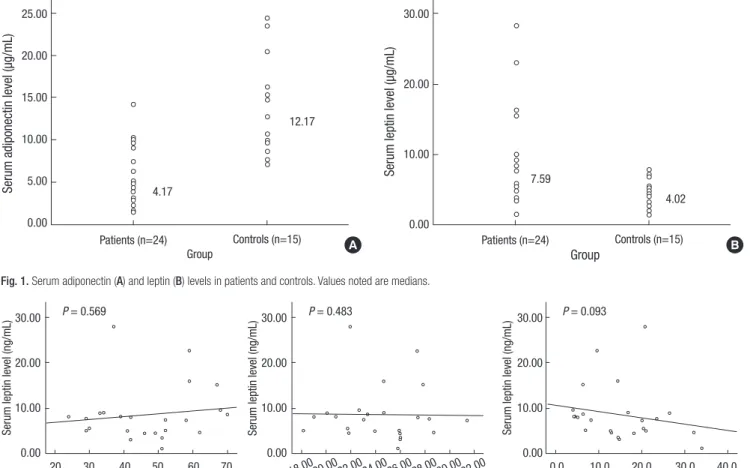
The mean ± SD serum leptin concentration in patients was sta- tistically significantly higher than that in controls (Fig. 1 and Ta- ble 3). Leptin levels of male patients with psoriasis were significantly higher than control males. Also, leptin levels of fe- male patients with psoriasis were significantly higher than con- trol females.
There was no significant difference in leptin levels of patients with a presence of nail involvement, familial history of psoria- sis, or psoriatic arthritis. However, leptin levels in vitamin D-de- ficient patients were statistically significantly higher than those of patients with sufficient vitamin D (Table 4). Serum leptin concentrations showed no correlations with age, BMI, or PASI Table 1. Demographic characteristics of patients (n = 24) and controls (n = 15)
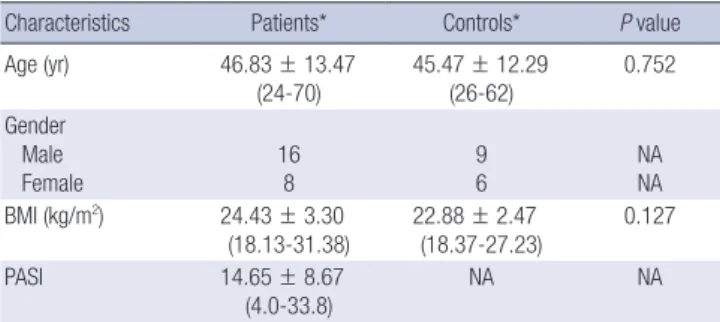
Characteristics Patients* Controls* P value
Age (yr) 46.83 ± 13.47
(24-70) 45.47 ± 12.29
(26-62) 0.752
Gender Male
Female 16
8 9
6 NA
NA BMI (kg/m2) 24.43 ± 3.30
(18.13-31.38)
22.88 ± 2.47 (18.37-27.23)
0.127
PASI 14.65 ± 8.67
(4.0-33.8) NA NA
*Patients and controls are in mean ± SD (range). BMI, body mass index; PASI, psori- asis area and severity index.
Table 2. Additional demographic characteristics of patients (n = 24)
Characteristics No. of patients
Yes No
Nail involvement 7 17
Familial history 5 19
Psoriatic arthritis 12 12
Vitamin D deficiency* 11 13
*Vitamin D deficiency; 25-hydroxy vitamin D < 20 ng/mL.
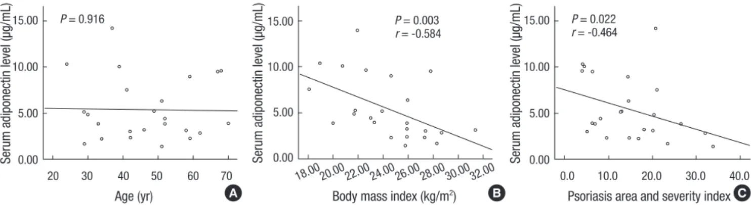
in patients with psoriasis (Fig. 2).
Serum adiponectin levels in relation to occurrence of psoriasis, patient characteristics, age, BMI, and PASI The mean ± SD serum adiponectin concentration in patients was statistically significantly lower than that in controls (Fig. 1 and Table 5). Adiponectin levels of male patients with psoriasis were significantly lower than control males. Also, adiponectin levels of female patients with psoriasis were significantly lower than control females.
There was no significant difference in adiponectin levels of patients with a presence of nail involvement, familial history of psoriasis, psoriatic arthritis, and vitamin D deficiency (Table 6).
Serum adiponectin concentrations showed no correlation with
age in patients with psoriasis. However, serum adiponectin con- centrations showed negative correlations with BMI and PASI in patients with psoriasis (Fig. 3).
DISCUSSION
Recent studies have demonstrated that the risk of psoriasis is di- rectly related to BMI, and that patients with obesity are likely to have more severe psoriasis (2, 7, 16). Psoriasis and obesity share similar mediators of inflammation, mainly TNF-α and IL-6, and the effectors of adipocytic and psoriatic inflammation, largely adipocytes and macrophages, which are derived from a com- Table 3. Serum leptin levels in patients (n = 24) and controls (n = 15) according to
gender
Characteristics Patients* (ng/mL) Controls* (ng/mL) P value
Total 8.65 ± 6.19
(1.16-27.94) 4.17 ± 2.12
(1.38-7.94) 0.001
Male 5.77 ± 2.30
(1.16-9.05)
2.75 ± 1.12 (1.38-4.53)
0.001
Female 14.41 ± 7.58
(7.49-27.94) 6.29 ± 1.26
(4.78-7.94) 0.001
*Patients and Controls are in Mean ± SD (range).
Table 4. Serum leptin levels according to patient characteristics Characteristics Concentrations* (ng/mL)
P value
Yes No
Nail involvement 6.28 ± 3.55
(1.16-15.19) 11.02 ± 7.43
(3.14-27.9) 0.078 Familial history 10.83 ± 8.04
(3.55-22.66)
8.08 ± 5.73 (1.16-27.94)
0.783 Psoriatic arthritis 10.04 ± 6.66
(3.55-22.66) 8.08 ± 6.10
(1.16-27.94) 0.576 Vitamin D deficiency 11.01 ± 7.94
(1.16-27.94) 6.65 ± 3.37
(3.14-15.98) 0.041
*Concentrations are in Mean ± SD (range).
Fig. 1. Serum adiponectin (A) and leptin (B) levels in patients and controls. Values noted are medians.
Serum leptin level (µg/mL)
Group 7.59
4.02 Patients (n=24) Controls (n=15) 30.00
20.00
10.00
0.00
Serum adiponectin level (µg/mL)
Group 4.17
12.17
Patients (n=24) Controls (n=15) 25.00
20.00 15.00 10.00 5.00
0.00
A B
Fig. 2. Correlation between serum leptin levels and age (A), BMI (B), and PASI (C).
P = 0.569
20 30 40 50 60 70 Age (yr)
30.00 20.00 10.00 0.00
Serum leptin level (ng/mL)
P = 0.483
Body mass index (kg/m2) 18.0020.00 22.00 24.00 26.00 28.00 30.0032.00 30.00
20.00 10.00 0.00
Serum leptin level (ng/mL)
P = 0.093
0.0 10.0 20.0 30.0 40.0 Psoriasis area and severity index 30.00
20.00 10.00 0.00
Serum leptin level (ng/mL)
A B C
mon mesothelial origin (17, 18). It has been documented that circulatory levels of TNF-α are significantly increased in obese as compared with non-obese subjects (19, 20). In 2005, Naldi and colleagues reported in a case-controlled study (2) that BMI is one of the risk factors associated with psoriasis. Although the underlying mechanisms may be complex, the “obesity of psori- asis” is thought to be a key link to cardiovascular diseases, in- cluding DM, stroke, heart disease, hypertension, and myocardi- al infarction (6, 21-23).
Adipose tissue is considered to be an important endocrine organ that contributes to the regulation of body metabolism and other vital functions related to inflammation and immune responses (22). It secretes multiple metabolically-active pro- teins termed adipokines. Some well-known adipokines include leptin, resistin, adiponectin, apelin, and visfatin (24).
Leptin hormone, the product of the obese gene, is an adipo- cyte-derived hormone which is a key factor in regulating a wide range of biological responses, including energy homeostasis, immune responses, and inflammatory processes (8). Leptin re- ceptor is expressed primarily in the hypothalamus, but is also expressed in various other tissues, including peripheral blood mononuclear cells, endothelial cells, and fibroblasts (25). Mice with either a leptin (ob ⁄ob) or leptin receptor (db ⁄db) deficien- cy develop severe obesity, diabetes, and impaired cell-mediat- ed immune responses (26, 27). Serum leptin levels are known to show a positive correlation with BMI and marked sexual di- morphism, with levels presenting two to three times higher in women than in men (28). The immunological and proliferative effects of leptin and immunopathogenesis of psoriasis have
many overlapping features.
Adiponectin is a collagen-like protein of 247 amino acids that circulates at relatively high serum concentrations (2–20 μg/mL) and regulates the metabolism of lipids and glucose (29). The mechanism of adiponectin action involves activation of multi- ple signaling pathways, which mediate its anti-inflammatory and anti-atherogenic functions (30). Low levels of adiponectin are associated with adverse metabolic states such as diabetes (22), metabolic syndrome, atherosclerotic cardiovascular dis- ease (31), and psoriasis (9, 10, 32, 33).
The purpose of this current study was to investigate the pos- sible roles of leptin and adiponectin in psoriasis pathogenesis.
We investigated serum leptin and adiponectin levels in patients with psoriasis and controls. The mean serum leptin concentra- tions in patients with psoriasis were statistically significantly higher than those in controls. Circulating leptin levels showed marked sexual dimorphism, which was two to three times higher in females than in males (11, 34). As sex appeared to be an important factor in relation to levels of serum adipokines, all comparisons were made among sex-stratified populations. Se- rum leptin levels in male and female psoriatic patients were significantly higher than those in gender-matched controls. In contrast, serum adiponectin levels in patients with psoriasis were significantly decreased compared with healthy controls.
Serum adiponectin levels in male and female psoriatic patients were significantly lower than those in gender-matched con- trols.
Table 5. Serum adiponectin levels in patients (n = 24) and controls (n = 15) accord- ing to gender
Characteristics Patients* (µg/mL) Controls* (µg/mL) P value
Total 5.42 ± 3.40
(1.43-14.23) 13.58 ± 5.66
(7.07-24.44) 0.000
Male 3.79 ± 1.68
(1.43-7.52)
10.12 ± 2.90 (7.07-14.88)
0.000
Female 8.68 ± 3.70
(2.30-14.23)
18.78 ± 4.71 (12.77-24.44)
0.000
*Patients and Controls are in Mean ± SD (range).
Table 6. Serum adiponectin levels according to patient characteristics Characteristics Concentrations* (µg/mL)
P value
Yes No
Nail involvement 4.55 ± 2.42
(1.43-9.52) 6.29 ± 4.07
(2.30-14.23) 0.41 Familial history 6.35 ± 3.61
(2.30-10.34) 5.18 ± 3.40
(1.43-14.23) 0.489 Psoriatic arthritis 6.88 ± 3.14
(2.30-10.34)
4.82 ± 3.40 (1.43-14.23)
0.099 Vitamin D deficiency 6.12 ± 4.30
(1.43-14.23) 2.43 ± 4.84
(2.36-10.34) 0.955
*Concentrations are in Mean ± SD (range).
P = 0.916
20 30 40 50 60 70 Age (yr)
15.00 10.00 5.00 Serum adiponectin level (µg/mL) 0.00
P = 0.022 r = -0.464
0.0 10.0 20.0 30.0 40.0 Psoriasis area and severity index 15.00
10.00 5.00 Serum adiponectin level (µg/mL) 0.00 P = 0.003
Body mass index (kg/m2) 18.0020.00 22.00 24.00 26.00 28.00 30.0032.00 15.00
10.00 5.00 Serum adiponectin level (µg/mL) 0.00
r = -0.584
Fig. 3. Correlation between serum adiponectin levels and age (A), BMI (B), PASI (C).
A B C
There were no significant differences in leptin levels with re- spect to the presence of nail involvement, familial history, or psoriatic arthritis. However, leptin levels in vitamin D-deficient patients were statistically significantly higher than those in vita- min D-sufficient patients. Several studies have been performed concerning the association between hypovitaminosis D and metabolic syndrome (35). Our results help to explain the link between leptin and the association of hypovitaminosis D with metabolic syndrome.
Serum leptin concentrations showed no correlation with age, BMI, or PASI in patients with psoriasis. Serum adiponectin con- centrations showed no correlation with age in patients with psoriasis. However, serum adiponectin concentrations showed a negative correlation with BMI and PASI in patients with psori- asis. This may be due to the fact that there are multiple factors involved in regulating serum leptin levels.
Several studies concerning the association between leptin and psoriasis showed results similar to ours (6, 8-14). These data support the view that leptin may be involved in the patho- genesis of psoriasis. Çerman et al. showed that serum leptin levels were significantly higher in patients with severe psoriasis than those of patients with BMI-matched mild to moderate psoriasis and controls (8). Serum leptin levels showed a posi- tive correlation with PASI score, suggesting that they might serve as a marker of severity in psoriasis patients (8).
The results of studies by Johnston et al. (7) and Aktan et al.
(15) did not support any possible relationship between serum leptin levels and psoriasis. However, Aktan et al. suggested that further studies investigating severe inflammatory forms of pso- riasis (15) were necessary due to the relatively small number of subjects (20) and relatively low mean PASI (6.2) scores in their patient group.
In conclusion, it was demonstrated that serum leptin con- centrations of patients with psoriasis were significantly higher than those of controls. In contrast, serum adiponectin levels of patients with psoriasis were significantly decreased compared with those of healthy controls. Therefore, leptin and adiponec- tin may play some role in the immunopathogenesis of psoriasis and might be used as biomarkers to assess the severity of psori- asis in Korean patients.
DISCLOSURE
The authors have no conflicts of interest to disclose.
ORCID
Yu Jin Oh http://orcid.org/0000-0001-8248-988X Hee Kyeong Lim http://orcid.org/0000-0002-3974-4571 Jeong Hwee Choi http://orcid.org/0000-0003-0270-9948 Jin Woo Lee http://orcid.org/0000-0003-0390-7954
Nack In Kim http://orcid.org/0000-0002-4810-7013
REFERENCES
1. Gaspari AA. Innate and adaptive immunity and the pathophysiology of psoriasis. J Am Acad Dermatol 2006; 54: S67-80.
2. Naldi L, Chatenoud L, Linder D, Belloni Fortina A, Peserico A, Virgili AR, Bruni PL, Ingordo V, Lo Scocco G, Solaroli C, et al. Cigarette smok- ing, body mass index, and stressful life events as risk factors for psoriasis:
results from an Italian case-control study. J Invest Dermatol 2005; 125:
61-7.
3. Otero M, Lago R, Lago F, Casanueva FF, Dieguez C, Gómez-Reino JJ, Gualillo O. Leptin, from fat to inflammation: old questions and new in- sights. FEBS Lett 2005; 579: 295-301.
4. Shehzad A, Iqbal W, Shehzad O, Lee YS. Adiponectin: regulation of its production and its role in human diseases. Hormones (Athens) 2012; 11:
8-20.
5. Hulthe J, Hultén LM, Fagerberg B. Low adipocyte-derived plasma pro- tein adiponectin concentrations are associated with the metabolic syn- drome and small dense low-density lipoprotein particles: atherosclerosis and insulin resistance study. Metabolism 2003; 52: 1612-4.
6. Saeki H, Shibata S, Tada Y, Karakawa M, Minatani Y, Tamaki K. Psoria- sis arthropathica associated with severe obesity showing high serum leptin level. J Dermatol 2009; 36: 364-6.
7. Johnston A, Arnadottir S, Gudjonsson JE, Aphale A, Sigmarsdottir AA, Gunnarsson SI, Steinsson JT, Elder JT, Valdimarsson H. Obesity in pso- riasis: leptin and resistin as mediators of cutaneous inflammation. Br J Dermatol 2008; 159: 342-50.
8. Herron MD, Hinckley M, Hoffman MS, Papenfuss J, Hansen CB, Callis KP, Krueger GG. Impact of obesity and smoking on psoriasis presenta- tion and management. Arch Dermatol 2005; 141: 1527-34.
9. Choi WJ, Park EJ, Kwon IH, Kim KH, Kim KJ. Association between psori- asis and cardiovascular risk factors in Korean patients. Ann Dermatol 2010; 22: 300-6.
10. Kim GW, Park HJ, Kim HS, Kim SH, Ko HC, Kim BS, Kim MB, Sim EK.
Analysis of cardiovascular risk factors and metabolic syndrome in kore- an patients with psoriasis. Ann Dermatol 2012; 24: 11-5.
11. Neimann AL, Shin DB, Wang X, Margolis DJ, Troxel AB, Gelfand JM.
Prevalence of cardiovascular risk factors in patients with psoriasis. J Am Acad Dermatol 2006; 55: 829-35.
12. Bonifati C, Carducci M, Cordiali Fei P, Trento E, Sacerdoti G, Fazio M, Ameglio F. Correlated increases of tumour necrosis factor-alpha, inter- leukin-6 and granulocyte monocyte-colony stimulating factor levels in suction blister fluids and sera of psoriatic patients: relationships with disease severity. Clin Exp Dermatol 1994; 19: 383-7.
13. Sterry W, Strober BE, Menter A; International Psoriasis Council. Obesity in psoriasis: the metabolic, clinical and therapeutic implications: report of an interdisciplinary conference and review. Br J Dermatol 2007; 157:
649-55.
14. Khabour OF, Wehaibi SH, Al-Azzam SI, Alzoubi KH, El-Akawi ZJ. Asso- ciation of adiponectin with hypertension in type 2 diabetic patients: the gender effect. Clin Exp Hypertens 2013; 35: 361-6.
15. Chen YJ, Wu CY, Shen JL, Chu SY, Chen CK, Chang YT, Chen CM. Pso- riasis independently associated with hyperleptinemia contributing to metabolic syndrome. Arch Dermatol 2008; 144: 1571-5.
16. Conde J, Gomez R, Bianco G, Scotece M, Lear P, Dieguez C, Gomez- Reino J, Lago F, Gualillo O. Expanding the adipokine network in carti- lage: identification and regulation of novel factors in human and mu- rine chondrocytes. Ann Rheum Dis 2011; 70: 551-9.
17. Cerman AA, Bozkurt S, Sav A, Tulunay A, Elbasi MO, Ergun T. Serum leptin levels, skin leptin and leptin receptor expression in psoriasis. Br J Dermatol 2008; 159: 820-6.
18. Tartaglia LA. The leptin receptor. J Biol Chem 1997; 272: 6093-6.
19. Krueger JG. The immunologic basis for the treatment of psoriasis with new biologic agents. J Am Acad Dermatol 2002; 46: 1-23.
20. Tanaka S, Inoue S, Isoda F, Waseda M, Ishihara M, Yamakawa T, Sugi- yama A, Takamura Y, Okuda K. Impaired immunity in obesity: sup- pressed but reversible lymphocyte responsiveness. Int J Obes Relat Metab Disord 1993; 17: 631-6.
21. Huang L, Li C. Leptin: a multifunctional hormone. Cell Res 2000; 10: 81- 92.
22. Dai Y, Pang J, Gong H, Fan W, Zhang TM. Roles and tissue source of adi- ponectin involved in lifestyle modifications. J Gerontol A Biol Sci Med Sci 2013; 68: 117-28.
23. Lee S, Zhang H, Chen J, Dellsperger KC, Hill MA, Zhang C. Adiponectin abates diabetes-induced endothelial dysfunction by suppressing oxida- tive stress, adhesion molecules, and inflammation in type 2 diabetic mice. Am J Physiol Heart Circ Physiol 2012; 303: H106-15.
24. Li L, Wu LL. Adiponectin and interleukin-6 in inflammation-associated disease. Vitam Horm 2012; 90: 375-95.
25. Nakajima H, Nakajima K, Tarutani M, Sano S. Clear association be- tween serum levels of adipokines and T-helper 17-related cytokines in patients with psoriasis. Clin Exp Dermatol 2013; 38: 66-70.
26. Russolillo A, Iervolino S, Peluso R, Lupoli R, Di Minno A, Pappone N, Di Minno MN. Obesity and psoriatic arthritis: from pathogenesis to
clinical outcome and management. Rheumatology (Oxford) 2013; 52:
62-7.
27. Gerdes S, Osadtschy S, Rostami-Yazdi M, Buhles N, Weichenthal M, Mrowietz U. Leptin, adiponectin, visfatin and retinol-binding protein-4 - mediators of comorbidities in patients with psoriasis? Exp Dermatol 2012; 21: 43-7.
28. Takahashi H, Tsuji H, Takahashi I, Hashimoto Y, Ishida-Yamamoto A, Iizuka H. Plasma adiponectin and leptin levels in Japanese patients with psoriasis. Br J Dermatol 2008; 159: 1207-8.
29. Matarese G, Sanna V, Di Giacomo A, Lord GM, Howard JK, Bloom SR, Lechler RI, Fontana S, Zappacosta S. Leptin potentiates experimental autoimmune encephalomyelitis in SJL female mice and confers suscepti- bility to males. Eur J Immunol 2001; 31: 1324-32.
30. Wang Y, Chen J, Zhao Y, Geng L, Song F, Chen HD. Psoriasis is associat- ed with increased levels of serum leptin. Br J Dermatol 2008; 158: 1134-5.
31. Al-Daghri NM, Al-Attas OS, Alokail MS, Alkharfy KM, Al-Othman A, Draz HM, Yakout SM, Al-Saleh Y, Al-Yousef M, Sabico S, et al. Hypovita- minosis D associations with adverse metabolic parameters are accentu- ated in patients with Type 2 diabetes mellitus: a body mass index-inde- pendent role of adiponectin? J Endocrinol Invest 2013; 36: 1-6.
32. Enany B, El Zohiery AK, Elhilaly R, Badr T. Carotid intima-media thick- ness and serum leptin in psoriasis. Herz 2012; 37: 527-33.
33. Abdel Hay RM, Rashed LA. Association between the leptin gene 2548G/
A polymorphism, the plasma leptin and the metabolic syndrome with psoriasis. Exp Dermatol 2011; 20: 715-9.
34. Kaur S, Zilmer K, Leping V, Zilmer M. The levels of adiponectin and leptin and their relation to other markers of cardiovascular risk in pa- tients with psoriasis. J Eur Acad Dermatol Venereol 2011; 25: 1328-33.
35. Aktan S, Rota S, Erdogan B, Ergin S, Kaptanoglu B, Bostanci M. A role of leptin in psoriasis? Turk J Med Sci 2007; 37: 135-8.