The Value of SPECT/CT in Localizing Pain Site and Prediction of Treatment Response in Patients with Chronic Low Back Pain
In many circumstances, causing sites of low back pain (LBP) cannot be determined only by anatomical imaging. Combined functional and morphological imaging such as bone scan with single-photon emission computed tomography/computed tomography (SPECT/CT) may be helpful in identifying active lesions. The purpose of this study was to evaluate the usefulness of bone SPECT/CT in localizing the pain site and the treatment of chronic LBP.
One hundred seventy-five patients suffering from chronic LBP who underwent SPECT/CT were included, retrospectively. All of the patients received multiple general treatments according to the symptoms, and some of them underwent additional target-specific treatment based on SPECT/CT. Numerical rating scale (NRS) pain score was used to assess the pain intensity. Of 175 patients, 127 showed good response to the given therapies, while the rest did not. Overall, 79.4% of patients with definite active lesions showed good response. Patients with mild active or no lesions on SPECT/CT had relatively lower response rate of 63.0%. Good response was observed by the treatment with the guidance of active lesions identified on SPECT/CT. SPECT/CT could be useful in identifying active lesions in patients with chronic LBP and guiding the clinicians to use adequate treatment.
Keywords: Low Back Pain; Tomography, Emission-Computed, Single-Photon;
Tomography, X-Ray Computed; Pain Management; Technetium-99m (Tc-99m), Methylene Diphosphonate (MDP)
Inki Lee,1* Hendra Budiawan,1,2,3* Jee Youn Moon,4* Gi Jeong Cheon,1,5 Yong Chul Kim,4 Jin Chul Paeng,1 Keon Wook Kang,1,5 June-Key Chung,1,5 and Dong Soo Lee1
1Department of Nuclear Medicine, Seoul National University Hospital, Seoul; 2Fellowship of Koh Chang Soon Program, Seoul National University College of Medicine, Seoul, Korea; 3Department of Nuclear Medicine, MochtarRiady Comprehensive Cancer Centre - Siloam Hospitals Semanggi, Jakarta, Indonesia; 4Department of Anesthesiology and Pain Medicine, Seoul National University Hospital, Seoul;
5Cancer Research Institute, Seoul National University, Seoul, Korea
*Inki Lee, Hendra Budiawan, and Jee Youn Moon contributed equally to this work.
Received: 9 May 2014 Accepted: 12 August 2014 Address for Correspondence:
Gi Jeong Cheon, MD
Department of Nuclear Medicine, Seoul National University College of Medicine, 101 Daehak-ro, Jongno-gu, Seoul 110-744, Korea
Tel: +82.2-2072-3386, Fax: +82.2-745-7690 E-mail: [email protected]
Funding: This work was supported by Research Settlement Fund for the new faculty of Seoul National University and a grant of the Korea Health Technology R&D Project through the Korea Health Industry Development Institute (KHIDI), funded by the Ministry of Health & Welfare, Republic of Korea (HI14C1072).
http://dx.doi.org/10.3346/jkms.2014.29.12.1711 • J Korean Med Sci 2014; 29: 1711-1716
INTRODUCTION
Low Back Pain (LBP) is a common health finding causing dis- ability that occurs in most people at some point in their lives. It was estimated that the incidence of a first-ever episode of LBP ranged from 6.3% to 15.4% a year in the general population (1).
LBP persists more than three months is considered as chronic (2).
Identifying the origin of pain site in chronic LBP patients is often not easy. There are myriad reasons causing chronic LBP such as disc herniation and/or disruption, spinal stenosis, mus- cular origin, facet joint pain syndrome, sacroiliac joint disease, and their combinations. Furthermore, in many cases, diagnos- tic evaluation using anatomical images which points out just anatomical degenerative diseases or change are not helpful to manage chronic LBP, since those kinds of changes can also oc-
cur in asymptomatic people (3-5).
A difficulty to find out an origin of chronic LBP has resulted in variable options of its treatment. A strategy to manage chron- ic low back pain was proposed consisting of three categories:
monotherapy, reductionism, and multidisciplinary treatment (6). While reductionism emphasizes target specific treatment based on pathoanatomical diagnosis using variable imaging devices, multidisciplinary treatment usually focuses the treat- ment of chronic LBP on its biopsychosocial rehabilitation (6).
There have been other strategies to alleviate chronic LBP and surgery would not be very beneficial and be avoided in most of these strategies (7).
Functional imaging such as a bone scan with single-photon emission computed tomography (SPECT) offered diagnostic value which was not provided by planar imaging and anatomi- cal imaging (8). Fusion of SPECT with computed tomography Anesthesiology & Pain
(SPECT/CT) increased the specificity of detecting lesions of the lower vertebral column and provided proper guidance to next step (9). Recently, it has been reported that bone scan with SPE- CT/CT was useful for identifying pain-inducing focus in the pa- tients with chronic foot and low back pain (10, 11). The reduc- tionism, which means treatment based on bone SPECT/CT find- ings, will be expected as an effective diagnostic tool in relieving pain (10, 11). In this study, the degree of treatment of low back pain according to the bone SPECT/CT finding was assessed.
We evaluated the usefulness of bone SPECT/CT in localizing the pain site for the treatment of chronic LBP.
MATERIALS AND METHODS Patients
Patients suffering from chronic low back pain and undergoing bone SPECT/CT from the second quarter of 2011 to the third quarter of 2013 were retrospectively included. Among them, multidisciplinary treatment with or without target-specific treat- ments were performed. Follow up was done until 3 months af- ter the date of the first treatment, which was performed not more than 1 month after bone SPECT/CT. Patients who had any oth- er causes of bone or joint abnormalities during or at the end of follow up were excluded.
Numerical rating scale (NRS) pain score of low back pain The intensity of low back pain was evaluated by an 11-pointed numerical rating scale (NRS) pain score with no pain scaled as 0 and worst pain as 10 at pre-treatment and during follow up time. The NRS have well documented its reliability, validity and sensitivity to treatments that are expected to affect pain (12, 13).
Evidence of temporary or permanent decrease of NRS score ≥ 2 during three month follow-up was considered as positive re- sponse to treatment (14, 15).
Treatment of low back pain
Patients were treated with multiple general treatments accord- ing to the symptoms. General treatments included: interven- tional muscle and nerve stimulation (IMNS), trigger point in- jection (TPI), needle transcutaneous electrical nerve stimula- tion (TENS), extracorporeal shock wave treatment (ESWT), medi- cation, exercise, epidural steroid injection (ESI), epidural adhe- siolysis, etc. Target-specific treatments such as facet joint injec- tion, sacroiliac joint injection, medial branch block, and radio- frequency medial branch neurotomy for facet joint lesions were performed according to the patients’ symptom and clinicians’
decision. In case of positive finding of SPECT/CT, the treatment level was decided based on the SPECT/CT results. The treat- ment was performed in accordance with the clinical symptom and physical examination.
Imaging acquisition
Bone planar imaging was done 3 hr after injection of approxi- mately 740-925 MBq (20-25 mCi) of technetium-99m methy- lene diphosphonate (Tc-99m MDP) intravenously followed by SPECT/CT of lumbar spine. SPECT acquisition was performed using low-energy high resolution collimator with 360° orbit, 120 steps (20 sec/step), and 128 × 128 matrix, while CT part of SPECT/
CT used 16-detector row helical CT with 100 kV, 40 mAs, 1 sec gantry rotation, and reconstructed section thickness of 2.5 mm.
Image interpretation and quantification
Locations/compartments of increased uptake were defined in 4 groups: facet joint, sacroiliac joint, combination, and no ac- tive lesion. The uptake positivity was scored by using 3-point scale (no uptake, mild increased uptake and definite positive uptake). The number of lesions in the same compartment and other compartments was also evaluated.
Statistical analyses
Positivity and intensity of uptake and gender were compared between responsive and non-responsive groups by chi-square test. Age, duration of disease, initial NRS, decreased NRS, num- ber of positive lesions, compartment were compared by Mann- Whitney test. Logistic regression analysis was performed to find out which parameters were significant in determining response.
A value of P < 0.05 was considered statistically significant. All statistical calculation was performed using SPSS software, re- lease 18 (Chicago, IL, USA).
Ethics statement
The study design and exemption of informed consent were ap- proved by the institutional review board of Seoul National Uni- versity Hospital (IRB No. 1404-149-001). Informed consent was waived by the board.
RESULTS
Patients’ characteristics
A total of 175 patients (74 male and 101 female, mean age of 61.0 ± 12.5 yr) suffering from chronic low back pain were includ- ed after excluding 6 patients with other causes of bone or joint abnormalities such as fibromyalgia (n = 2), myalgia (n = 1), pe- ripheral nervous disease (n = 1), peripheral vascular disease (n = 1), and diabetic polyneuropathy (n = 1). All patients were underwent bone SPECT/CT imaging for the purpose of diagno- sis and treatment decision. One hundred forty-nine patients were found out to have active lesions on bone SPECT/CT, while 26 patients did not. All patients underwent non-target specific treatment (medication, exercise, TPI, IMNS, TENS, ESWT, ESI or combination of them), while 74 patients got 1 or more addi- tional target-specific treatments such as facet joint injection
(n = 47), sacroiliac joint injection (n = 17), medial branch block (n = 49), radiofrequency medial branch neurotomy (n = 9) or combination of them (n = 16) according to the patients’ symp- tom and clinicians’ decision. Bone SPECT/CT revealed active lesions of facet joints in 56 (32.0%), sacroiliac joints in 7 (4.0%), combination of the above findings (mostly with facet joint com- ponent) in 86 (49.1%), and no active lesions in 26 (14.9%) pa- tients, respectively. Detail characteristics are summarized in Table 1.
Overall response to pain relief treatment
Positive response to treatment was observed in 127 of total 175 patients (72.6%), while non-response to treatment in 48 patients (27.4%). Patients with facet joint lesions showed positive respon- ses in 43 patients of 56 (76.8%). Patients with sacroiliac joint le-
sions showed response in 5 patients of 7 (71.4%). Patients with at least 2 compartments combination lesions showed 72.1% of positive response in 62 patients of 86 (Table 1).
In positive-uptake group including mild or definite positive uptake in SPECT/CT, 73.8% (110 of 149 patients) showed respon- se, while 65.4% (17 of 26 patients) showed response for treatment in no-uptake group (Fig. 1). The response for treatment is slight- ly higher in the positive-uptake group compared to that of the no-uptake group. This did not show statistical significance (P = 0.373). When positive uptake in the SPECT/CT was divided into mild and definite uptake based on tracer intensity, the treatment response of definite uptake group was significantly higher than mild uptake group (63.0% vs. 79.4%, respectively, P = 0.017) (Fig. 1).
Number of active lesions had no role in determining response rate (Table 2). There was no difference in response rate between single and multiple lesions groups (P = 0.809). Response rates among different compartments in which active lesions located (facet joint, sacroiliac joint and combination) were not signifi- cantly different (P = 0.815) (Table 3).
In univariate logistic regression analysis, initial NRS and defi- nite positive uptake were regarded as significant predictors for Table 1. Distribution of patient based on responsiveness to treatment given after bone
SPECT/CT imaging
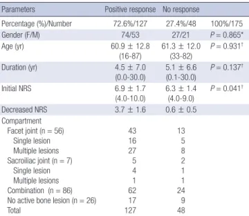
Parameters Positive response No response
Percentage (%)/Number 72.6%/127 27.4%/48 100%/175
Gender (F/M) 74/53 27/21 P = 0.865*
Age (yr) 60.9 ± 12.8
(16-87)
61.3 ± 12.0 (33-82)
P = 0.931†
Duration (yr) 4.5 ± 7.0
(0.0-30.0) 5.1 ± 6.6
(0.1-30.0) P = 0.137†
Initial NRS 6.9 ± 1.7
(4.0-10.0) 6.3 ± 1.4
(4.0-9.0) P = 0.041†
Decreased NRS 3.7 ± 1.6 0.6 ± 0.5
Compartment Facet joint (n = 56) Single lesion Multiple lesions Sacroiliac joint (n = 7) Single lesion Multiple lesions Combination (n = 86) No active bone lesion (n = 26) Total
43 16 27 5 4 1 62 17 127
13 5 8 2 1 1 24 9 48
*Fisher’s exact test; †Mann-Whitney Test. NRS, numerical rating scale.
Fig. 1. Treatment response according to the intensity of lesions in SPECT/CT. The pa- tients with definite uptake in SPECT/CT have better treatment response compared to the patients with no or mild uptake in SPECT/CT. *P = 0.017 (Pearson chi square).
Treatment response (%)
(-) uptake / (+) uptake (-) or mild uptake / definite uptake 100
80 60 40 20 0
*
Table 2. Treatment response according to the number of lesions in SPECT/CT Response
Number of active lesions
Total Single Multiple in single
compartment Multiple in multi- ple compartment Response (-)
Response (+)
6 20
9 28
24 62
39 110 Total
Response percentage (%) 26
76.9 37
75.7 86
72.1 149
P = 0.809*
*Fisher’s exact test (single lesion vs. multiple lesions).
Table 3. Treatment response according to the compartment
Response Compartment
Facet joint Sacroiliac joint Combination Response (-)
Response (+) 13
43 2
5 24
62 Total
Response percentage (%)
56 76.8
7 71.4
86 72.1 Compartment; facet joint, sacroiliac joint and combination (P = 0.815, Fisher’s exact test).
Table 4. Univariate and multivariate logistic regression analysis of treatment response
Variables Univariate Multivariate
P Odds ratio P Odds ratio
Gender 0.810 1.086 0.979
Age 0.859 0.998 0.454
Duration 0.554 0.986 0.506
Uptake 0.018* 2.264* 0.028* 2.477*
Initial NRS 0.025* 1.274* 0.032* 1.264*
Number of lesions 0.424 1.076 0.990
*P values indicate statistically significant differences. NRS, numerical rating scale.
the treatment (P = 0.025, 95% CI, 1.031-1.574 and P = 0.018, 95%
CI, 1.152-4.448 respectively). In multivariate logistic regression analysis, both definite uptake was initial NRS were also regard- ed as significant predictors (P = 0.028, odds ratio, 2.477; 95% CI, 1.103-5.566 and P = 0.032, odds ratio, 1.264; 95% CI, 1.021-1.565 respectively). Other parameters (gender, age, duration of low back pain and number of lesions) were not significant (Table 4).
In no active lesion with response group, the clinical diagnosis after the end of follow up was internal derangement of disc, failed back surgery syndrome, and spinal stenosis of lumbar spine.
While, in no active lesion without response group the clinical diagnosis was spinal stenosis of lumbar spine and facet joint syndrome.
Initial NRS before treatment between responsive and non- responsive group was different (P = 0.041). The score of respon- sive group was higher than that of non-responsive group. No significant difference is observed in NRS before treatment be- tween definite uptake and other subgroup in responsive group (P = 0.901) and non-responsive group (P = 0.635). NRS decrease after treatment between uptake and non-uptake subgroup in both responsive group and non-responsive group were also not significantly different (P = 0.841 and 0.345, respectively) (Table 5).
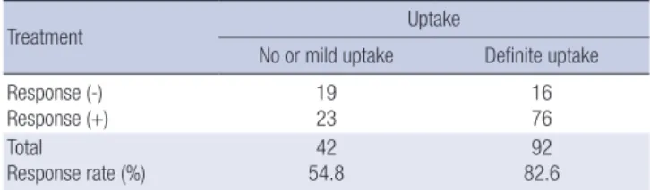
Response to treatment of regional target-specific pain control according to the active sites on bone SPECT/CT The percentage of patients having target specific treatment in the definite positive uptake group was 90.2% (92 patients of to- tal 102 patients). Of 73 patients with no or mild uptake on SPECT/
CT, 44 patients were treated with target specific management (60.3%). Overall, 134 of 175 patients had target specific treat- ment. In the 134 patients with target specific treatment, 99 pa- tients showed positive response for the treatment (73.9%). How- ever 27 of 41 patients (65.9%) having only general treatment also showed positive response. No significant difference was observed between both groups (P = 0.596).
Considering the patients with target specific treatment (134 patients), the group of definite uptake showed 82.6% of positive response (76 of 92 patients). On the contrary, the groups of no or mild uptake showed 54.8% of positive response (23 of 42 pa- tients). These results showed that the treatment response rate of the definite uptake group was significantly higher than that of no or mild uptake group (P = 0.001) (Table 6).
DISCUSSION
In this study, the active lesion on bone SPECT/CT and initial NRS were found as the parameter that can predict outcome of treatment. The patients with definite positive finding on SPECT/
CT had good response for the treatment compared to those with no or mild uptake. The higher the initial NRS, the better outcome for the treatment had the patient. Other parameters such as gen- der, age, duration of back pain, number of active lesions and lo- cation had no role for the disease prognosis.
Patients with definite facet joint uptake on SPECT/CT show- ed good responses. This result is consistent with the previously reported result. Koh et al. reported that 79% of patients with pos- itive facet joint lesions on SPECT/CT had good response to the treatment (16). This finding is already well known. Holder et al.
(17) stated that the real value of bone SPECT was to find out suit- able patients to undergo facet joint injection procedure. This statement was based on the high negative predictive value of bone SPECT (100%), while negative predictive value of planar imaging was 93%. Other studies stated similar findings. Dolan et al. (18) showed that bone SPECT improved the finding of met- abolically active facet joints, directing to local steroid injection and better clinical outcome. McDonald et al. (19) also found that SPECT/CT was useful in showing the localization of active lesions in all 37 patients and facet joint injections were perform- ed based on these findings. These studies suggest that SPECT/
CT finding in patients with low back pain may be a prognostic factor. Our results were in accordance with these findings. The patients with definite uptake in SPECT/CT would have a favor- able prognosis. Hence, the physicians should consider the SPECT/
CT finding for the therapeutic decision of the patient with low back pain.
Patients with sacroiliac (SI) joint lesions also showed good response to treatment in our study. In clinical setting, determin- ing sacroiliac joint as the cause of low back pain is not easy. One of the reasons was the uncertainty of the innervations of the joint (20). Different authors suggested different innervations, such as from sacral dorsal rami (21, 22), lumbosacral nerve root (23), and sacroiliac ligament (24). Murata et al. (25) illustrated sensory nerve fibers supplying the dorsal side originating from the dorsal root ganglions of the lower lumbosacral levels (from L4 to S2), while the ventral side from the dorsal root ganglions Table 5. Numerical rating scale change in patients with low back pain
Groups Initial After treatment
Response group With uptake Without uptake
6.9 ± 1.7 (range: 4-10) 6.9 ± 1.6 (range: 4-10) 6.9 ± 1.6 (range: 4-10)
3.8 ± 1.6 (range: 2-8) 3.8 ± 1.6 (range: 2-8) 3.8 ± 1.7 (range: 2-8) Non-response group
With uptake Without uptake
6.3 ± 1.4 (range: 4-9) 6.4 ± 1.3 (range: 4-8.5) 6.2 ± 1.6 (range: 4-9)
0.5 ± 0.5 (range: 0-1.5) 0.5 ± 0.5 (range: 0-1.5) 0.5 ± 0.5 (range: 0-1.5) P values > 0.05.
Table 6. Treatment response according to the target specific pain control
Treatment Uptake
No or mild uptake Definite uptake Response (-)
Response (+)
19 23
16 76 Total
Response rate (%) 42
54.8 92
82.6 P = 0.001, Pearson chi square.
of the lumbar to sacral levels (from L1 to S2). Erosions and scle- rosis are the findings of radiologic imaging of sacroiliitis due to inflammatory spondyloarthropathy. Inflammatory spondylo- arthropathy should be present for imaging to detect painful SI joint syndrome (20). Slipman et al. (26) reported that abnormal bone scan findings were only found in small number (13%) of patients who were responsive to SI joint injection, despite the high specificity of 100%. Therefore, we suggest that SPECT/CT may be a useful tool for the diagnosis and treatment of patients with sacroiliac joint lesions.
It is well known that active lesion (increased uptake) reflects bone remodeling. Immunohistochemical analysis revealed that osteoblastic activity in facet joints was found in both ankylosing spondylitis and osteoarthritis cases suggesting that bone remo- deling/formation had a role in physiological repair function (27). Ratcliffe et al. (28) also found similar finding that bone re- modeling was proved to be increased in patient with osteoar- thritis. Woo et al. (29) showed that in patients with ankylosing spondylitis, response to treatment with etanercept was followed by increase of bone formation markers (bone alkaline phospha- tase, osteocalcine) and decrease of matrix metalloproteinase-3 (MMP-3), although with unchanged bone resorption marker (C-telopeptide of type-I collagen/CTX). The above studies and this current study showed that the response rate in patients with definite positive uptake was significantly higher than in those of no or mild uptake on bone scan. Thus, we can postu- late that active lesion found on bone scan, which reflects bone remodeling (formation), is more potential to be responsive to treatment than lesion without uptake (30, 31).
In this current study, we proved that bone SPECT/CT was useful in the management of patients with chronic low back pain. In cases with facet joint arthropathy, bone SPECT/CT could be used to guide the clinician to use target specific treatment such as medial branch block. In some cases, bone SPECT/CT showed the abnormality of the involved bone and guide to use other modality to investigate bone and the related tissue (spinal cord, nerve, disc, etc.) in more detail to lead to target specific treatments if needed. For temporary pain control, in many cas- es non-target specific treatments were enough. This may ex- plain the non-different response rates between target specific and non-target specific treatments in all sub-groups, although this may also be caused by the too-low number of patients in each sub-group.
Limitation of this study was the small number of patients suf- fering from other than facet joint arthropathy, as well as no-up- take lesions. The patient groups had heterogeneous lesions in low back and underwent variable combined treatment. This was also limitation of this retrospective study. Further study fo- cusing on each compartment abnormality and each treatment may be needed for validation. The efficacy of the treatment was assessed during the 3 months after the initial treatment. In this
study, long term effect of the treatment was not evaluated. The long term correlation between the SPECT/CT finding and treat- ment is needed.
Bone SPECT/CT is useful in identifying active lesions in pa- tients with chronic low back pain and guiding the clinicians to use target specific treatment of pain control. In cases of active lesions identified on SPECT/CT, good response of pain control can be expected by the guidance of adequate location in target specific treatment. It also guided the use of other modalities to investigate bone and related tissues (spinal cord, nerve, disc, etc) in more detail.
In conclusion, treatment management based on bone SPECT/
CT findings can be considered to be effective on pain control in patients with chronic low back pain.
DISCLOSURE
The authors declare no competing financial interest.
ACKNOWLEGEMENTS
Fellowship of Koh Chang Soon Program is an international train- ing project of Seoul National University College of Medicine in order for the promotion of nuclear medicine practice in devel- oping countries, following the immortal spirit of the late profes- sor Chang Soon Koh; Innovative Thinking and Harmonization.
ORCID
Inki Lee http://orcid.org/0000-0002-2051-3597
Hendra Budiawan http://orcid.org/0000-0002-8645-5916 Jee Youn Moon http://orcid.org/0000-0001-5551-7750 Gi Jeong Cheon http://orcid.org/0000-0002-1360-5186 Yong Chul Kim http://orcid.org/0000-0002-8461-5322 Jin Chul Paeng http://orcid.org/0000-0002-7464-9342 Keon Wook Kang http://orcid.org/0000-0003-2622-9017 June-Key Chung http://orcid.org/0000-0002-6866-8571 Dong Soo Lee http://orcid.org/0000-0001-9013-4835
REFERENCES
1. Hoy D, Brooks P, Blyth F, Buchbinder R. The Epidemiology of low back pain. Best Pract Res Clin Rheumatol 2010; 24: 769-81.
2. Frymoyer JW. Back pain and sciatica. N Engl J Med 1988; 318: 291-300.
3. Powell MC, Wilson M, Szypryt P, Symonds EM, Worthington BS. Preva- lence of lumbar disc degeneration observed by magnetic resonance in symptomless women. Lancet 1986; 2: 1366-7.
4. Jensen MC, Brant-Zawadzki MN, Obuchowski N, Modic MT, Malkasian D, Ross JS. Magnetic resonance imaging of the lumbar spine in people without back pain. N Engl J Med 1994; 331: 69-73.
5. Jarvik JJ, Hollingworth W, Heagerty P, Haynor DR, Deyo RA. The Longi- tudinal Assessment of Imaging and Disability of the Back (LAIDBack)
Study: baseline data. Spine (Phila Pa 1976) 2001; 26: 1158-66.
6. Bogduk N, McGuirk B. Medical management of acute chronic low back pain : an evidence-based approach. Amsterdam; Boston: Elsevier, 2002, p173-174.
7. Last AR, Hulbert K. Chronic low back pain: evaluation and management.
Am Fam Physician 2009; 79: 1067-74.
8. Ryan PJ, Evans PA, Gibson T, Fogelman I. Chronic low back pain: com- parison of bone SPECT with radiography and CT. Radiology 1992; 182:
849-54.
9. Even-Sapir E, Flusser G, Lerman H, Lievshitz G, Metser U. SPECT/mul- tislice low-dose CT: a clinically relevant constituent in the imaging algo- rithm of nononcologic patients referred for bone scintigraphy. J Nucl Med 2007; 48: 319-24.
10. Kretzschmar M, Wiewiorski M, Rasch H, Jacob AL, Bilecen D, Walter MA, Valderrabano V. 99mTc-DPD-SPECT/CT predicts the outcome of imaging-guided diagnostic anaesthetic injections: a prospective cohort study. Eur J Radiol 2011; 80: e410-5.
11. Carstensen MH, Al-Harbi M, Urbain JL, Belhocine TZ. SPECT/CT im- aging of the lumbar spine in chronic low back pain: a case report. Chiro- pr Man Therap 2011; 19: 2.
12. Dworkin RH, Turk DC, Farrar JT, Haythornthwaite JA, Jensen MP, Katz NP, Kerns RD, Stucki G, Allen RR, Bellamy N, et al. Core outcome mea- sures for chronic pain clinical trials: IMMPACT recommendations. Pain 2005; 113: 9-19.
13. Turk DC, Melzack R. Handbook of pain assessment. 3rd ed. New York:
Guilford Press, 2011, p19-44.
14. Manejias EM, Hu J, Tatli Y, Lutz GE. Lumbar zygapophysial joint radio- frequency denervation: a long-term clinical outcome study. HSS J 2008;
4: 180-7.
15. Sloman R, Wruble AW, Rosen G, Rom M. Determination of clinically meaningful levels of pain reduction in patients experiencing acute post- operative pain. Pain Manag Nurs 2006; 7: 153-8.
16. Koh WU, Kim SH, Hwang BY, Choi WJ, Song JG, Suh JH, Leem JG, Shin JW. Value of Bone Scintigraphy and Single Photon Emission Computed Tomography (SPECT) in Lumbar Facet Disease and Prediction of Short- term Outcome of Ultrasound Guided Medial Branch Block with Bone SPECT. Korean J Pain 2011; 24: 81-6.
17. Holder LE, Machin JL, Asdourian PL, Links JM, Sexton CC. Planar and high-resolution SPECT bone imaging in the diagnosis of facet syndrome.
J Nucl Med 1995; 36: 37-44.
18. Dolan AL, Ryan PJ, Arden NK, Stratton R, Wedley JR, Hamann W, Fo-
gelman I, Gibson T. The value of SPECT scans in identifying back pain likely to benefit from facet joint injection. Br J Rheumatol 1996; 35: 1269- 73.
19. McDonald M, Cooper R, Wang MY. Use of computed tomography-sin- gle-photon emission computed tomography fusion for diagnosing pain- ful facet arthropathy. Technical note. Neurosurg Focus 2007; 22: E2.
20. Tuite MJ. Facet joint and sacroiliac joint injection. Semin Roentgenol 2004; 39: 37-51.
21. Grob KR, Neuhuber WL, Kissling RO. [Innervation of the sacroiliac joint of the human]. Z Rheumatol 1995; 54: 117-22.
22. Fortin JD, Kissling RO, O’Connor BL, Vilensky JA. Sacroiliac joint inner- vation and pain. Am J Orthop (Belle Mead NJ) 1999; 28: 687-90.
23. Frymoyer JW, Ducker TB. The Adult spine : principles and practice. New York: Raven Press, 1991, p143-150.
24. Vilensky JA, O’Connor BL, Fortin JD, Merkel GJ, Jimenez AM, Scofield BA, Kleiner JB. Histologic analysis of neural elements in the human sac- roiliac joint. Spine (Phila Pa 1976) 2002; 27: 1202-7.
25. Murata Y, Takahashi K, Yamagata M, Takahashi Y, Shimada Y, Moriya H.
Origin and pathway of sensory nerve fibers to the ventral and dorsal sides of the sacroiliac joint in rats. J Orthop Res 2001; 19: 379-83.
26. Slipman CW, Sterenfeld EB, Chou LH, Herzog R, Vresilovic E. The value of radionuclide imaging in the diagnosis of sacroiliac joint syndrome.
Spine (Phila Pa 1976) 1996; 21: 2251-4.
27. Appel H, Maier R, Loddenkemper C, Kayser R, Meier O, Hempfing A, Sieper J. Immunohistochemical analysis of osteoblasts in zygapophyseal joints of patients with ankylosing spondylitis reveal repair mechanisms similar to osteoarthritis. J Rheumatol 2010; 37: 823-8.
28. Ratcliffe A, Seibel MJ. Biochemical markers of osteoarthritis. Curr Opin Rheumatol 1990; 2: 770-6.
29. Woo JH, Lee HJ, Sung IH, Kim TH. Changes of clinical response and bone biochemical markers in patients with ankylosing spondylitis taking etan- ercept. J Rheumatol 2007; 34: 1753-9.
30. Chun IK, Cheon GJ, Paeng JC, Kang KW, Chung JK, Lee DS. Detection and Characterization of Parathyroid Adenoma/Hyperplasia for Preop- erative Localization: Comparison Between (11)C-Methionine PET/CT and (99m)Tc-Sestamibi Scintigraphy. Nucl Med Mol Imaging 2013; 47:
166-72.
31. Hong CM, Ahn BC, Choi SY, Kim DH, Lee SW, Kwon TG, Lee J. Implica- tions of three-phase bone scintigraphy for the diagnosis of bisphospho- nate-related osteonecrosis of the jaw. Nucl Med Mol Imaging 2012; 46:
162-8.