Corresponding author:
Seung-Ju YangDepartment of Biomedical Laboratory Science, Konyang University, 158 Gwanjeodong-ro, Seo-gu, Daejeon 35365, Korea
E-mail:
[email protected]ORCID:
https://orcid.org/0000-0001-9261-2749ORIGINAL ARTICLE
Metformin or α-Lipoic Acid Attenuate Inflammatory Response and NLRP3 Inflammasome in BV-2 Microglial Cells
Hye-Rim Choi 1 , Ji Sun Ha 1 , In Sik Kim 2 , Seung-Ju Yang 1
1
Department of Biomedical Laboratory Science, Konyang University, Daejeon, Korea
2
Department of Biomedical Laboratory Science, School of Medicine, Eulji University, Daejeon, Korea
BV-2 미세아교세포에서 메트포르민 또는 알파-리포산의 염증반응과 NLRP3 인플라마솜 약화에 관한 연구
최혜림 1 , 하지선 1 , 김인식 2 , 양승주 1
1
건양대학교 임상병리학과,
2을지대학교 임상병리학과
ARTICLE INFO ABSTRACT
Received
August 18, 2020Revised 1
st August 25, 2020Revised 2
nd August 31, 2020Accepted
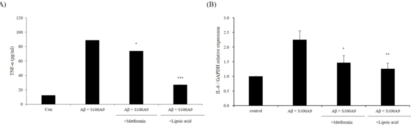
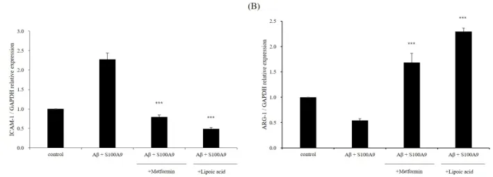
August 31, 2020Alzheimer’s disease (AD) is a chronic and progressive neurodegenerative disease that can be described by the occurrence of dementia due to a decline in cognitive function. The disease is characterized by the formation of extracellular and intracellular amyloid plaques. Amyloid beta (Aβ) is a hallmark of AD, and microglia can be activated in the presence of Aβ. Activated microglia secrete pro-inflammatory cytokines. Furthermore, S100A9 is an important innate immunity pro-inflam- matory contributor in inflammation and a potential contributor to AD. This study examined the effects of metformin and α-LA on the inflammatory response and NLRP3 inflammasome activation in Aβ- and S100A9-induced BV-2 microglial cells. Metformin and α-LA attenuated inflammatory cytokines, such as tumor necrosis factor-α (TNF-α) and interleukin-6 (IL-6). In addition, metformin and α-LA inhibited the phosphorylation of JNK, ERK, and p38. They activated the nuclear factor kappa B (NF-κ B) pathway and the NOD-like receptor pyrin domain containing 3 (NLRP3) inflammasome. Moreover, metformin and α-LA reduced the marker levels of the M1 phenotype, ICAM1, whereas the M2 phenotype, ARG1, was increased. These findings suggest that metformin and α-LA are therapeutic agents against the Aβ- and S100A9-induced neuroinflammatory responses.
Copyright Ⓒ 2020 The Korean Society for Clinical Laboratory Science. All rights reserved.
Key words α-lipoic acid Amyloid beta Metformin
NLRP3 inflammasome S100A9
INTRODUCTION
Alzheimer’s disease (AD) is a neurodegenerative disease, in which the onset and progression of dementia is determined by synapse loss and neuronal death. It is
pathologically characterized by abnormal protein accumulation, including extracellular and intracellular aggregation of amyloid beta (Aβ) protein and hyper- phosphorylated tau protein [1]. Inflammation plays a fundamental role in AD progression since microglia are able to be activated in the presence of Aβ [2]. When microglia are active in AD, they play a dual role. On the one hand, chronically activated microglia contribute to the release of pro-inflammatory cytokines, which initiates a pro-inflammatory cascade and subsequently
Korean Society for Clinical Laboratory Science
![For any α ∈ K and R ∈]0](data:image/gif;base64,R0lGODlhAQABAIAAAP///wAAACH5BAEAAAAALAAAAAABAAEAAAICRAEAOw==)