Biomedical Science Letters 2019, 25(1): 40~53 https://doi.org/10.15616/BSL.2019.25.1.40 eISSN : 2288-7415
A Comparison of Genospecies of Clinical Isolates in the Acinetobacter spp.
Complex Obtained from Hospitalized Patients in Busan, Korea
Gyu-Nam Park1,*, Hye-Sook Kang2,* *, Hye-Ran Kim3,* * *, Bo-Kyung Jung1,*, Do-Hee Kim4,* and Kyung-Soo Chang1,†,* * *
1Department of Clinical Laboratory Science, College of Health Sciences, Catholic University of Pusan, Busan 46252, Korea
2Department of Laboratory Medicine, Maryknoll Medical Center, Busan 48972, Korea
3Department of Clinical Laboratory Science, College of Health and Therapy, Daegu Haany University, Gyeongsangbuk-Do 38610, Korea
4Department of Laboratory Medicine, Busan Veterans Hospital, Busan 46996, Korea
Of the Acinetobacter spp., A. baumannii (genospecies 2) is the most clinically significant in terms of hospital-acquired infections worldwide. It is difficult to perform Acinetobacter-related taxonomy using phenotypic characteristics and routine laboratory methods owing to clusters of closely related species. The ability to accurately identify Acinetobacter spp. is clinically important because antimicrobial susceptibility and clinical relevance differs significantly among the different genospecies. Based on the medical importance of pathogenic Acinetobacter spp., the distribution and characterization of Acinetobacter spp. isolates from 123 clinical samples was determined in the current study using four typically applied bacterial identification methods; partial rpoB gene sequencing, amplified rRNA gene restriction analysis (ARDRA) of the intergenic transcribed spacer (ITS) region of the 16~23S rRNA, the VITEK® 2 system (an automated microbial identification system) and matrix-assisted laser desorption/ionization-time of flight mass spectrometry (MALDI-TOF MS).
A. baumannii isolates (74.8%, 92/123) were the most common species, A. nosocomialis (10.6%, 13/123) and A. pittii isolates (7.5%, 9/123) were second and third most common strains of the A. calcoaceticus-A. baumannii (ACB) complex, respectively. A. soli (5.0%, 6/123) was the most common species of the non-ACB complex. RpoB gene sequencing and ARDRA of the ITS region were demonstrated to lead to more accurate species identification than the other methods of analysis used in this study. These results suggest that the use of rpoB genotyping and ARDRA of the ITS region is useful for the species-level identification of Acinetobacter isolates.
Key Words: Acinetobacter spp., Genomic species, RpoB genotyping, ARDRA of the ITS region, MALDI-TOF MS, Colony morphology
INTRODUCTION
The genus Acinetobacter belongs to the order Pseudomona-
dales and family Moraxellaceae. It comprises Gram-negative, non-motile, oxidase-negative, glucose non-fermenting, strictly aerobic and catalase-positive coccobacilli rods. To date, more than 40 Acinetobacter species have been described. Of these,
Original Article
Received: December 18, 2018 / Revised: January 10, 2019 / Accepted: January 14, 2019
*Graduate student, **Researcher, ***Professor.
†Corresponding author: Kyung-Soo Chang. Department of Clinical Laboratory Science, College of Health Sciences, Catholic University of Pusan, Busan 46252, Korea.
Tel: +82-51-510-0565, Fax: +82-51-510-0568, e-mail: [email protected]
○CThe Korean Society for Biomedical Laboratory Sciences. All rights reserved.
○CCThis is an Open Access article distributed under the terms of the Creative Commons Attribution Non-Commercial License (http://creativecommons.org/licenses/by-nc/3.0/) which permits unrestricted non-commercial use, distribution, and reproduction in any medium, provided the original work is properly cited.
A. baumannii is the most clinically significant because it is closely associated with nosocomial infections. It is also most frequently found in clinical samples. A. baumannii is asso- ciated with a wide range of infections, including bacteraemia, meningitis, pneumonia, urinary tract infection, ventilator associated pneumonia, and wound infections. In recent years, infection due to other Acinetobacter spp. that belong to the A. calcoaceticus-A. baumannii (ACB) complex (including A.
calcoaceticus, A. nosocomialis and A. pittii) has raised major concerns worldwide (Vaneechoutte et al., 1995; Cisneros et al., 2002; Lee et al., 2011; Visca et al., 2011; Tien et al., 2012;
Almasaudi, 2018).
The rapid and accurate identification of pathogens, and in particular, Acinetobacter spp., is a clinically important objective in clinical microbiology. This is primarily because different genospecies have differing biological characteristics and pathogenicity, and this can be relevant when seeking to ensure treatment efficacy. However, identification at the in- dividual species level of Acinetobacter is difficult because Acinetobacter spp. are very similar phenotypically, while being closely related genetically. As a result, the traditional approaches used to for identify non-baumannii Acinetobacter species often result in the latter being falsely identified as A.
baumannii. Therefore, the development of reliable identifi- cation methods has been the focus of recent studies. Several genotypic methods have been found to be effective when seeking to differentiate the Acinetobacter spp. isolates from one another. Of the molecular techniques employed, sequ- ence analysis of the 16~23S rRNA or rpoB genes is widely used in clinical microbiology laboratories. However, these approaches are not sufficiently polymorphic to successfully differentiate between the different Acinetobacter species. A number of genomic fingerprinting methods have been propo- sed for genospecies typing. Amplified rRNA gene restriction analysis (ARDRA) and amplified fragment length polymorp- hism (AFLP) analysis are examples of useful genomic finger- printing techniques that are used for clinical identification.
However, these methods are laborious, especially when a large group of isolates is under consideration (Koeleman et al., 1998; Cisneros et al., 2002; Lee et al., 2014; Wang et al., 2014).
Matrix-assisted laser desorption/ionization time-of-flight
mass spectrometry (MALDI-TOF MS), a rapid, accurate, and low-cost technique, is successfully utilized to achieve microbial characterization and identification. This technology is applied to identify diverse microorganisms, including bac- teria, fungi and viruses in clinical microbiology laboratories (Croxatto et al., 2012; Dingle and Butler-Wu, 2013; Jeong et al., 2016).
The study objective was to compare the identification accuracy of four typically applied bacterial identification methods; partial rpoB gene sequencing, ARDRA of the ITS region of the 16~23S rRNA gene, the VITEK® 2 system (an automated microbial identification system), and MALDI- TOF MS. Although the epidemiology of pathogenic Acineto- bacter spp. has been described, there are few data on patho- genic Acinetobacter spp. in Korea. Motivated by on the medical importance of pathogenic Acinetobacter spp., an evaluation was performed in the current study of the iso- lation frequency and characterization of Acinetobacter spp.
clinical isolates from various clinical samples collected from general hospitals in Busan, Korea, between 2013 and 2015.
MATERIALS AND METHODS Bacterial isolates
One hundred and twenty-three non-duplicate clinical iso- lates of Acinetobacter strains, which were collected from two general hospitals in Busan, Korea (between March 2013 and February 2015); 68 isolates from sputum (55.3%), 17 isolates from blood (13.8%), 14 isolates from urine (11.4%), 7 isolates bronchial fluid (5.7%), 6 isolates from wound (4.9%), 5 isolates from foley catheter (4.1%), and 3 isolates from pus and ear, respectively (2.4%). All the isolates were identified as Acinetobacter spp. using an automated microbial identification system (VITEK®2; bioMerieux, Marcy l'Etoile, France) utilized in conjunction with the VITEK®2 GN-ID card. Reference strain American Type Culture Collection [ATCC] 19606 (A. baumannii, genospecies 2) was purchased.
The clinical isolates and reference strain were stored at -70℃ in a freezer prior to use.
Cultures and isolation
The clinical isolates were recovered by using blood agar
that containing 5% sheep blood. The agar plates were in- cubated in the dark at 37℃ under a humid conditions for 24~48 hrs. The plates were examined daily to search for the appearance of pure colonies. The colonies were sub-cultured on MacConkey agar for the pure isolation of Acinetobacter isolates.
Bacterial genomic DNA extraction of the isolates
The clinical isolates were incubated in a brain-heart in- fusion (BHI) broth at 37℃ for 18 hrs for genotyping. The genomic DNA of each of the 123 Acinetobacter clinical isolates was extracted using the Accuprep® Genomic DNA extraction kit (Bioneer, Daejeon, Korea). Bacterial genomic DNA extraction was performed according to the manufac- turer's instructions. The extracted genomic DNA was stored at -20℃ prior to further molecular analysis.
The rpoB gene amplification and sequencing
The rpoB gene amplification and sequencing was perfor- med using a specific oligonucleotide primer set for geno- species identification of the clinical isolates (Table 1). PCR was performed using the following optimum conditions:
initial denaturation at 94℃ for 5 min, followed by 35 cycles of 94℃ for 1 min (denaturation), 55℃ for 30 sec (annealing), and 72℃ for 90 sec (extension), and a final extension at 72℃ for 7 min. The expected amplification size (of the PCR product) was approximately 380 bp. The PCR products were analyzed by electrophoresis (100 V for 25 min) with 1.5% agarose gel that contained ethidium bromide (EtBr) and visualized under ultraviolet (UV) light. The amplified genomic DNA of the isolates in the gel was purified using the Accuprep® Gel Purification Kit (Bioneer, Daejeon, Korea).
The purified PCR products were sequenced via the sequ- encing service provided by a professional molecular analysis facility (CosmoGENTECH Inc., Seoul, Korea). After sequ- encing the DNA, the sequences were compared to available
reference data in the Genbank database using Basic Local Alignment Search Tool (BLAST) to determine species iden- tification.
Amplified rRNA gene restriction analysis of the ITS region
ARDRA of the ITS region of the 16~23S rRNA was per- formed to identify genospecies of the isolates. The sequences of the primers were 5'-TGG CTC AGA TTG AAC GCT GGC GGC-3' (forward) and 5'-TAC CTT GTT ACG ACT TCA CCC CA-3' (reverse).
PCR was performed using the following optimum con- ditions: initial denaturation at 94℃ for 5 min, followed by 35 cycles of 94℃ for 1 min (denaturation), 55℃ for 1 min (annealing), and 72℃ for 90 sec (extension), and a final extension at 72℃ for 7 min. The length of the amplified product was approximately 1,300 bp. Five restriction en- zymes, AluI (AGCT), HhaI (GCGC), HaeIII (GGCC), MboI (GATC), and MSPI (CCGG) (Enzynomics, Daejeon, Korea), were used for DNA digestion, performed according to the manufacturer's instructions. The restriction fragment pattern of each sample was separated by 2% agarose gel electro- phoresis (100 V for 1 hr) in 1 × Tris-bortate EDTA buffer and visualized under UV illumination.
Phylogenetic and genetic characterization analysis
The rpoB gene sequences obtained from positive Acineto- bacter spp. samples were analyzed to determine the geno- species. Multiple sequence alignment and sequence com- parisons, including reference sequences that corresponding with genospecies in the GenBank database, were made using the Clustal W (ver. 2.1) software.
Phylogenetic analysis was performed using the Maximum- Likelihood (ML) method in MEGA 6.0 software. Nucleotide distances (p-distance) and 1,000 replications were employed in the bootstrap analysis.
Table 1. Primer pair used in the experiment for rpoB gene amplification
Gene Sequence (5' to 3') Product size (bp) Reference
rpoB Forward : TAC CGT AAA GAC TTG AAA GAA G
380 La Scola et al., 2006 Reverse : CAA CAC CTT TGT TCC CGT GA
MALDI-TOF MS analysis
Measurements were performed on VITEK® MS instru- ment equipped with both IVD 2.0 and RUO databases (bio- Merieux, Marcy l'Etoile, France). The isolates were identified on the VITEK® MS system in accordance with the manu- facturer's instructions. Escherichia coli (E. coli) ATCC 8739 was used as the calibration strain.
RESULTS
The diversity of Acinetobacter spp. colony morphology on MacConkey agar plates
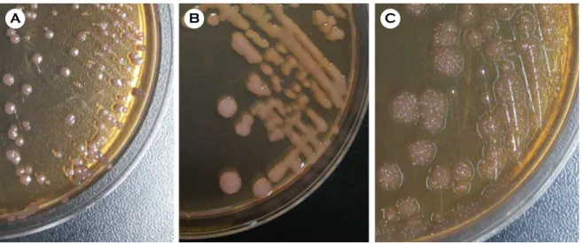
Colony morphology examination is an important method of bacteria identification. MacConkey agar is commonly used for the detection of Acinetobacter spp. in clinical cul- tures. In the current study, the isolates were classified as three different morphotypes according to colony color (pigmen- tation) and shape (including form, elevation, and margin) on MacConkey agar plates (Fig. 1). One-hundred one isolates (Group A, 82.1%) were observed to be small in size (< 3 mm), smooth (the entire margin), round (circular form), and raised colonies on the MacConkey agar plates following incubation for 24 hrs. Sixteen isolates (Group B, 13.0%) were seen to be of a medium size (3~5 mm), smooth (the entire margin),
shiny, round (circular form), and characterized by slightly mucoid colonies. Six isolates (Group C, 4.9%) were of a medium size, rough, irregular and characterized by flat col- onies.
None of the Acinetobacter spp. isolates produced any pig- ment. Non-lactose fermenting colonies (colorless or light lavender color) were observed. The colonies of all isolates were Gram-negative (data not shown).
Phenotypic identification of Acinetobacter spp. isolates using the VITEK®2 system
VITEK®2, an automated microbial identification system, is frequently used to rapidly identify pathogens in clinical laboratories around the world. The clinical isolates in the current study were identified using the VITEK®2 system in conjunction with the GN-ID card. All the isolates (n=123, 100.0%) were identified as A. baumannii (genospecies 2) (Table 2).
Genotypic identification of Acinetobacter spp. isolates using the rpoB gene sequencing
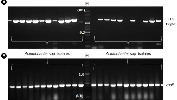
Fig. 2 shows the PCR amplification results for the rpoB gene. One-hundred and twenty three Acinetobacter clinical isolates were analyzed using the rpoB gene genotyping (Supplementary data 1). Five Acinetobacter spp. were identi- Fig. 1. Characterization of colonies of Acinetobacter spp. clinical isolates on MacConkey agar plates after incubation 24 hrs. (A) Group A isolates appeared as colorless small size smooth colonies with circular margin. (B) Group B isolates appeared as colorless medium size mucoid colonies. (C) Group C isolates appeared as colorless medium size rough colonies.
A B C
fied: A. baumannii (genospecies 2), 92 isolates (74.8%); A.
nosocomialis (genospecies 13TU), 13 isolates (10.6%); A.
calcoaceticus or A. pittii (genospecies 1 or 3), 12 isolates (9.7%, 12 isolates were incorrectly identified as A. calcoa- ceticus or A. pittii); and A. soli, 6 isolates (4.9%) isolates (Table 2).
The rpoB gene sequencing correctly identified 117 iso- lates (95.1%) belonging to ACB-complex. Only 6 isolates (4.9%) belonging to non-ACB complex.
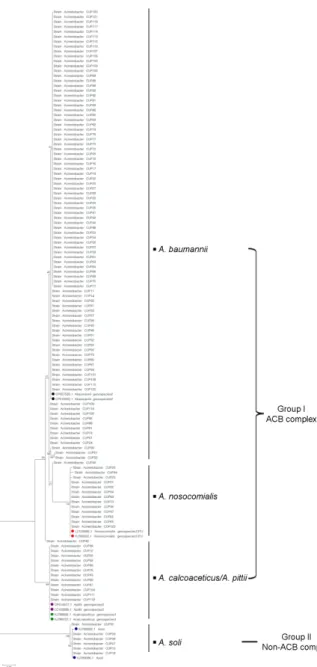
Phylogenetic analysis using partial rpoB gene sequences
The sequences of part of the rpoB gene (approximately Table 2. Detailed results of the identification of 123 Acinetobacter clinical isolates obtained using the rpoB gene sequencing, ITS region
sequencing, the MALDI-TOF MS based VITEK®MS, and the VITEK®2
Groups
Final Identification (No. of isolates)
Colony type (n)
Identification results
Gene sequencing MALDI-TOF MS (VITEK® MS) VITEK®2
system (n)
rpoB (n) ITS (n) IVD 2.0 (n) RUO v 4.13 (n)
ACB complex
A. calcoaceticus
(3) A (3) A. calcoaceticus/
A. pittii (3)
A. calcoaceticus/
A. pittii (3)
A. baumannii complex (3)
Acinetobacter spp. (3)
A. baumannii (3) A. bauamnnii
(92)
A (76) B (16)
A. baumannii (92)
A. baumannii (92)
A. baumannii complex (92)
A. baumannii (92)
A. baumannii (92) A. pittii
(9) A (9) A. calcoaceticus/
A. pittii (9)
A. calcoaceticus/
A. pittii (9)
A. baumannii complex (9)
Acinetobacter spp. (9)
A. baumannii (9) A. nosocomialis
(13) A (13) A. nosocomialis (13)
A. nosocomialis/
A. calcoaceticus (13)
A. baumannii complex (13)
A. baumannii (13)
A. baumannii (13) Non-ACB
complex
A. soli
(6) C (6) A. soli
(6)
A. soli/
A. baumannii (6)
No ID (6)
No ID (6)
A. baumannii (6) ACB: A. calcoaceticus-A. baumannii complex, ITS: internal transcribed spacer, MALDI-TOF MS: matrix-assisted laser desorption/ionization time-of-flight mass spectrometry.
A
B
Fig. 2. ITS region and rpoB gene amplification of Acinetobacter spp. isolates by PCR. Lanes M; 100 bp molecular marker (Bioneer, Seoul), the others; PCR products of Acinetobacter spp. isolates. (A) ITS region was amplified specific fragments of 1,300 bp in Acinetobacter spp.
isolates. (B) rpoB gene was amplified specific fragments of 400 bp in Acinetobacter spp. isolates.
380 bp) from all Acinetobacter isolates were aligned and edited, and the following sequences were used as reference strains: A. calcoaceticus (GenBank accession number: KJ- 788852 and KJ788727), A. baumannii (GenBank accession number: CP027530 and CP018332), A. pittii (GenBank acces- sion number: CP014477 and LC102689), A. nosocomialis
(GenBank accession number: LC102686 and KJ789022) and A. soli (GenBank accession number: KJ789086 and KJ- 789092). The Acinetobacter clinical isolates were separated into two major groups (group I and II) based on the phylo- genetic tree constructed using ML. Group I comprised A.
calcoaceticus, A. baumannii, A. pittii and A. nosocomialis, in which Acinetobacter genospecies 1~3 were observed to be closely grouped. This clustering was supported by high bootstrap value of 99%. Group II comprised A. soli (a boots- trap values of 99%) (Fig. 3).
Genotypic identification of Acinetobacter spp. isolates using ITS region sequencing
One-hundred and twenty three Acinetobacter clinical iso- lates were analyzed using ITS region genotyping. PCR amplification results for the ITS region are provided in Fig. 2.
As a result of ITS region sequencing, the 5 Acineto- bacter spp. were as follows: A. baumannii (genospecies 2), 92 isolates (74.8%); A. nosocomialis or A. calcoaceticus (genospecies 13TU or 1), 13 isolates (10.6%, 13 isolates were incorrectly identified as A. nosocomialis or A. calcoaceticus);
A. calcoaceticus, A. pittii or A. baumannii (genospecies 1, 3 or 2), 12 isolates (9.7%, 12 isolates were incorrectly identified as A. calcoaceticus, A. pittii or A. baumannii); and A. soli or A. baumannii, 6 isolates (4.9%, isolates were incorrectly identified as A. soli or A. baumannii) isolates (Table 2). Thus, the findings using ITS region sequencing were relatively less accurate than those obtained using rpoB gene sequencing.
Molecular analysis of Acinetobacter spp. isolates by ARDRA
For genospecies identification of isolates, ARDRA of the ITS region was applied as previously described by Vaneechoutte et al. (Vaneechoutte et al., 1995). ITS region of 16~23S rRNA gene was amplified for a total of 123 isolates belonging to the 5 species of the genus Acinetobacter and amplified product was restricted independently with a total of 5 different endonucleases (AluI, HhaI, HaeIII, MboI, and MSPI). Each enzyme generated up to 5 fragments per iso- lates.
Fig. 4 shows an overview of different restriction patterns observed. Digestion of the ITS region by AluI resulted in 2 Fig. 3. A phylogenetic tree based on rpoB gene sequences shows
the relationship of the 123 Acinetobacter clinical isolates. Numbers on branches indicate percentages of the number of times that the node was supported in 1,000 replicates of bootstrap analysis. Scale bar, 0.02 substitutions per site (Supplementary data 2).
different patterns. Genospecies 2, 3, and 13TU (A. baumannii, A. pittii, and A. nosocomialis) isolates were digested into three different size fragments. Genospecies 1 (A. calcoace- ticus) isolates were digested into two different size fragment.
Digestion by HhaI resulted in 2 different patterns. Geno- species 1 and 3 isolates were digested into four different size fragments. Genospecies 2 and 13TU isolates were digested into three different size fragments. Digestion by MboI re- sulted in 2 different patterns. Genospecies 1, 2, 13TU isolates were four different size fragments. Genospecies 3 isolates were digested into four different size fragments. Genospecies 3 isolates were digested into three different size fragments.
Combination of the patterns obtained after separate restric- tion with AluI, HhaI and MboI enabled us to identify the 4 genospecies of Acinetobacter spp. isolates. Therefore, four species (1, 2, 3 and 13TU) of Acinetobacter isolates were determined by three endonucleases (AluI, HhaI and MboI).
However, A. soli isolates were not differentiated from other species of Acinetobacter isolates, in this study.
After analysis for ARDRA of ITS region, the 5 Acineto- bacter spp. were identified correctly: A. baumannii (geno-
species 2), 92 isolates (74.8%); A. nosocomialis (genospecies 13TU), 13 isolates (10.6%); A. pittii (genospecies 3), 9 iso- lates (7.3%); A. soli, 6 isolates (4.9%) isolates; and A. calcoa- cetius (genospecies 1), 3 isolates (2.4%).
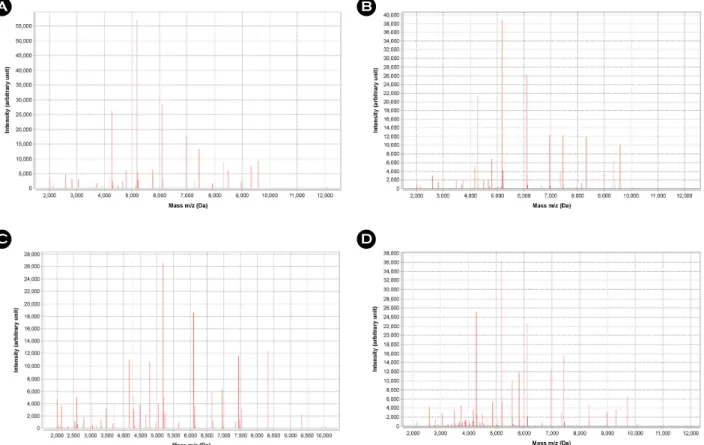
MALDI-TOF MS analysis using the VITEK® MS for species identification of Acinetobacter isolates
MALDI-TOF MS is a useful method of identifying bac- teria and has several benefits. The identification results ob- tained from the use of two distinct test system (VITEK®- MS: IVD 2.0 and RUO) that were applied to the 123 isolates are summarized in Table 2. One-hundred and seventeen isolates (95.1%) identified as A. baumannii complex using the VITEK®-MS IVD 2.0 system. Six isolates (4.9%) were not identified. One-hundred and five isolates (85.4%) were identified as A. baumannii using the VITEK®-MS RUO system. Twelve isolates (9.7%) of Acinetobacter spp. identi- fied at the genus level. Six isolates (4.9%) were not identified.
Each group showed very similar peak profiles, but there were some significant differences between both groups (Fig. 5).
Distribution of Acinetobacter spp. isolates by various clinical samples
A. baumannii accounted for 74.8% of the 123 Acineto- bacter spp. isolates (n=92), and non-baumannii Acineto- bacter spp. accounted for 25.2% (n=31). The distribution of Acinetobacter spp. isolates by clinical samples is depicted in Table 3. A. baumannii was most frequently isolated in all 123 clinical samples (sputum, blood, urine, foley tip, pus discharge, bronchial fluid, wound and ear discharge). A. baumannii only was isolated in pus discharge, the tips of Foley catheters, and wound and bronchial fluid samples (100.0%). All of the A. soli isolates (n=6, 100.0%) were isolated in sputum samples.
DISCUSSION
The proportions of Acinetobacter spp. isolates has been reported in several previous studies. According to these studies, A. baumannii (genospecies 2) is known as the most common species of Acinetobacter cause of nosocomial in- fections. However, the growing number of new infections Fig. 4. ARDRA patterns of Acinetobacter clinical isolates obtained
after restriction of amplified ITS region with three different enzymes (AluI, HhaI, and MboI). Lanes M; molecular size marker, 1~10;
clinical isolates.
from non-baumannii Acinetobacter species is increasingly being recognized, and non-baumannii Acinetobacter species have been identified as important contributors to nosocomial infection (Lee et al., 2011; Visca et al., 2011; Antunes et al.,
2014). The distribution of Acinetobacter spp. isolates, collec- ted from two general hospitals, in Busan, Korea, was evalu- ated in the current study. A. baumannii isolates (74.8%, 92/
123) were the most common species, A. nosocomialis isolates Table 3. The distribution of Acinetobacter species isolates by clinical sample type
Isolates Sample type
Acinetobacter species: n (%)
Total (%) A. baumannii A. calcoaceticus A. nosocomialis A. pittii A. soli
Sputum 45 (66.2) 2 (2.9) 10 (14.7) 5 (7.4) 6 (8.8) 68 (55.3)
Urine 9 (64.3) 1 (7.1) 1 (7.1) 3 (21.4) - 14 (11.4)
Blood 15 (88.2) - 1 (5.9) 1 (5.9) - 17 (13.8)
Bronchial fluid 7 (100.0) - - - - 7 (5.7)
Foley tip 5 (100.0) - - - - 5 (4.1)
Wound discharge 6 (100.0) - - - - 6 (4.9)
Pus discharge 3 (100.0) - - - - 3 (2.4)
Ear discharge 2 (66.7) - 1 (33.3) - - 3 (2.4)
Total (%) 92 (74.8) 3 (2.4) 13 (10.6) 9 (7.3) 6 (4.9) 123 (100.0)
Fig. 5. Peak profiles of four representative Acinetobacter species isolates generated by MALDI-TOF MS based VITEK MS. The x-axis shows the m/z values and the y-axis indicates of the peaks expressed in arbitrary intensity units. (A) Peak profile of A. baumannii isolate.
(B) Peak profile of A. nosocomialis isolate. (C) Peak profile of A. pittii isolate. (D) Peak profile of A. soli isolate.
A B
D C
(10.6%, 13/123) and A. pittii isolates (7.5%, 9/123) were the second and third common strains of ACB-complex, respect- ively (Table 2). A different Acinetobacter species distribution to that reported in other studies was determined in the cur- rent research. According to Lee et al., A. baumannii (74.9%), A. nosocomialis (12.3%) and A. bereziniae (3.4%, Geno- species 10; non-ACB complex) were the common species of Acinetobacter from 495 clinical isolates (collected from 2005 to 2012) in Korea (Lee et al., 2014). By contrast, A.
bereziniae was not isolated, and A. soli (5.0%, 6/123) was the most common non-ACB complex species, in the present study. (Endo et al., 2014) demonstrated that A. soli accounted for a high proportion of Acinetobacter isolates (27.1%) that were the cause of bacteremia in a Japanese tertiary hospital (Endo et al., 2014). According to Khosravi et al., A. baumannii (66.0%), A. calcoaceticus (4.5%) and Acinetobacter geno- species 16 (4.0%; non-ACB complex) were the most cited species of Acinetobacter identified in 197 clinical isolates (collected from 2011 to 2013) in Iran (Khosravi et al., 2015).
These results show that the distribution of Acinetobacter spp.
varies depending on geographical, institutional and epid- emiolgical differences.
The genus Acinetobacter was originally suggested by Brisou and Prevot (1954). Since then, more than 30 Acineto- bacter species have been described. The identification of Acinetobacter at the species level is difficult. The ACB- complex species, in particular, are known to be a common cause of nosocomial infections. A. baumannii, A. calcoa- ceticus, A. pittii and A. nosocomialis are phenotypically very similar and genetically closely related (Vaneechoutte et al., 1995; Lee et al., 2011; Visca et al., 2011; Tien et al., 2012;
Almasaudi, 2018). The accurate identification of bacterial pathogens is essential within a clinical laboratory. Various phenotypic and genotypic methods have been developed and validated to ensure the correct identification of different Acinetobacter spp. the rpoB gene sequencing. ARDRA of the ITS region was demonstrated to have greater accuracy than other methods (i.e., ITS region sequencing, MALDI- TOF MS and VITEK®2 system) in species identification in the current research. However, the A. pittii and A. calcoace- ticus isolates (7.5%, 12/123) were not correctly differentiated from one another using rpoB gene sequencing and phylo-
genetic analysis. MALDI-TOF MS is presently the pre- dominant method used to identify microorganisms in an increasing number of clinical laboratories. In the current study, the results obtained using the MALDI-TOF MS based VITEK®MS system were superior to those obtained using a commercial biochemical approach (i.e., VITEK®2). How- ever, 31 isolates (25.2%) could not be identified at the species level. The rate for correct identification at the species level, misidentification, and the lack of identification was 74.8%
(92/123), 20.3% (25/123) and 4.9% (6/123), respectively.
Only A. baumannii isolates were correctly identified at the species level using VITEK® MS RUO mode. In the present study, isolates of A. soli, which are not included in VITEK® MS database, were not identified at all. The system database should be further expanded and optimized with respect to the species that were misidentified. Similarly, rare or recently named species should be added to the database in order to ensure more accurate identification when using MALDI- TOF-based VITEK® MS. Jeong et al. (2016) demonstrated that MALDI-TOF MS was a useful method of correctly identifying Acinetobacter isolates at the species level fol- lowing updates to the database (Jeong et al., 2016).
Traditionally, colony formation of microorganisms and its morphological analysis (size, shape, surface and color) with a naked eye have been applied for detection and identifi- cation of microbial species (Maeda et al., 2017). In the cur- rent study, colonies of 123 Acinetobacter spp. isolates were observed on MacConkey agar, and colony based bacterial strain typing was performed. Three types of colonies were identified; (1) small size (< 3 mm), smooth, round shape and raised colonies (Group A), (2) medium size (3~5 mm), smooth, round shape and mucoid colonies (Group B), and (3) medium-large size (5~10 mm), rough, irregular shape and flat colonies (Group C). Group C was identified as A. soli only (n=6, 100.0%). Like this result, colony morphological type was related with Acinetobacter spp., and considered to be one of useful species screening method in the clinic.
In conclusion, our study demonstrated that the rpoB gene sequencing and ARDRA of ITS region of 16~23S rRNA seem to be very useful genotypic methods for the differen- tiation of Acinetobacter spp. at the species level. As a result, 123 Acinetobacter spp. isolates were identified correctly: A.
baumannii (genospecies 2), 92 isolates (74.8%); A. nosocom- ialis (genospecies 13TU), 13 isolates (10.6%); A. pittii (geno- species 3), 9 isolates (7.3%); A. soli, 6 isolates (4.9%) iso- lates; and A. calcoacetius (genospecies 1), 3 isolates (2.4%).
ACKNOWLEDGEMENT
This paper was supported by a research fund offered from the Catholic University of Pusan, Republic of Korea in 2015.
CONFLICT OF INTEREST
No potential conflict of interest relevant to this article was reported.
REFERENCES
Almasaudi SB. Acinetobacter spp. as nosocomial pathogens: Epid- emiology and resistance features. Saudi J Biol Sci. 2018. 25:
586-596.
Antunes LC, Visca P, Towner KJ. Acinetobacter baumannii: evolu- tion of a global pathogen. Pathog Dis. 2014. 71: 292-301.
Cisneros JM, Rodriguez-Bano J. Nosocomial bacteremia due to Acinetobacter baumannii: epidemiology, clinical features and treatment. Clin Microbiol Infect. 2002. 8: 687-693.
Croxatto A, Prod'hom G, Greub G. Applications of MALDI-TOF mass spectrometry in clinical diagnostic microbiology. FEMS Microbiol Rev. 2012. 36: 380-407.
Dingle TC, Butler-Wu SM. Maldi-tof mass spectrometry for micro- organism identification. Clin Lab Med. 2013. 33: 589-609.
Endo S, Yano H, Kanamori H, Inomata S, Aoyagi T, Hatta M, Gu Y, Tokuda K, Kitagawa M, Kaku M. High frequency of Acinetobacter soli among Acinetobacter isolates causing bac- teremia at a tertiary hospital in Japan. J Clini Microbiol. 2014.
52: 911-915.
Jeong S, Hong JS, Kim JO, Kim KH, Lee W, Bae IK, Lee K, Jeong SH. Identification of Acinetobacter Species Using Matrix- Assisted Laser Desorption Ionization-Time of Flight Mass Spectrometry. Ann Lab Med. 2016. 36: 325-334.
Khosravi AD, Sadeghi P, Shahraki AH, Heidarieh P, Sheikhi N.
Molecular methods for identification of Acinetobacter species by partial sequencing of the rpoB and 16SrRNA genes. J Clin Diagn Res. 2015. 9: DC09-13.
Koeleman JG, Stoof J, Biesmans DJ, Savelkoul PH, Vandenbroucke-
Grauls CM. Comparison of amplified ribosomal DNA restric- tion analysis, random amplified polymorphic DNA analysis, and amplified fragment length polymorphism fingerprinting for identification of Acinetobacter genomic species and typing of Acinetobacter baumannii. J Clin Microbiol. 1998. 36: 2522 -2529.
La Scola B, Gundi VA, Khamis A, Raoult D. Sequencing of the rpoB gene and flanking spacers for molecular identification of Acinetobacter species. J Clin Microbiol. 2006. 44: 827-832.
Lee MJ, Jang SJ, Li XM, Park G, Kook JK, Kim MJ, Chang YH, Shin JH, Kim SH, Kim DM, Kang SH, Moon DS. Comparison of rpoB gene sequencing, 16S rRNA gene sequencing, gyrB multiplex PCR, and the VITEK2 system for identification of Acinetobacter clinical isolates. Diagn Microbiol Infect Dis.
2014. 78: 29-34.
Lee K, Yong D, Jeong SH, Chong Y. Multidrug-resistant Acineto- bacter spp.: increasingly problematic nosocomial pathogens.
Yonsei Med J. 2011. 52: 879-891.
Maeda Y, Dobashi H, Sugiyama Y, Saeki T, Lim TK, Harada M, Matsunaga T, Yoshino T, Tanaka T. Colony fingerprint for dis- crimination of microbial species on lensless imaging of micro- colonies. PLos One. 2017. 12: e0174723.
Tien N, You BJ, Chang HL, Lin HS, Lee CY, Chung TC, Lu JJ, Chang CC. Comparison of genospecies and antimicrobial re- sistance profiles of isolates in the Acinetobacter calcoaceticus- Acinetobacter baumannii complex from various clinical speci- mens. Antimicrob Agents Chemother. 2012. 56: 6267-6271.
Vaneechoutte M, Dijkshoorn L, Tjernberg I, Elaichouni A, de Vos P, Claeys G, Verschraegen G. Identification of Acinetobacter genomic species by amplified ribosomal DNA restriction analysis. J Clin Microbiol. 1995. 33: 11-15.
Visca P, Seifert H, Towner KJ. Acinetobacter infection-an emerging threat to human health. IUBMB Life. 2011. 63: 1048-1054.
Wang J, Ruan Z, Feng Y, Fu Y, Jiang Y, Wang H, Yu Y. Species distribution of clinical Acinetobacter isolates revealed by dif- ferent identification techniques. PLoS One. 2014. 9: e104882.
https://doi.org/10.15616/BSL.2019.25.1.40
Cite this article as: Park GN, Kang HS, Kim HR, Jung BK, Kim DH, Chang KS. A Comparison of Genospecies of Clinical Isolates in the Acinetobacter spp. Complex Obtained from Hospitalized Patients in Busan, Korea.
Biomedical Science Letters. 2019. 25: 40-53.