저작자표시-비영리-변경금지 2.0 대한민국 이용자는 아래의 조건을 따르는 경우에 한하여 자유롭게 l 이 저작물을 복제, 배포, 전송, 전시, 공연 및 방송할 수 있습니다. 다음과 같은 조건을 따라야 합니다: l 귀하는, 이 저작물의 재이용이나 배포의 경우, 이 저작물에 적용된 이용허락조건 을 명확하게 나타내어야 합니다. l 저작권자로부터 별도의 허가를 받으면 이러한 조건들은 적용되지 않습니다. 저작권법에 따른 이용자의 권리는 위의 내용에 의하여 영향을 받지 않습니다. 이것은 이용허락규약(Legal Code)을 이해하기 쉽게 요약한 것입니다. Disclaimer 저작자표시. 귀하는 원저작자를 표시하여야 합니다. 비영리. 귀하는 이 저작물을 영리 목적으로 이용할 수 없습니다. 변경금지. 귀하는 이 저작물을 개작, 변형 또는 가공할 수 없습니다.
의학 박사학위 논문
microRNA 181a-5p Reprogramed
Glucose and Lipid Metabolism in
NonSmall Cell Lung Cancer
아 주 대 학 교 대 학 원
의 학 과
mRNA-181a-5p reprogrammed
glucose and lipid metabolism in non
small cell lung cancer
지도교수 임 상 현
이 논문을 의학 박사학위 논문으로 제출함
2020년 2월
아주대학교 대학원
의 학 과
김 정 태
김정태의 의학 박사학위 논문을 인준함
,
심사위원장 박 태 준 인
심사위원 임 상 현 인
심사위원 김 영 진 인
심사위원 신 승 수 인
심사위원 김 장 희 인
아 주 대 학 교 대 학 원
2020년 1월 9일
i
Abstract
microRNAs play an important role in cancer metabolisms. miRNA-181a-5p has decreased expression, particularly in non small cell lung cancer tissues and mesenchymal like lung cancer cells. Epithelial-mesenchymal transition mechanisms and cancer metabolism are regulated by the same signaling pathway. Functional analysis was performed to identify the functional role of miRNA-181a-5p in cancer metabolism and to find the target gene. To define the role of miRNA-181a-5p in cancer metabolism, we performed LDH assay, glucose uptake assay, Oil Red O staining, and mitochondrial ATP synthase inhibitor assay. The target gene of miRNA-181a-5p was determined by luciferase reporter assay, qRT-PCR, and western blot analyses. The Functional effect of miRNA-181a-5p on cancer metastasis was measured by migration and invasion assays. Experimental results showed that overexpression of miRNA-181a-5p reduced aerobic glycolysis and adipocytes and decreased cancer cell invasion and migration. Expression of SIRT1 and ACSL4 was reduced by the binding of miRNA-181a-5p to the 3’-UTR. These results suggest that miRNA-181a-5p might participate in the reprogramming of cancer metabolism.
Key Words: microRNA, Aerobic glycolysis, lipid metabolism, cancer metabolism, Lung cancer
ii
Table of Contents 1) List of Text
I. Introduction
A. Introduction
1. The relation between Glucose, Lipid metabolism and Cancer cell 2. The role of miRNA in glucose and lipid metabolism
3. The role of miRNA in Cancer Cell
4. miRNA-181a-5p and Non Small Cell Lung Cancer 5. Hypothesis
II. Subjects
A. Materials and Methods
1. Cell culture and growth conditions 2. Glucose-uptake assay3. Lactate dehydrogenase assay
4. Mitochondrial ATP synthase Inhibition assay
5. Measurement of extracellular acidification rate (ECAR) after treatment of miRNA-181a-5p
6. Lipid Staining
7. Invasion and migration assays 8. TaqMan miRNA expression assay 9. miRNA mimic and siRNA transfection 10. qRT-PCR
11. Western blot
iii
13. Luciferase reporter assays 14. Statistical analyses
B. Results
1. Function of miRNA-181a-5p in aerobic glycolysis 2. miRNA-181a-5p reduces total lipid in lung cancer cell
3. miRNA-181a-5p inhibits lung cancer cell migration and invasion in vitro
4. SIRT1 and ACSL4 transcriptions are repressed by binding of miRNA-181a-5p to the 3'-UTR
5. Quantitative analysis of miRNA-181a-5p, SIRT1, and ACSL4 expression in lung cancer cell lines
6. miRNA-181a-5p regulates SIRT1 and ACSL4 expression at mRNA and protein levels
C. Discussion
iv
2) List of Figure
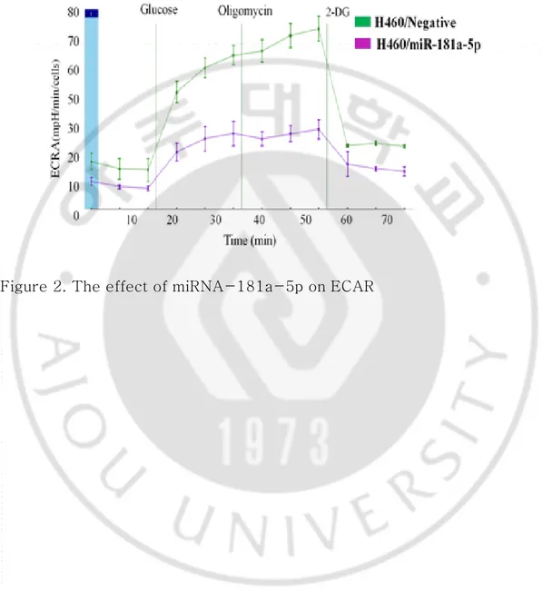
Figure 1A, 1B, 1C. miRNA-181a-5p decreases glucose uptake, LDH release and ATP in lung cancer cells---17 Figure 2. The effect of miRNA-181a-5p on ECAR --- 18 Figure 3. Lipid content in A549 cells treated with miRNA-181a-5p and siRNA for ACSL4 --- 19 Figure 4A, 4B. miRNA-181a-5p inhibits tumor cell invasion and
migration in vitro--- 20
Figure 5A, 5B. siRNA-SIRT1 and siRNA-ACSL4 inhibits tumor cell invasion and migration in vitro --- 21 Figure 6. Predicted miRNA-181a-5p binding sites within the 3'-UTR of SIRT1 and ACSL4 mRNA --- 22 Figure 7. The signal from a luciferase reporter was significantly
decreased at one miRNA-181a-5p target site at the 3-UTR of SIRT1 and ACSL4--- 23 Figure 8. Effect of miRNA-181a-5p on p53, p27, and p21 expression in lung cancer cells --- 24 Figure 9. Real-time (RT)-PCR analysis of miRNA-181-5p, SIRT1, and ACSL4 expression in lung cancer cell lines --- 25 Figure 10A, 10B. The effect of miRNA-181a-5p on SIRT1 mRNA and ACSL4 mRNA expression in lung cancer cells--- 26 Figure 11. Effect of miRNA-181a-5p on SIRT1 and ACSL4 protein expression in lung cancer cells---27
1
I. Introduction
A. Introduction
1. The relation between Glucose, Lipid metabolism and Cancer cell
Cancer cells need a lot of nutrients to increase the biosynthesis of the various cell constituents required for cell proliferation, energy production and cell division (1). The reprogramming of glucose and lipid metabolism, a process that creates nutrients and energy, is one of the key strategies for cancer cells to live or metastasize. Along with metabolic reprogramming, by-products of aerobic metabolism, such as free radicals, damage cells and cause changes in DNA (2). Thus, metabolic reprogramming may trigger tumorigenesis (3) and help cancer cells evade immune surveillance, survival advantage and metastasis (4). Genetic changes of oncogene or tumor suppressor gene also affect cellular signaling, which also leads to reprogramming of glucose and lipid metabolism. Targeting metabolic reprogramming is a promising and rapidly rising direction for anti-cancer therapy (5).
2. The role of miRNA in glucose and lipid metabolism
The importance of genetic control mechanisms by microRNA (miRNA) in the regulation of tissue metabolism has already been emphasized. Recent studies have shown that the reprogramming process associated with the activity of oncogene and tumor suppressor gene is regulated by miRNA. Aerobic glycolysis in cancer cells has also been reported to be regulated or increased by miRNA through the regulation of transcription factors or signaling systems (6). Some recent studies have revealed a new role for miRNAs in lipid metabolism regulation. For example, miRNA-33 inhibits
2
the expression of ABCA1 and ABCGA1 (ABC binding cassette transporter) and is known to promote the synthesis of high-density lipoprotein (HDL) through the release of cholesterol accumulated in tissue cells in blood vessels (7). As such, miRNA are involved in glucose and lipid metabolism. In this experiment, the effects of miRNA-181a-5p on glucose and lipid metabolism in cancer cells were studied.
3. The role of miRNA in Cancer Cell
Recent studies showed that miRNA-181a-5p is involved in the metastasis and expression of various cancer cells. The upregulation of matrix metalloproteinase MMP-14(MT1-MMP) is associated with poor prognosis in cancer patients. miRNA-181a-5p is downregulated in human breast and colon cancers and miRNA-181a-5p reduced MMP-14 expression, whereas miRNA-181a-5p attenuation elevated MMP-14 expression. As a result, miRNA-181a-5p mediated reduction of MMP-14 levels was sufficient to decrease cancer cell migration and invasion (8). Also, the expression of miRNA-181a-5p was decreased in endometrial carcinoma (9). In non-small cell lung cancer, miRNA-181a-5p inhibited the growth and metastasis of cancer cells by inhibiting the expression of Kras oncogene (10).
4. miRNA-181a-5p and Non Small Cell Lung Cancer
Previous studies have shown that miRNA-181a-5p has less expression in lung cancer tissue than in normal cells (11-13). To identify the association between epithelial-mesenchymal transition mechanism(EMT) of miRNA and non small cell lung cancer, we investigated miRNA of mesenchymal-like lung cancer and expressed miRNA-181a-5p in mesenchymal-like lung cancer cell lines and found that miRNA-181a-5p
3
is down-regulated in mesenchymal-like lung cancer cell lines (14). Recent evidence also suggests that EMT-related transcriptomic alterations are related to metabolic reprogramming in cancer. Metabolic changes may allow cancer to adapt to environmental stressors, supporting the irregular molecular demand for rapid proliferation of cancer cells. Also, it has been demonstrated that expressions of key molecules in signaling pathways of EMT are also main regulators in dysregulated metabolic alteration (15). These results suggest that miRNA-181a-5p might be involved in metastasis and metabolism of lung cancer cells.
5. Hypothesis
Experiments were made with the following hypothesis.
(A) miRNA-181a-5p plays an important role in cancer metabolism of non-small lung cancer cells. To confirm this, LDH, glucose uptake assay, and mitochondrial ATP synthase inhibitor assay were confirmed (16, 17). The expression of miRNA-181a-5p reduces aerobic glycolysis and lipid metabolism.
(B) Sirtuin 1 (SIRT1) and acyl-CoA synthetase long-chain family member 4 (ACSL4), which are known to play an important role in cancer metabolism, were selected as target genes for miRNA-181a-5p through a review of the literature (18-20). To determine whether miRNA-181a-5p could regulate SIRT1 and ACSL4, we performed luciferase assays.
(C) Overexpression of miRNA-181a-5p reduces cancer invasion and migration. The expression of SIRT1 and ACSL4 was inhibited by binding of miRNA-181a-5p to the 3’-UTR.
non-4
small cell metabolism and metastasis.
II. Subjects
A. Materials and Methods
1. Cell culture and growth conditionsNon-small lung cancer cells (HCC358, HCC827, HCC1438, H292, H262, H522, A549, H460 and H1299) were maintained in RPMI 1640 medium (GIBCO BRL, Rockville, MD) supplemented with 10% fetal bovine serum (FBS) and antibiotics (100 units/ml penicillin and 100 mg/ml streptomycin).
2. Glucose-uptake assay
H460 and H1299 cells were transfected with miRNA-181a-5p mimic (Ambion) and miRNA-mimic-Negative control#1 (Ambion). At 48 hours after transfection, glucose uptake rate was measured using Glucose Uptake Assay Kit Colorimetric (Abcam, Cambridge, UK) according to the manufacturer’s instructions.
3. Lactate dehydrogenase assay
At 72 hours after transfection with miRNA-181a-5p mimic (Ambon) or miRNA-mimic-Negative control#1 (Ambon) in H460 and H1299 cells, CytoTox 96® Non-Radioactive Cytotoxicity Assay (Promega, Madison, WI)
was performed to quantitatively measure lactate dehydrogenase (LDH) levels according to the manufacturer’s instructions.
4. Mitochondrial ATP synthase Inhibition assay
Following miRNA transfection on day 3, H460 and H1299 cells were treated with oligomycin A for 30 minutes. Cellular ATP changes after treatment were measured with CellTiter-Glo® Luminescent Cell Viability
5
Assay (Promega) according to the manufacturer’s instructions.
5. Measurement of extracellular acidification rate (ECAR) after treatment of miRNA-181a-5p
The ECAR of H460 cells were measured using the XFp extracellular flux analyzer (Agilent Technologies, Santa Clara, CA) as follows: 100,000 cells were plated per well into an XFp cell culture microplate. Experiments were conducted by the manufacturer’s protocol. The assay was performed in triplicate.
6. Lipid Staining
To observe fat droplets in lung cancer, A549 cells were stained with Oil Red O(Sigma) for 10 minutes on day 3 following transfection with miRNA mimics or siRNA. Cells were washed twice with Phosphate Buffered Saline (PBS) and fixed with 10% formalin for 60 minutes. Cells stained with hematoxylin for 30 seconds were imaged under a light microscope (Olympus, Tokyo, Japan).
7. Invasion and migration assays
The invasion assay was performed in triplicates using 48-well microchemotaxis chambers (Neuro. Probe, Inc., Gaithersburg, MD) and 8-μm pore membranes (Neuro. Probe, Inc., Gaithersburg, MD) pre-coated with 10 μg/ml Matrigel (BD Bioscience). Cells (1X10⁴) in 50 µl of serum-free medium were placed in the upper chamber. The lower chamber was filled with 26-27 µl of medium supplemented with 10% FBS. After incubation at 37˚C for 24 hours, cells that migrated to the lower surface of the membrane were stained with a Diff-Quick kit and counted under a microscope. The migration assay was done using the same procedure using membranes coated with 5 μg/ml collagen IV
6
(TRAVIGEN, Gaithersburg, MD). 8. TaqMan miRNA expression assay
qRT-PCR analysis for miRNAs was performed in triplicates using MicroRNA assay kit (Applied Biosystems, Foster City, CA) according to the manufacturer’s instructions.
9. miRNA mimic and siRNA transfection
Cells were seeded into 6-well plates at a density of 1.2×10⁵ cells/well.
On the next day, cells were transfected with 30 nMmiR-181a-5p mimic
(Ambion Austin, TX) and miRNA-mimic-Negative control#1
(Ambion)with Lipofectamine™ RNAiMAX (Invitrogen, Carlsbad, CA) according to the manufacturers’ instructions. Cells were also transfected with specific Silencer® Select siRNA for SIRT1 (Ambion), ACSL4 (Ambion), and Silencer® Select Negative Control No. 1 siRNA (Ambion)
using Lipofectamine™ RNAiMAX (Invitrogen). 10. qRT-PCR
Total RNA was isolated with TRIzol solution (Ambion) according to the manufacturer’s instructions. First strand cDNA was synthesized using oligo (dT) primer and SuperScript III First-strand Synthesis System (Invitrogen). PCR was run for 40 cycles of denaturation at 95 ˚C for 15 seconds, annealing at 56 ˚C for 15 seconds, and elongation at 72 ˚C for 15 seconds. Gene expression was quantified by the comparative CT method, with CT values normalized to that of housekeeping gene ß-actin. After amplification, melting curve analysis was performed to ensure the specificity of products.
11. Western blot
7
Biotechnology, Gyeonnggi-do, Korea) at 72 h after transfection. An equal amount of proteins was resolved on 8% SDS-PAGE gels (Laemmli, 1970). Primary antibodies used for the analysis included mouse anti-SIRT1(1:1000, Abcam, Cambridge, UK), ACSL4antibody (1:1000; Santa Cruz Biotechnology, Santa Cruz, CA), and b-actin antibodies (1:2000; Santa Cruz Biotechnology).
12. Approach to search the potential target genes of miRNA-181a-5p SIRT1 and ACSL4 were selected as candidate targets of miRNA-181a-5p based on literature search and in silicoprogram (TargetScanHuman 7.2).
13. Luciferase reporter assays
To verify that miRNA-181a-5p could regulate SIRT1 and ASCL4 gene directly, we generated a Renilla luciferase reporter plasmid cloned downstream to a segment of SIRT1 and ASCL4 3'-UTR containing putative miRNA-181a-5p binding sequences that were predicted to have two binding site [position 2345-2367 bp of SIRT1 3'-UTR (NM_012238) and position 2399-2421 bp of ASCL4 3'-UTR (NM_004458)] using TargetScanHuman 7.2. These constructs were then co-transfected into NCI-H1299 cells with miRNA-181a-5p mimic or mimic-Negative control and Renilla/firefly reporter plasmid. After 48h, renilla/firefly luciferase activity was measured using a Lumat LB9501 instrument (Berthold, Bad Wildbad, Germany). Results were normalized against the activity of firefly luciferase. All experiments were performed in triplicates. 14. Statistical analyses
Statistical differences between groups were analyzed using Student’s t-test when two groups were compared. Statistical significance was
8
considered when P value was less than 0.05
.
B. Results
1. Function of miRNA-181a-5p in aerobic glycolysis
The biological function of miRNA-181a-5p in aerobic glycolysis was investigated. It has already been reported that miR-181a-5p expression was low in H460 and H1299 cell lines (mesenchymal-like lung cancer cells) (14). Thus, experiments were carried out using H460 and H1299 cell lines. Ectopic expression of miRNA-181a-5p in human lung cancer cells H460 and H1299 efficiently reduced glucose consumption (Figure 1A). At the same time, the production of LDH was also reduced (Figure 1B). Several studies have already reported that the expression of LDH has increased in cancer cells. Cancer cells rely on glycolysis to produce lactic acid, as is well known for the Warburg effect. This fermentative glycolysis is catalyzed by LDH and generates energy to promote cell growth and replication. LDH is an enzyme that acts in the final stage of glycolysis and is involved in the interconversion of pyruvate and lactic acid. In the case of cancer cells, it mainly produces lactic acid, and the reduction of glycolysis means a decrease in lactic acid and also a decrease in LDH. Therefore, the expression level of LDH is closely related to glycolysis. When the expression of LDH decreases, we can predict the decrease of glycolysis (21).
Cancer cells are known to produce ATP from aerobic glycolysis and oxidative phosphorylation (OXPHOS). Based on this information, we investigated the involvement of aerobic glycolysis and oxidative phosphorylation in ATP production in association with miRNA-181a-5p.
9
To determine the proportion of ATP produced by OXPHOS versus glycolysis, we limited OXPHOS ATP synthesis by treating lung cancer cells with oligomycin A. We found that the relative ATP production was reduced in lung cancer cells treated with miRNA-181a-5p and oligomycin A. This indicates that miRNA-181a-5p treatment could reduce a proportion of ATP from glycolysis (Figure 1C), suggesting that miR-181a-5p could inhibit aerobic glycolysis in lung cells. To determine the inhibitory effect of miRNA-181a-5p on the glycolysis of non small cell lung cancers, the extracellular acidification rate (ECAR) was measured. Overexpression of miRNA-181a-5p significantly reduced glycolysis rate and glycolytic capacity of H460 cells (Figure 2).
2. miRNA-181a-5p reduces total lipid in lung cancer cell
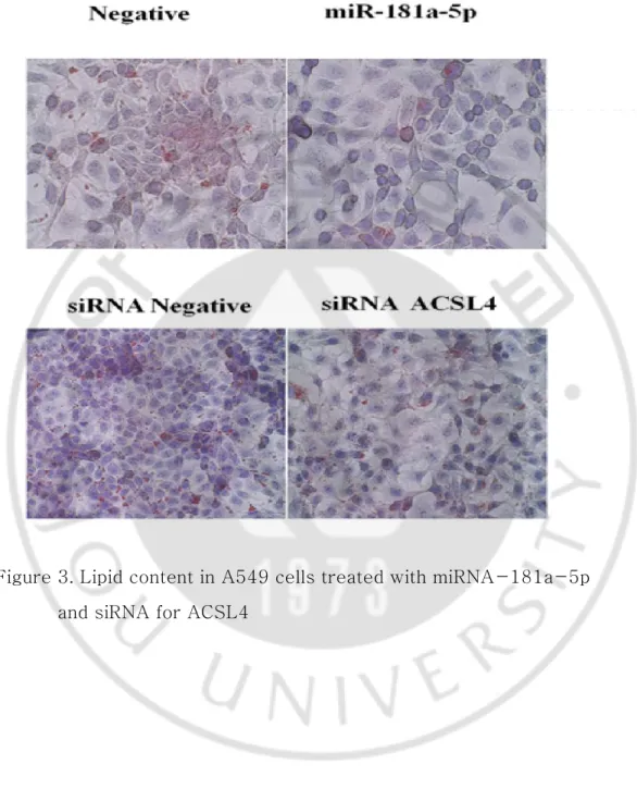
Oil Red O staining of lung cancer cells shows a reduction in fat accumulation in cells treated with miRNA-181a-5p. ASCL4 siRNA also induced a decrease in lipid droplets in lung cancer cells than control group (Figure 3). These results indicate that miRNA-181a-5p could reduce lipid metabolism in lung cancer by regulating ASCL4.
3. miRNA-181a-5p inhibits lung cancer cell migration and invasion in vitro To observe the effect of overexpression of miRNA-181a-5p on lung cancer progression, the invasion and migration assays were performed using a lung cancer cell line. Cells transfected with miRNA-181a-5p mimic displayed showed a significant reduction of invasion and migration (Figures 4A, 4B). To confirm that the effect of miRNA-181a-5p in lung cancer cells occurred through SIRT1 and ACSL4 repression, siRNA was used to transfect H1299 cells. SIRT1 and ACSL4 siRNA decreased both invasion and migration (Figures 5A, 5B).
10
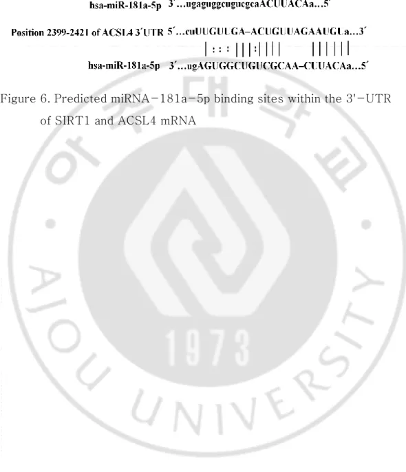
4. SIRT1 and ACSL4 transcriptions are repressed by binding of miRNA-181a-5p to the 3'-UTR
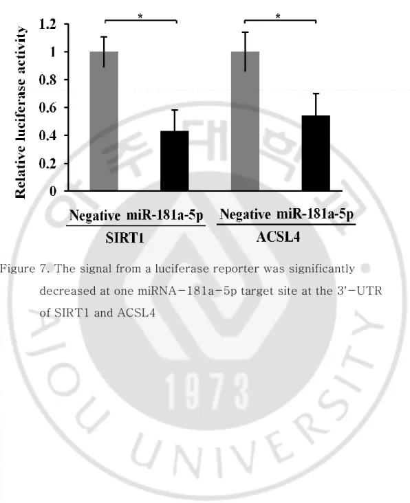
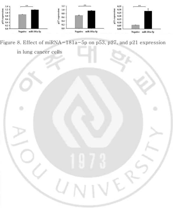
The Rellina luciferase reporter plasmid was generated to verify that miRNA-181a-5p directly regulates SIRT1 and ACSL4 genes. It was cloned downstream to each segment of SIRT1 and ACSL4 3'-UTR containing the putative miRNA-181a-5p binding sequence (Figure 6). Figure 7 shows significantly lower luciferase activity on the side treated by miRNA-181a-5p mimics as compared with negative miRNA (P<0.05). This result indicates that miRNA-181a-5p can directly inhibit the transcription of SIRT1 and ACSL4 by directly binding to the 3'-UTR. Indirectly, relative expression levels of p53, p27, and p21 mRNA undergoing regulation by SIRT1 in H460 cell lines were increased by miRNA-181a-5p (Figure 8).
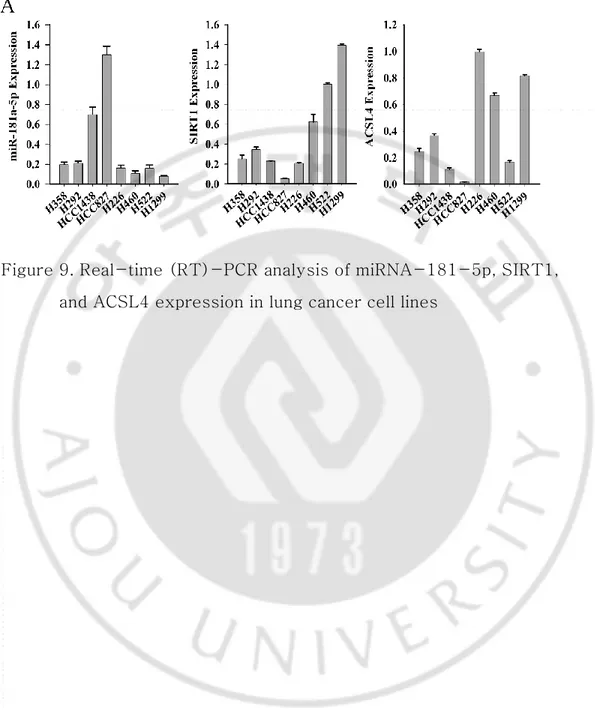
5. Quantitative analysis of miRNA-181a-5p, SIRT1, and ACSL4 expression in lung cancer cell lines
Quantitative reverse transcription-PCR (qRT-PCR) was performed to examine the expression levels of miRNA-181a-5p, SIRT1, and ACSL4. In H358, H292, H1438, and H827 cell lines (epithelial-like lung cancer cells), miRNA-181a-5p was highly expressed, whereas SIRT1 and ACSL4 showed low expression. However, miRNA-181a-5p exhibited low expression while SIRT1 and ACSL4 had high expression in H226, H460, H522, and H1299 cell lines (mesenchymal-like lung cancer cells) (14, 22) (Figure 9). At the cellular level, the expression of SIRT1 and ACSL4 decreased in cells with high miRNA-181a-5p expression, whereas the expression of SIRT1 and ACSL4 increased in cells with reduced miRNA-181a-5p expression (Figure 10A, 10B).
11
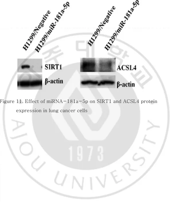
6. miRNA-181a-5p regulates SIRT1 and ACSL4 expression at mRNA and protein levels
miRNAs can inhibit the expression of target genes through translational repression or degradation of target transcripts. To assess the functional role of miRNA-181a-5p in the downregulation of endogenous SIRT1 and ACSL4 expression, H460 and H1299 cells were transfected with miRNA-181a-5p mimics and expression levels of targets genes were then measured by qRT-PCR and Western blot analysis. In the presence of overexpression of miRNA-181a-5p, the relative expression levels of SIRT1 and ACSL4 were decreased compared to those in the control group (Figure 11).
C. Discussion
Carcinogenesis is a complex process through multistep molecular changes of oncogenes, tumor suppressors, and metabolic pathways. Changes in the metabolic processes are an evolutionary process that seeks to create an environment in which cancer can continue to grow and change to an environment suitable for tumor growth (23). Warburg observed that tumor cells shifted from oxidative phosphorylation to aerobic glycolysis more than 90 years ago (24). Since then, research on cancer has focused on understanding the molecular details of cancer metabolism. Recently, many research studies have been conducted to understand the cellular pathways needed for energy production and synthesis of molecules to maintain the cell replication for uncontrolled cell growth. Based on this researches, cancer metabolism has become a new promising therapeutic targeting strategy [25].
miRNA-181a-12
5p in lung cancer metabolism in this study. First, we confirmed that miRNA-181a-5p regulated aerobic glycolysis and lipid metabolism in lung cancer. miRNAs, such as miRNA-33/33, miRNA 122, and miRNA 378/378, have been reported to be involved in lipid metabolism like lipid synthesis, in fatty acid oxidation, the formation, and secretion of lipoproteins (26). miRNA-181a-5p was found to suppress migration and invasion in lung cancer cell lines. Next, we showed that miRNA-181a-5p directly regulated the expression of SIRT1 and ACSL4 by directly binding to its 3'-UTR.
Moreover, induced overexpression of miRNA-181a-5p in lung cancer reduced LDH production, glucose uptake, and glycolysis. In addition, Oil Red O staining demonstrated that miRNA-181a-5p induced downstream regulation of lipid metabolism. Finally, our study showed that miRNA-181a-5p inhibited the migration and invasion of lung cancer by targeting SIRT1 and ACSL4.
Metastatic disease is responsible for more than 90% of all cancer-related deaths. “Rewiring” the metabolic programming is an important way for cancer to overcome nutrient and energy depletion. Aerobic metabolism from glucose to lactate in tumor cell is an important process for increasing lactate production. Lactate production results in normal cell death via caspase-mediated activation of p53-dependent apoptotic pathway, but cancer cells can export lactate by monocarboxylate transporters, resulting in acidification of microenvironment (26). This extracellular acidification of microenvironment creates a good microenvironment for the activation of proteases, which can induce extracellular matrix degradation and facilitate tumor cells to metastasis
13
(27-29).
p53 is directly involved in some glucose metabolism pathways, including glucose uptake, glycolysis, and oxidative phosphorylation. In particular, expression of glucose transporters GLUT1 and GLUT4 is directly inhibited by p53. p53 inhibits the glycolytic pathway by upregulating the expression of TP53-induced glycolysis and apoptosis regulator (TIGAR), an enzyme that inhibits the expression of glycolytic activator fructose-2,6-bisphosphate. p53 induces oxidative phosphorylation by the expression of cytochrome C oxidase assembly protein(SCO2) (25, 30). p53 also inhibits pyruvate dehydrogenase kinase 2 (PDK2), which inactivates the pyruvate dehydrogenase complex (PDC), converts pyruvate to acetyl-CoA for cellular respiration. It is also known to counteract the Warburg effect by attenuating aerobic glycolysis and promoting oxidative phosphorylation through multiple mechanisms (31, 32). NAD-dependent Class III histone deacetylase SIRT1 is a protein that is critically involved in cellular metabolism by deacetylating p53 (33, 34). In addition, p53, a non-histone target of SIRT1, plays an important role in the formation and aging of SIRT1-mediated tumors, and SIRT1 was generally considered as a potential tumor promoter to inhibit the tumor suppressor gene p53 (35). Therefore, as shown in his study, the decrease of SIRT1 by miRNA-181a-5p showed an increase of p53.
Cancer cells require changes not only in glucose metabolism, but also in lipid metabolism. Lipids including triacylglycerides, phosphoglycerides, sterols, and sphingolipids play very important roles in cellular and organismal levels. Fatty acids are an important part of the synthesis of
14
Phosphoglycerides, sterols, and sphingolipids are the important structural substances that form biological membranes (36). These alternations of lipid metabolism in cancer affect a series of cellular processes, including cell growth, proliferation, differentiation, and motility (37). Many lipids are synthesized from fatty acids. However, they must be activated by acyl-CoA synthetase (ACS) enzymes to be biologically active. Mammals have five ACS isoforms (ACSL1, ACSL3, ACSL4, ACSL5, and ACSL6) (38). ACSL4 has increased expression in some colon cancers. Overexpression of ACSL4 promotes tumor cell survival by preventing apoptosis through depletion of un-esterified arachidonic acid, which produces a proapoptotic signal. Chemical inhibition of ACSL1, ACSL3, and ACSL4 by triacsin C causes apoptotic cell death in cancer of the lung, colon, and brain (20, 39, 40). Although the increased metabolism in cancer cells compared to normal benign tissues, metabolism in tumor is characteristically different between individual patients. Due to genetic and environmental factors, cancer metabolism of non small cell lung cancer is heterogeneous. EGFR-mutated adenocarcinomas are less biologically active cancers with lower glucose metabolism than wild type tumors (41). Therefore, lung cancer with active metabolism requires targeting treatment centered on metabolic reprogramming (42, 43).
III. Conclusion
In summary, this study showed that miRNA-181a-5p could reprogram cancer metabolism in non small cell lung cancer. In addition, it was confirmed that miRNA-181a-5p is a regulator of SIRT1 and ACSL4 expression. Thus, miRNA-181a-5p has shown the potential of the new
15
therapeutic candidates to mitigate cancer progression in metabolically active lung cancer
.
16
Figure 1A, 1B, 1C. miRNA-181a-5p decreases glucose uptake, LDH release and ATP in lung cancer cells
17
18
Figure 3. Lipid content in A549 cells treated with miRNA-181a-5p and siRNA for ACSL4
19
Figure 4A, 4B. miRNA-181a-5p inhibits tumor cell invasion and migration in vitro
20
Figure 5A, 5B. siRNA-SIRT1 and siRNA-ACSL4 inhibits tumor cell invasion and migration in vitro
21
Figure 6. Predicted miRNA-181a-5p binding sites within the 3'-UTR of SIRT1 and ACSL4 mRNA
22
Figure 7. The signal from a luciferase reporter was significantly decreased at one miRNA-181a-5p target site at the 3'-UTR of SIRT1 and ACSL4
23
Figure 8. Effect of miRNA-181a-5p on p53, p27, and p21 expression in lung cancer cells
24
Figure 9. Real-time (RT)-PCR analysis of miRNA-181-5p, SIRT1, and ACSL4 expression in lung cancer cell lines
25
Figure 10A, 10B. The effect of miRNA-181a-5p on SIRT1 mRNA and ACSL4 mRNA expression in lung cancer cells
26
Figure 11. Effect of miRNA-181a-5p on SIRT1 and ACSL4 protein expression in lung cancer cells
27
References
1. Keenan MM1, Chi JT(2015) Alternative fuels for cancer cells. The Cancer Journal. 21:49–55.
2. Vogelstein B, Kinzler KW (2004) Cancer genes and the pathways they control. Nat Med10:789-799.
3. Jang M, Kim SS, Lee J (2013) Cancer cell metabolism: implications for therapeutic targets. Experimental & Molecular Medicine. 45, e45. 4. Xie H, Hanai J, Ren JG, Kats L, Burgess K et al (2014) Targeting lactate dehydrogenase-A inhibits tumorigenesis and tumor
progression in mouse models of lung cancer and impacts tumor initiating cells. Cell metabolism. 19: 795-809.
5. Phan LM, Yeung S-CJ, Lee M-H (2014) Cancer metabolic reprogramming: importance, main features, and potentials for precise targeted anti-cancer therapies. Cancer Biology & Medicine.11: 1-19.
6. Tomasetti M, Amati M, Santarelli L, Neuzil J (2016) MicroRNA in Metabolic Re-Programming and Their Role in Tumorigenesis. International Journal of Molecular Sciences. 17: 754
28
intron inhibits cholesterol export and fatty acid oxidation. J Biol Chem 285(44):33652-61
8. Yiyi Li, CemKuscu, Anna Banach, Qian Zhang, Ashleigh Pulkoski- Gross et al (2015) miR-181a-5p Inhibits Cancer Cell Migration and Angiogenesis via Downregulation of Matrix Metalloproteinase -14. Cancer Res. Jul 1;75(13)
9. Yu J, Jiang L, Gao Y, Sun Q, Liu B, Hu Y et al (2019) LncRNA CCAT1 negatively regulates miRNA-181a-5p to promote endometrial carcinoma cell proliferation and migration. ExpTher Med. 2019 May;17(5):4259-4266
10. Ma Z, Qui X, Wang D, Li Y, Zhang B et al (2015) miR-181a-5p inhibits cell proliferation and migration by targeting Kras in non- small lung cancer A549 cells. ActaBiochimBiophys Sin Aug; 47(8):630-8
11. Son JW, Kim YJ, Cho HM, Lee SY, Jang JSet al (2009) MicroRNA Expression Profiles in Korean Non-Small Cell Lung Cancer.
TubercRespir Dis. 67: 413-421.
12. Volinia S, Calin GA, Liu CG, Ambs S, Cimmino A et al (2006) A microRNA expression signature of human solid tumors defines
29
cancer gene targets. ProcNatlAcadSci U S A.103:2257-61 13. Yu SL1, Chen HY, Chang GC, Chen CY, Chen HWet al (2008)
MicroRNA Signature Predicts Survival and Relapse in Lung Cancer. Cancer Cell. 13: 48-57.
14. Park DH, Jeon HS, Lee SY, Choi YY, Lee HW et al (2015) MicroRNA-146a inhibits epithelial mesenchymal transition in non-small cell lung cancer by targeting insulin receptor substrate 2. International journal of oncology. 47: 1545-1553.
15. Huang R, Zong X (2017) Aberrant cancer metabolism in epithelial mesenchymal transition and cancer metastasis: Mechanisms in cancer progression. Crit Rev OncolHematol. 115:13-22
16. Lain S1, Hollick JJ, Campbell J, Staples OD, Higgins Met al. (2008) Discovery, In Vivo Activity, and Mechanism of Action of a Small- Molecule p53 Activator. Cancer Cell. 13: 454-463.
17. Yamamoto H, Schoonjans K, Auwerx J (2007) Sirtuin Functions in Health and Disease. Molecular Endocrinology. 21: 1745-1755. 18. Lu J, Zhang M, Huang Z, Sun S, Zhang Yet al (2015) SIRT1 in B[a]P-induced lung tumorigenesis. Oncotarget. 6: 27113-27129. 19. Lewin TM, Kim J-H, Granger DA, Vance JE, Coleman RA (2001)
30
Acyl-CoA Synthetase Isoforms 1, 4, and 5 Are Present in
Different Subcellular Membranes in Rat Liver and Can Be Inhibited Independently. Journal of Biological Chemistry.276: 24674-24679. 20. Wu X, Deng F, Li Y, Daniels G, Du X et al (2015) ACSL4 promotes prostate cancer growth, invasion and hormonal resistance.
Oncotarget. 6: 44849-44863.
21. Sheng SL, Liu JJ, Dai YH, Sun XG, Xiong XP, Huang G (2012) Knockdown of lactate dehydrogenase A suppresses tumor growth and metastasis of human hepatocellular carcinoma. FEBS J. 2012 Oct;279(20):3898-910
22. Thomson S, Petti F, Sujka-Kwok I, Epstein D, Haley JD (2008) Kinase switching in mesenchymal-like non-small cell lung cancer lines contributes to EGFR inhibitor resistance through pathway redundancy. Clinical & Experimental Metastasis. 25: 843-854. 23. McCleland ML, Adler AS, Deming L, Cosino E, Lee L et al (2013) Lactate Dehydrogenase B Is Required for the Growth of KRAS- Dependent Lung Adenocarcinomas. Clinical Cancer Research. 19: 773-84
31
interpretations of an established concept. Free RadicBiol Med. 79:253-63
25. Cairns RA, Harris IS, Mak TW(2011) Regulation of cancer cell metabolism. Nat Rev Cancer.1: 85-95.
26. BhutiaYangzom d, Babu E, Ganapathy V (2016) Re-programming tumour cell metabolism to treat cancer: no lone target for
lonidamine. Biochemical Journal. 473: 1503-1506.
27. Han T, Kang D, Ji D, Wang X, Zhan Wet al (2013) How does cancer cell metabolism affect tumor migration and invasion? Cell AdhMigr.7: 395-403.
28. Sciacovelli M, Frezza C. (2017) Metabolic reprogramming and epithelial-to-mesenchymal transition in cancer. The FEBS journal. 284: 3132-3144
29. Li X, Yu X, Dai D, Song X, Xu W (2016) The altered glucose metabolism in tumor and a tumor acidic microenvironment
associated with extracellular matrix metalloproteinase inducer and monocarboxylate transporters. Oncotarget 7: 23141-23155.
30. Zhao L, Mao Y, Zhao Y, Cao Y, Chen X. (2016) Role of
32
clinical significance. Oncotarget. 7: 1572-31585.
31. Berkers CR, Maddocks OD, Cheung EC, Mor I, Vousden KH (2013) Metabolic Regulation by p53 Family Members. Cell Metabolism. 18: 617-633.
32. Sinthupibulyakit C, Ittarat W, St. Clair WH, St. Clair DK (2010) p53 protects lung cancer cells against metabolic stress.
International journal of oncology. 37: 1575-1581.
33. Sarma P, Bag I, Ramaiah MJ, Kamal A, Bhadra Uet al (2015) Bisindole-PBD regulates breast cancer cell proliferation via SIRT-p53 axis. Cancer Biology & Therapy. 16: 1486-1501. 34. Grbesa I, Pajares MJ, Martínez-Terroba E, Agorreta J, Mikecin AM et al (2015) Expression of Sirtuin 1 and 2 Is Associated with Poor Prognosis in Non-Small Cell Lung Cancer Patients. PLoS ONE. 10:e0124670.
35. Guan Y, Rao Z, Chen C. (2017) miR-30a suppresses lung cancer progression by targeting SIRT1. Oncotarget. 9:4924-4934
36. Rohrig F, Schulze A. (2016) The multifaceted roles of fatty acid synthesis in cancer. Nat Rev Cancer. 16: 732-749.
33
FEBS journal. 279: 2610-2623.
38. Soupene E, Kuypers FA. Mammalian (2008) Long-Chain Acyl- CoA Synthetases. Experimental Biology and Medicine (Maywood). 233:507-521.
39. Currie E, Schulze A, Zechner R, Walther TC, Farese RV (2013) Cellular Fatty Acid Metabolism and Cancer. Cell metabolism. 18: 153-161.
40. Xia H, Lee KW, Chen J, Kong SN, Sekar K et al (2017)
Simultaneous silencing of ACSL4 and induction of GADD45B in hepatocellular carcinoma cells amplifies the synergistic
therapeutic effect of aspirin and sorafenib. Cell Death Discov. 11:17058
41. Takamochi K, Mogushi K, Kawaji H, Imashimizu K, Fukui M et al (2017) Correlation of EGFR or KRAS mutation status with (18)F- FDG uptake on PET-CT scan in lung adenocarcinoma. PLoS ONE. 12: e0175622.
42. Hensley CT, Faubert B, Yuan Q, Lev-Cohain N, Jin Eet al (2016) Metabolic Heterogeneity in Human Lung Tumors. Cell. 164: 681- 694
34
43. Moreno P1, Jiménez-Jiménez C, Garrido-Rodríguez M, Calderón- Santiago M, Molina S et al. (2018) Metabolomic profiling of human lung tumor tissues: nucleotide metabolism as a candidate for therapeutic interventions and biomarkers. Mol Oncol.doi: 10.1002/1878-0261.12369
35
논문 요약
miRNA는 암 대사에 있어 중요한 역할을 한다. miRNA-181a-5p는 특히 비 소세포 폐암조직과 mesenchymal like lung cancer 세포에서 발현이 감소되어 있다. Epithelial-mesenchymal transition mechanism과 암 대사는 같은 신호 전달체계에 의해 조절된다. 암 대사에 있어서 miRNA-181a-5p의 기능적 역 할을 확인하기 위해 기능적 분석을 시행하였으며 표적유전자를 찾았다. 암 대 사에 있어서 miRNA-181a-5p의 역할을 정의하기 위해 젖산 탈 수소화 효소 분석(LDH assay), 당 흡수 분석(glucose uptake assay), Oil Red O 염색, 미 토콘드리아 ATP 합성 억제 분석(mitochondrial ATP synthase inhibitor assay)을 시행하였다. miRNA-181a-5p의 표적유전자를 발광 효소 보고 분 석(luciferase reporter assay), qRT-PCR, western blot 분석을 통해 결정하 였다. 암 전이에 대한 miRNA-181a-5p의 기능적 효과는 이동(migration)과 침범(invasion)분석으로 측정하였다. 실험 결과 miRNA-181a-5p의 과 발현 은 호기성 당 분해와 지방세포를 감소시켰으며 암 세포의 침입과 이동을 감소 시켰다. SIRT1과 ACSL4의 발현은 miRNA-181a-5p가 3’-UTR에 결합함 으로써 감소하였다. 이 결과는 miRNA-181a-5p가 암 대사의 재조정과정에 참여한다는 것을 의미한다.