저작자표시 2.0 대한민국 이용자는 아래의 조건을 따르는 경우에 한하여 자유롭게 l 이 저작물을 복제, 배포, 전송, 전시, 공연 및 방송할 수 있습니다. l 이차적 저작물을 작성할 수 있습니다. l 이 저작물을 영리 목적으로 이용할 수 있습니다. 다음과 같은 조건을 따라야 합니다: l 귀하는, 이 저작물의 재이용이나 배포의 경우, 이 저작물에 적용된 이용허락조건 을 명확하게 나타내어야 합니다. l 저작권자로부터 별도의 허가를 받으면 이러한 조건들은 적용되지 않습니다. 저작권법에 따른 이용자의 권리는 위의 내용에 의하여 영향을 받지 않습니다. 이것은 이용허락규약(Legal Code)을 이해하기 쉽게 요약한 것입니다. Disclaimer 저작자표시. 귀하는 원저작자를 표시하여야 합니다.
Clinical and Histological Characteristics of Melanocytic
Nevus in External Auditory Canals and Auricles
by
Oak-Sung Choo
Major in Medicine
Department of Medical Sciences
The Graduate School, Ajou University
Clinical and Histological Characteristics of Melanocytic
Nevus in External Auditory Canals and Auricles
by
Oak-Sung Choo
A Dissertation Submitted to
the Graduate School of Ajou University
in Partial Fulfillment of the Requirements of
the Degree of Master of Medicine
Supervised by
Yun-Hoon Choung M.D., Ph.D.
Major in Medicine
Department of Medical Sciences
The Graduate School, Ajou University
This certifies that the dissertation of Oak-Sung Choo
is approved.
SUPERVISORY COMMITTEE
________________________
Yun-Hoon Choung
________________________
Chol Ho Kim
________________________
Hyun Jun Kim
The Graduate School, Ajou University
November 8th, 2013
- i -
- ABSTRACT -
Clinical and Histological Characteristics of Melanocytic Nevus in
External Auditory Canals and Auricles
Nevi, which consist of nevus cells arising from external auditory canals (EACs) and auricles, are rare and their characteristics are not thoroughly understood. The purpose of this study was to analyze the clinicopathological characteristics of melanocytic nevus (MN) in EACs and auricles. Medical records were reviewed in 35 cases with junctional, compound and intradermal nevi treated in Ajou University Hospital, Korea between 2001 and 2011. Patient demographics; location, shape and diameter of nevi; and pathologic results were analyzed according to the location, EACs (23 cases) and auricles (12 cases). Female predominance was shown in both EAC (60.9%, 14 cases) and auricular (75%, nine cases) nevi. The mean age of EAC nevi (37.1 years) was younger than that of auricular nevi (42.2 years). The chief complaint was a symptomless mass in both groups, mostly in dome-like gross appearances. The mean diameter of EAC and auricular nevi was 9.6 (3 – 16) mm and 12.2 (3 – 25) mm, respectively. Histological findings chiefly presented intradermal nevi in EACs (78.3%) and auricles (83.3%) which showed preference to older patients, in contrast to the compound type. All nevi including five cases with skin grafts were completely excised without any recurrence within the follow-up period (average 5.3 months). A possible dysplastic nevus was detected in only one case. All MNs in EACs or auricles reveal similar characteristics. Early and complete excision is recommended to avoid skin graft, functional problems and the risk of malignant melanoma.
Key words: Melanocytic nevus external auditory canal auricle external ear
- ii -
TABLE OF CONTENTS
ABSTRACT ... i
TABLE OF CONTENTS ... ii
LIST OF FIGURES ...iii
LIST OF TABLES ... iiv
I. INTRODUCTION ... 1
II. MATERIALS AND METHODS ... 2
III. RESULTS A. EAC nevi……….…...3
B. Auricular nevi………5
C. Analysis of characteristics according to age……….…8
IV. DISCUSSION ... 9
V. CONCLUSION ... 14
REFERENCES ... 15
- iii -
LIST OF FIGURES
Fig. 1. Otoscopic findings of external auditory canal nevi ……….………4 Fig. 2. Gross findings auricular nevi………6 Fig. 3. A case of auricular nevus located on the cavum concha………...7
- iv -
LIST OF TABLES
Table 1. Characteristics of patients with nevus of the EAC……….….4 Table 2. Characteristics of patients with nevus of auricle……….6 Table 3. Characteristics of the EAC nevi patients according to age………..…...…8
- 1 -
I. INTRODUCTION
Melanocytic nevus (MN), clinically one of the most common benign skin neoplasms, is also known as pigmented nevus, nevocytic nevus or common mole. MN is a well
circumscribed benign malformation of the skin and mucosa, which is composed of melanocytic nevus cells representing progressive stages of melanocytic migration and proliferation [1]. General features of MN include homogeneous surface and coloration pattern, round and oval shape, regular outlines, and relatively sharp borders [2].
Melanocytic nevi is classified into acquired and congenital melanocytic nevi according to the time of appearance. In England and Austria, 1–3% of infants are diagnosed with congenital MN [3]. However, most of the cases are known to be acquired nevi which begin to appear throughout childhood and puberty with peak in the 4th decade [4]. The incidence of acquired MN is possibly related to genetic susceptibility and environmental factors including excessive exposure to sunlight. In most cases, complete excision is recommended immediately after diagnosis of congenital MN due to its potential of malignant
transformation, especially in large-sized nevus. In contrary, no definite malignant potential has been proven in most acquired MN. Therefore, excision of all MNs is not mandatory unless malignant tendency is noticed such as asymmetrical shape, recent change in color and size, or irregular surface. Additional reasons for excision of benign appearing lesions may include cosmetic concerns, continual irritation or associated secondary inflammation.
Although MNs may be discovered in all areas in human body, external auditory canals (EACs) and auricles are relatively uncommon sites. Moreover, MNs cannot be found easily, especially in EAC, because of their restricted visibility to the naked eye. Previously, few case reports on MNs in EACs or auricles have been presented in English literatures [5-11]. To our knowledge, our present study maintains the largest case data in the English literatures for the clinicopathological evaluation of EAC and auricular MNs.
The aim of this study was to present clinical and pathological features of MNs arising from EACs and auricles, and to review previously published literatures for further understanding of MNs.
- 2 -
II. MATERIALS AND METHODS
From 2001 to 2011, 35 patients diagnosed as MN of EACs or auricles were treated at Ajou University Hospital, Suwon, Korea. The medical records of the otolaryngology unit were reviewed retrospectively for cases that included junctional, compound and intradermal nevus in pathologic reports. This study was approved by the Institutional Review Board of the Ajou University School of Medicine. The patients were divided into EAC nevi (n = 23) and auricular nevi (n = 12), and analysis was processed according to age, chief complaint, location, gross shape, size and histopathology. The locations and gross shapes of the EAC lesions were identified with a telescope for documentation of tympanic membranes. The gross shapes were classified into flat, slightly elevated, papillomatous, dome-shaped and pedunculated lesions. Temporal bone computed tomography (TBCT) and pure tone audiometry (PTA) were performed in the cases which implied ear symptoms or suspicious lesions such as EAC cholesteatoma in telescopic findings. Surgical excisions via transcanal approach were performed mostly under local anesthesia in EAC nevi. In auricular nevi, excisions were done meticulously preserving the underlying perichondrium. In cases of large skin defects left after excision, partial thickness skin grafts were applied simultaneously using skins from the thigh or post-auricular areas.
- 3 -
III. RESULT
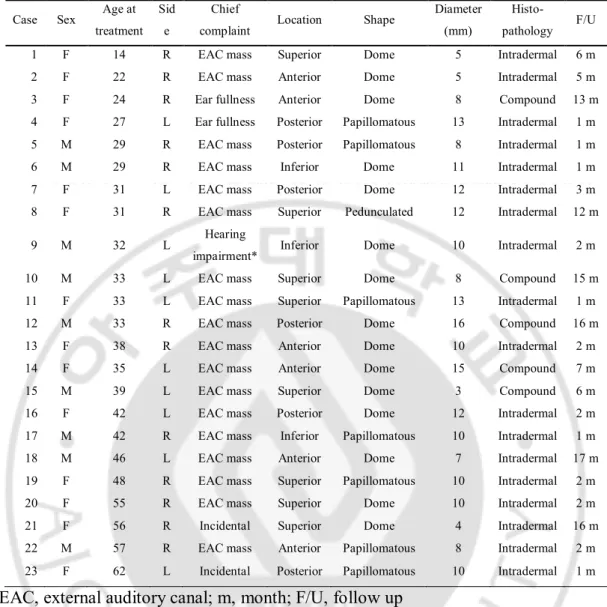
A. EAC neviA total of 23 EAC nevi were excised via the transcanal approach (Table 1). There were 9 males and 14 females. The mean age was 37.1 years, ranging from 14 to 62 years. The right ears were affected in 13 patients, and the left in 10 patients. Main chief complaints of the patients were incidentally detected EAC masses during ear picking. Itching sensation was also reported in one of the EAC cases, as was difficulty in removal of ear wax. Only three patients complained of ipsilateral ear symptoms (two patients with ear fullness and one patient with hearing impairment). However, PTA s showed normal hearing levels in all these patients. Additionally, two patients with EAC nevi were found incidentally during evaluation of tinnitus of the contralateral ear. In all EAC cases, preoperative photo documentation was done with a rigid telescope and the lesion sites within the EAC were classified according to four quadrants: EAC lesions located in the superior (34.8%, eight cases), inferior (13%, three cases), anterior (26.1%, six cases) and posterior (26.1%, six cases) quadrants. The shapes of the lesions were flat, slightly elevated, papillomatous, dome-shaped and pedunculated lesions [4]. The shapes of lesions were mostly dome-shaped (65.2%, 15 cases) and papillomatous (30.4%, seven cases), with one case of pedunculated lesion (4.3%) (Fig. 1). The mean diameter of EAC lesions measured 9.6 mm, ranging from 3 to 16 mm. One single case, Case 20, was operated under general anesthesia to perform ipsilateral mastoidectomy simultaneously due to chronic otitis media (Table 1). In all other cases, surgical treatments were done under local anesthesia. The excision of EAC nevus usually did not require skin graft, however, partial thickness skin graft from thigh was performed to cover skin defect in case 7 due to the broad base of the lesion. Histopathological findings presented 18
intradermal and 5 compound nevi. None of the cases revealed junctional nevus in our study. The mean follow-up duration was 6 months, ranging from 1 to 17 months without any recurrence in all cases.
- 4 -
Fig. 1. Otoscopic findings of external auditory canal nevi. Various gross
morphologies including dome-shaped (case 12 in Table 1, A), papillomatous (case 19
in Table 1, B) and pedunculated (case 8 in Table 1, C) appearance were evident.
- 5 -
Table 1. Characteristics of patients with nevus of the EAC.
EAC, external auditory canal; m, month; F/U, follow up
*Patient 9 complained of hearing impairment but pure tone audiometry showed a
normal hearing level.
Case Sex Age at treatment
Sid e
Chief
complaint Location Shape
Diameter (mm)
Histo-pathology F/U 1 F 14 R EAC mass Superior Dome 5 Intradermal 6 m 2 F 22 R EAC mass Anterior Dome 5 Intradermal 5 m 3 F 24 R Ear fullness Anterior Dome 8 Compound 13 m 4 F 27 L Ear fullness Posterior Papillomatous 13 Intradermal 1 m 5 M 29 R EAC mass Posterior Papillomatous 8 Intradermal 1 m 6 M 29 R EAC mass Inferior Dome 11 Intradermal 1 m 7 F 31 L EAC mass Posterior Dome 12 Intradermal 3 m 8 F 31 R EAC mass Superior Pedunculated 12 Intradermal 12 m 9 M 32 L Hearing
impairment* Inferior Dome 10 Intradermal 2 m 10 M 33 L EAC mass Superior Dome 8 Compound 15 m 11 F 33 L EAC mass Superior Papillomatous 13 Intradermal 1 m 12 M 33 R EAC mass Posterior Dome 16 Compound 16 m 13 F 38 R EAC mass Anterior Dome 10 Intradermal 2 m 14 F 35 L EAC mass Anterior Dome 15 Compound 7 m 15 M 39 L EAC mass Superior Dome 3 Compound 6 m 16 F 42 L EAC mass Posterior Dome 12 Intradermal 2 m 17 M 42 R EAC mass Inferior Papillomatous 10 Intradermal 1 m 18 M 46 L EAC mass Anterior Dome 7 Intradermal 17 m 19 F 48 R EAC mass Superior Papillomatous 10 Intradermal 2 m 20 F 55 R EAC mass Superior Dome 10 Intradermal 2 m 21 F 56 R Incidental Superior Dome 4 Intradermal 16 m 22 M 57 R EAC mass Anterior Papillomatous 8 Intradermal 2 m 23 F 62 L Incidental Posterior Papillomatous 10 Intradermal 1 m
- 6 -
B. Auricular nevi
The clinical characteristics of the patients with auricular nevi are summarized Table 2. There were three male (25%) and nine female (75%) patients. The mean surgical treated age was 42.1 years, ranging from 30 to 50 years. The nevi were affected more on the right side (75%, nine cases) than the left (25%, three cases). All chief complaints were visible masses in and around auricles, except for case 6, who reported hypesthesia on the lesion. Concerning the location of auricular lesions, there were two cases in cymba concha (16.7%), two cases in cavum concha (16.7%), two cases in the pre-auricular area (16.7%), four cases in the post-auricular area (33.3%) and two cases in the infra-post-auricular area (16.7%). Only four cases (33.3%) of intra-auricular lesions were present in our study. Physical examination revealed that most patients (75%, nine cases) had dome-shaped lesions, two patients (16.7%) showed papillomatous lesions and one patient had a pedunculated lesion (8.3%) (Fig. 2). The mean diameter of lesions was 12.2 mm, ranging from 3 to 25 mm. The mean diameter of auricular nevi (11.1 mm) was larger than that of EAC (9.6 mm). In histopathological findings, there were 10 intradermal nevi (83.3%), two compound nevi (16.7%) and no junctional nevus. Six patients (50%) were operated under local anesthesia and the others (50%) under general anesthesia. Five of the general anesthesia cases were performed with other surgeries (one parotidectomy, four mastoidectomies) and partial thickness skin grafts were proceeded in the cases 2, 3, 11 (using post-auricular skin) and 4 (using thigh skin, Fig. 3). The mean follow-up duration was 4.3 months and there was no evidence of recurrence during the follow-up period.
- 7 -
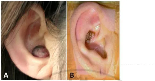
Fig. 2. Gross findings auricular nevi. The dome-like nevus was located in the
cavum concha (case 3 in Table 2, A) and papillomatous nevus was found in the
cymba concha (case 11 in Table 2, B).
Table 2. Characteristics of patients with nevus of auricle.
Case Sex Age at treatment Side
Chief
complaint Location Shape
Diameter (mm)
Histo-pathology F/U
1 F 30 L Mass Post-auricular Dome 8 Compound 3 m
2 F 33 R Auricle mass Cymba concha Dome 10 Compound 2 m 3 F 38 R Auricle mass Cavum concha Dome 15 Intradermal 2 m 4 F 38 R Auricle mass Cavum concha Dome 25 Intradermal 3 m
5 F 39 R Mass Pre-auricular Dome 6 Intradermal 2 m
6 M 43 R Hypesthesia Post-auricular Dome 6 Intradermal 1 m 7 F 44 R Mass Pre-auricular Papillomatous 3 Intradermal 2 m 8 F 45 L Mass Infra-auricular Dome 7 Intradermal 2 m 9 F 46 R Mass Post-auricular Dome 8 Intradermal 13 m 10 M 46 L Mass Post-auricular Dome 14 Intradermal 9 m
11 F 48 R Auricle mass Cymba concha Papillomatous 11
Intradermal, Possibility of dysplastic
nevus
10 m
12 M 50 R Mass Infra-auricular Pedunculated 20 Intradermal 3 m
- 8 -
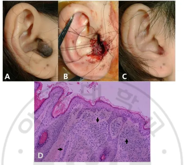
Fig. 3. An auricular nevus (case 4 in Table 2) located on cavum concha. (A)
preoperative and (B) postoperative findings. (C) Partial thickness skin graft from
thigh was performed. (D) Histopathological finding shows intradermal nevus
including nests of nevus cells in the dermis (arrows). (H&E stain, magnification X
200).
- 9 -
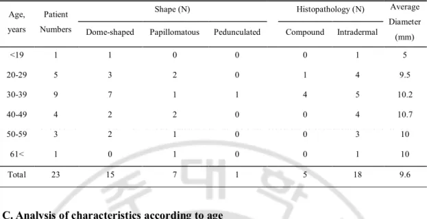
Table 3. Characteristics of the EAC nevi patients according to age.
C. Analysis of characteristics according to age
The EAC nevi were classified according to age (Table 3). Mostly, nevi were found in patients in their 30s (39.1%, nine of 23) and fewer nevi were discovered in younger and older patients. All age groups showed higher incidence of dome-like shaped nevi than papillomatous nevi, except for those in their 60s and older. Histopathologically, high
tendency of compound nevus was only recognized in younger patients. The average diameter of nevi increased (from 5 mm to 10.7 mm) with age and reached a plateau (10 mm) after the age of 50.
Characteristics of auricular nevi according to age are described in Table 4. No patients were younger than 30 or older than 60 in the auricular MN group. Auricular nevi were most common in patients in their 40s (50%, six of 12), followed by those in their 30s (41.7%, five of 12). In all age, dome-like shaped, intradermal nevi were dominantly noted. The only two compound nevi (16.7%, two of 12) were found in their 30s, the youngest group. The diameter of auricular nevi in patients in their 50s reached up to 20 mm.
Age, years
Patient Numbers
Shape (N) Histopathology (N) Average Diameter (mm) Dome-shaped Papillomatous Pedunculated Compound Intradermal
<19 1 1 0 0 0 1 5 20-29 5 3 2 0 1 4 9.5 30-39 9 7 1 1 4 5 10.2 40-49 4 2 2 0 0 4 10.7 50-59 3 2 1 0 0 3 10 61< 1 0 1 0 0 1 10 Total 23 15 7 1 5 18 9.6
- 10 -
IV. DISCUSSION
Nevus, which means “birthmark” in Latin, is the medical term for a sharply-circumscribed and chronic skin lesion. The MN is defined as a benign neoplasm consisting of nests of melanocytic cells located in the epidermis, dermis and, rarely, subcutaneous tissue.
Histologically, nevi are subcategorized into junctional nevus in epidermis, intradermal nevus in dermis and compound nevus in both areas according to the location of clusters of
melanocytic cells. In general, lighter pigmentations are seen in elevated-shaped nevus whereas darker pigments are commonly revealed in flat-formed nevus. Therefore, darker pigmentations are observed in flat junctional nevi, and lighter brown to black-colored lesions are appeared in more elevated compound nevi. Furthermore, intradermal nevi are elevated lesions without pigmentation.
The presumptive pathogenesis of MN includes proliferation within the epidermis as junctional nests, and the subsequent migration or diminish of nevus cells into the papillary dermis where proliferation results in clusters of cells [12]. In compound nevus, the
discontinuation of proliferating in the epidermis and normalization of the overlying epidermis may result in transformation into intradermal nevus [13].
Causes of MNs involve age, race, genetic susceptibility and environmental factors. In a study with monozygotic and dizygotic twins [14], the number of nevi correlated strongly between monozygotic twin pairs compared to dizygotic twin pairs. Pope et al. studied boys and girls of different ethnicity with diverse history of sun exposure. They reported that MNs appeared more commonly in white boys with abundant sun exposure than in girls of other ethnicities with less sun exposure [15]. In the present study, the male-to-female ratio in those with EAC nevi was 9 : 14 and was 3 : 9 in those with auricular nevi. The predominance of sex varies in previous studies. Female predominance (2: 9) was reported in those with EAC nevi in one study [5], whereas another study revealed male predominance (12: 9) in auricular nevi [11].
Although auricular nevus can be discovered more easily than EAC nevus, the mean age of treatment was older in the auricular nevi group (41.6 years) than the EAC nevi group (37.3 years). Similar to other nevi in the body, common figures of auricular nevi may lead to disregard of lesion until alteration in appearances. Other reasonable explanation to this
- 11 -
matter may include higher concerns for the restricted range of vision in EAC nevi. From a functional point of view, EAC nevus may cause conductive hearing loss due to obstruction of the ear canal, leading to recurrent episodes of external otitis [6]. MN exceeding 12mm in diameter is able to obstruct the EAC resulting in EAC cholesteatoma with bony erosion of EAC medial to the nevus [5]. In our study, most EAC nevi patients complained of palpable masses while ear picking. Only three patients claimed ear symptoms such as ear fullness and hearing impairment, and two cases were found incidentally.
Preoperative TBCTs were performed in seven cases, however, no signs of EAC
cholesteatoma were developed even in large nevi (case 4, 11, 12 and 14) exceeding 12 mm. PTAs showed normal findings in all cases. Based on our study, preoperative evaluations such as TBCT and PTA may not be essential before surgical treatments. Similar to EAC nevi, only one patient suffered from hypesthesia among the auricular cases, and there was no specific symptom except palpable masses.
EAC nevi are located in various sites, and have been previously classified in four categories in terms of quadrants. Lee et al. reported that EAC nevi were more frequently located at posterior (33.3%, four of 12) and inferior (33.3%, four of 12) quadrants, and that the inferior nevi had the largest average diameter (8.5 mm) [5]. Oh et al. reported that the anterior quadrant (40%, four of 10) was most frequent, and the largest average diameter (12 mm) was noted in the posterior quadrant [7]. In our study, the superior quadrant lesions (34.8%, eight of 23) were most common, followed in order by anterior (26.1%, six of 23), posterior (26.1%, six of 23) and inferior (13%, three of 23) quadrants lesions. The average diameter of lesions was largest (11.8 mm) in the posterior quadrant. In comparison of our cases with previous studies [5, 7], no common patterns were evident regarding the most common quadrant and the largest average diameter. Twelve cases of auricular nevi were categorized as intra-auricular lesions (33%, four of 12) or peri-intra-auricular lesions (67%, eight of 12). Intra-auricular nevi appeared with the same frequency in the cymba concha (50%, two of four) and the cavum concha (50%, two of four). Around the auricles, the nevi of the post-auricular area (50%, four of eight) were most common and same number (25%, two of eight) was apparent in the infra-auricular and pre-auricular areas. In calculation of the average diameter according to locations of external nevi, the largest average size was 20 mm in the cavum concha. Finally, comparison of the overall diameter between EAC and auricular lesions
- 12 -
resulted in longer overall average diameter (12.2 mm) and maximum diameter (case 4, 25 mm) in the auricle.
The majority of dome-shaped and papillomatous lesions are discovered in intradermal nevi indicating a correlation of gross morphology and histopathological findings [13]. In our study (Tables 1 and 2), gross morphologic findings indicated that dome-shaped and
papillomatous lesions were mostly observed in EAC nevi (15 of 23, seven of 23, respectively) and auricular nevi (nine of 23, two of 23, respectively). Most histopathologic findings were intradermal and compound in EAC nevi (18 of 23, five of 23, respectively) and auricle nevi (10 of 12, two of 12, respectively). In EAC nevi (Table 3), the histopathologic ratio of intradermal to compound according to age showed an increasing tendency with age, consistent with the involution of compound nevi to intradermal nevi with aging [16].
Dysplastic nevus is defined as a nevus that is larger than 6 mm in diameter, usually with irregular edge and pigmentation [17]. On histological examination, the majority of dysplastic nevi are variants of acquired compound nevi, characterized by specific cytology, architecture and host response. Among the present cases, in case 11 of auricular nevi, the possibility of dysplastic nevus could not be excluded in the histopathologic report.A single dysplastic nevus without familial or personal history implies low potential to develop into melanoma despite the ongoing debates. Therefore, explanation of possible risks and outcomes, notification of precautious signs, and regular follow-up are required to patients. For the clinicians, excisional biopsy with at least 2 mm margin of normal skin and pathologic confirmations are indispensable if melanoma cannot be ruled out [17].
As most of MNs are benign and do not require treatments, an interdisciplinary approach with dermatologists is recommended to increase the diagnostic accuracy. The
otolaryngologist alone may have difficulty in concluding the severity of the lesion due to wide spectrum of clinicopathologic appearance of MNs. Dermatologists are able to differentiate the lesions more easily to exclude the presence of nonmelanocytic pigmented lesions using dermoscopy (epiluminescence or incident-light microscopy) by visualizing a skin lesion in depth than naked eyes [18].
Surgical excision is recommended in MNs for histopathologic confirmation in cases with cosmetic concerns, chronic irritation or lesions with suspicious clinical features such as atypical appearing central nevi, presence of an asymmetric halo, eccentric placement of a
- 13 -
melanocytic nevi, and familial or personal history. Differential diagnoses of acquired nevi include freckles, seborrheic keratoses, dermatofibroma and early malignant melanoma. Malignant melanoma is the most important differential disease that is suspicious in
circumstance(Malignant including the history of recent rapid growth, sensation change, the presence of irregular outline, several shades of brown and black, and the number and size of nevi.
In EAC acquired nevi, some authors have insisted that all EAC nevi must be removed in all cases [8] or if symptoms are evident [6]. Unlike to MNs in other region, large nevi can cause complications like EAC cholesteatoma, inflammations and hearing loss [5] and regular follow-up by clinician is necessary due to invisibility. As regards of surgical skills, Fraser et al. reported an excision technique with underlay temporalis fascia graft via endaural incision, which resulted in less granulation tissue formation and better healing [9]. In the nevi
involving more than 180° of EAC, meatoplasty is required for the prevention of
postoperative EAC stenosis [19]. In addition, after incomplete removal, MN may reappear as recurrent lesions (pseudomelanoma) that clinically and pathologically resemble malignant melanoma in situ regardless of the initial pathology [20].
Although auricular MNs are not problematic functionally, aesthetic and psychological concerns do exist. There have been several reports of congenital auricular MNs [10, 21]. Congenital auricular MNs are larger than acquired MNs and have a higher potential of malignant transformation, even though the exact rate of malignant transformation is
uncertain. Concerning the frequency of malignant melanoma of auricle, malignant melanoma occurs more often (over 68%) on the periphery (helix and antihelix) of the auricle [22], is more frequent in men [23] and in those in their late 50s [24]. However, these site- and age-specific features were not evident in our cases. How then should we treat the acquired auricular nevi? Above all, the characteristics of malignant melanomas arising from auricle are important. In some reports, malignant melanoma on auricle showed a worse prognosis compared with melanoma on the face and neck [25]. The 5-year survival rate of malignant melanoma according to location was 78% for the face, 58% for the neck and 33% for the ear [25]. Although there is no exact consensus regarding treatment in acquired auricular nevi, Benmeir et al. suggested that every auricular nevi should be removed as soon as possible because of aggressiveness of malignant melanoma [26]. Saad et al. reported that the MNs of
- 14 -
the auricular region exhibit some histologic features commonly found in melanomas. They recommended the pathologist must aware the different histologic appearances of nevi in different anatomical regions [11]. We agree with this point of view. There is no reason to avoid excision of acquired auricular nevi showing different characteristics. If excision is delayed, the possibility of requiring skin graft will be increased and the aesthetic outcomes will become worse even though the pathologic result is benign. Therefore, we suggest that nevus in the EAC and auricle should be completely excised earlier.
- 15 -
V. CONCLUSION
Melanocytic nevi arising from the EAC and the auricle are uncommon with scarce
information on their characteristic features. In this report, we studied the clinicopathological features of 23 patients with EAC MNs and 12 patients with auricular MNs. The gross appearances are predominantly dome-like and papillomatous and histopathologic findings mostly showed intradermal and compound type in that order. We recommend earlier
excisional biopsy of EAC and auricular nevi based on functional aspects, aesthetic concerns and possibilities of malignant transformation.
- 16 -
REFERENCES
1. Adler N, Margulis A, Bauer BS (2009) Congenital pigmented nevi of the auricle: clinical experience and approach to treatment. Plast Reconstr Surg 124:1932-1939 2. Alves RV, Brandao FH, Aquino JE, Carvalho MR, Giancoli SM, Younes EA (2005)
Intradermal melanocytic nevus of the external auditory canal. Braz J Otorhinolaryngol 71:104-106
3. Benmeir P, Baruchin A, Weinberg A, Nahlieli O, Neuman A, Wexler MR (1995) Rare sites of melanoma: melanoma of the external ear. J Craniomaxillofac Surg 23:50-53 4. Cohen BJ, Melisi J, Cohen MH (1990) Ear preservation in the surgical treatment of
auricular melanoma. Head Neck 12:346-351
5. Easton DF, Cox GM, Macdonald AM, Ponder BA (1991) Genetic susceptibility to naevi--a twin study. Br J Cancer 64:1164-1167
6. Fraser L, Smith WK (2009) Excisional technique for intradermal nevi of the external auditory canal. J Otolaryngol Head Neck Surg 38:501-503
7. Grichnik JM, Rhodes AR, Sober AJ (2008) Benign Neoplasias and Hyperplasias of Melanocytes. In: Wolff K, Goldsmith LA, Katz SI, Gilchrest BA, Paller AS, Leffell DJ (ed) Fitzpatrick’s Dermatology in general medicine, 7th edn. Mc Graw Hill Medical, New York, pp 1104-1109
8. Harrison SL, MacKie RM, MacLennan R (2000) Development of melanocytic nevi in the first three years of life. J Natl Cancer Inst 92:1436-1438
9. Herschorn A (2012) Dermoscopy for melanoma detection in family practice. Can Fam Physician 58:740-745
10. Kazikdas KC, Onal K, Kuehnel TS, Ozturk T (2006) An intradermal nevus of the external auditory meatus. Eur Arch Otorhinolaryngol 263:253-255
11. Langrock ML, Hohenleutner U (2009) Congenital nevus of the left ear: partial earlobe resection and full-thickness skin graft. J Dtsch Dermatol Ges 7:472-473
12. Lee FP (2006) Pigmented nevus of the external auditory canal. Otolaryngol Head Neck Surg 135:124-128
13. Lloyd KM (1990) Bulky, benign tumor of the external auditory canal. Arch Dermatol 126:589-590
- 17 -
14. Lund HZ, Stobbe GD (1949) The natural history of the pigmented nevus: factors of age and anatomic location. Am J Pathol 25:1117-1155
15. Mackie RM (2004) Disorders of the cutaneous melanocyte. In: Burns TF, Rook ATod. Rook's textbook of dermatology. Blackwell Science, Mass, pp 38.5-38.9
16. Naeyaert JM, Brochez L (2003) Clinical practice. Dysplastic nevi. N Engl J Med 349:2233-2240
17. Oh JI, Kim HS, Choi KY, Cho SJ, Kim CW (2010) Characteristics of Melanocytic Nevus Arising from the External Auditory Canal. Korean J Otorhinolaryngol-Head Neck Surg 53:84-88
18. Pariser RJ (1998) Benign neoplasms of the skin. Med Clin North Am 82:1285-1307 19. Pope DJ, Sorahan T, Marsden JR, Ball PM, Grimley RP, Peck IM (1992) Benign
pigmented nevi in children. Prevalence and associated factors: the West Midlands, United Kingdom Mole Study. Arch Dermatol 128:1201-1206
20. Saad AG, Patel S, Mutasim DF (2005) Melanocytic nevi of the auricular region: histologic characteristics and diagnostic difficulties. Am J Dermatopathol 27:111-115 21. Sexton M, Sexton CW (1991) Recurrent pigmented melanocytic nevus. A benign lesion,
not to be mistaken for malignant melanoma. Arch Pathol Lab Med 115:122-126 22. Stegmaier OC (1959) Natural regression of melanocytic nevus. J Invest Dermatol
32:413-419
23. Stegmaier OC, Becker SW Jr (1960) Incidence of melanocytic nevi in young adults. J Invest Dermatol 34:125-129
24. Sylven B, Hamberger CA (1950) Malignant melanoma of the external ear. Report of 36 cases treated between 1928-1944. Ann Otol Rhinol Laryngol 59:631-647
25. Ward NO, Acquarelli MJ (1968) Malignant melanoma of the external ear. Cancer 21:226-233
26. Wanebo HJ, Cooper PH, Young DV, Harpole DH, Kaiser DL (1988) Prognostic factors in head and neck melanoma. Effect of lesion location. Cancer 62:831-837
- 18 -
- 국문 요약 -
이개 및 외이도에서 발생하는 색소 세포성 모반의 조직학적 및 임상적 특징 아주대학교 대학원 의학과 추옥성 (지도교수: 정연훈) 배경 및 목적: 멜라닌세포성 모반은 조직내 위치에 따라 경계(junctional), 복합 (compound), 진피내(intradermal) 모반으로 분류되는 멜라닌세포 증식으로 인한 양 성종양으로 악성종양과 감별을 요하는 질환이며 외이도를 포함한 외이구조에서 는 아주 드문 질환이다. 본 연구는 지난 15년간 외이에서 발생한 멜라닌세포성 모반 20예에 대한 임상 및 조직학적 특징을 분석해 보는 데 있다. 연구 대상 및 디자인: 1995년 1월부터 2009년 7월까지 아주대학병원 이비인후과 에서 외이도 종양으로 진단받고 수술받은 환자 중 멜라닌세포성 모반으로 확진 된 20예를 대상으로 하였다. 환자의 나이, 성별, 주증상, 종양위치, 수술방법, 병 리소견, 재발여부 등을 환자 chart를 중심으로 후향적으로 분석하였다. 결과: 20예의 평균연령은 35 세 (7 - 62)이고, 남녀 3 : 7, 좌우 3 : 7 이었다. 주호소 증상은 심미적 외이변형과 모반의 크기증가 이었으며 청력이상 증상은 없었다. 모반의 위치는 외이개 8예, 외이도 12예 였으며, 평균 크기는 1.0 x 1.25 cm 로 (0.3 x 0.2 – 2.5 x 2.0 cm) 였다. 국소마취 15건, 전신마취 5건으로 1 mm 경 계로 연골막 손상 없이 직상방까지 nevus 절제를 시행하였고, 피부결손 부위가 큰 2예서 skin graft를 시행하였다. 병리조직소견상 복합모반 5예, 진피내모반 15예 였으며, 그 중 1예는 20년전 생검을 시행한 예가 있는 경우로 조직검사상 모반성 이형성 소견을 보였으며, 그외 악성조직이 발견된 예는 없었다. 평균 2개월 ( 2 주 - 15개월) 추적관찰 중에 현재 재발 된 예는 없었다.- 19 -
결론: 복합모반, 진피내모반을 포함한 멜라닌세포성 모반도 외이에 적지않게 발
견되며, 악성의 변화가 있을 수 있어 발견즉시 절제하는 것이 좋을 것으로 사료 된다.