INTRODUCTION
The requirements for bone plate have been increasing because of industrial accidents and an aging society, especial-ly for an efficient filling of a local bone replacement. Over
the last several decades, the bone plate was fabricated by using metal on a metal system, polymer on metal and ceramic on ceramic, etc. However, the metal on metal system is highly likely to take a heavy toll on a surgical subject such as a later injury due to wearied metal nano-particles and reoperation for removing implants (Ferguson et al. 1996; Nie et al. 2000; Poole et al. 2005; Spoerke et al. 2005; Khan et al. 2006; Sargeant and Goswami 2006; Brown et al. 2007). The search
─ ─ 147 ──
Fabrication and Materials Properties of High-Density
Polyethylene (HDPE)/Biphasic Calcium Phosphate (BCP)
Hybrid Bone Plates
Sun-Young Jo, Min-Ho Youn, Youn-Mook Lim*, Hui-Jeong Gwon, Jong-Seok Park and Young-Chang Nho
Radiation Research Division for Industry & Environment, Advanced Radiation Technology Institute, Korea Atomic Energy Research Institute, Jeongeup 580-185, Korea
Abstract -- Biphasic calcium phosphate-reinforced high-density polyethylene (BCP/HDPE) hybrid composite is a new orthopedic biomaterial, which was made to simulate a natural bone composition. Calcium phosphate systems and HDPE hybrid composites have been used in biomedical applications without any inflammatory response. Differences in natural bone of both materials have motivated the use of coupling agents to improve their interfacial interactions. The composites were prepared using medical grade BCP powder and granular polyethylene. This material was produced by replacing the mineral component and collagen soft tissue of the bone with BCP and HDPE, respec-tively. As expected, increased volume fraction of either reinforcement type over 0~~50 vol.% resulted in a increased Vickers hardness and Young’s modulus. Thus, BCP particle-reinforced HDPE composites possessed improved material and mechanical properties. BCP particles-reinforc-ed composites were anisotropic due to an alignment of the particles in the matrix during a process-ing. On the other hand, bending and tensile strength was dramatically changed in the matrix. To change the material and mechanical properties of HDPE/BCP composites, the process of a blending was used, and its effect on the microstructure and mechanical proprieties of HDPE/BCP composites were investigated by means of FT-IR/ATR spectroscopy, XRD, FE-SEM, Vickers Hardness Testing Machine, Universal Testing Machine, Mercury Porosimeter and Ultrasonic Flaw Detector at room temperature. For the evaluation of the cell viability and proliferation onto the external surface of HDPE/BCP hybrid plates with a HaCaT cell line, which is a multipotent cell line able to differentiate towards different phenotypes under the action of biological factors, has been evaluated with in vitro studies and quantified by colormetric assays. These findings indicate that the HDPE/BCP hybrid plates are biocompatible and non-toxic.
Key words : Biphasic calcium phosphate, High-density polyethylene, Hybrid, Bone plate
* Corresponding authors: Youn-Mook Lim, Tel. +82-63-570-3065, Fax. +82-63-570-3079, E-mail. [email protected]
for materials that comply with the mechanical and biological requirements for a high wears resistance and permanent surgery operation has been ongoing. In other words, impor-tance of artificial biomaterials having a good biocompatibil-ity as a bone is increasing because of the entry of an aging population in the society together with various kinds of acci-dental and diseases related skeletal problems and with the rapid development of the health and medical treatment tech-nology (Rahman and Lau 1998; Aksakal et al. 2004; Schm-alzried 2007; Alhassan and Goswami 2008; Goswami and Alhassan 2008). These biomaterials demand a diversity of shapes like block, granule and a specific part of a body such as leachy forms depending on the affected region and must have a good biocompatibility and resemblance of mechani-cal properties, especially the fracture toughness and elastic modulus. Many researchers have focused on a biomaterial development and its practical application (Haaren and Hey-ligers 2003; Slonaker and Goswami 2004; Chen et al. 2007; Goodman 2007; Grubl et al. 2007; You et al. 2007; Samuel et al. 2008).
One of the calcium phosphate systems, biphasic calcium phosphate (BCP) bioceramics consisting in hydroxyapatite (HAp, Ca10(PO4)2(OH)2) and β-tricalcium phosphate (β-TCP, β-Ca3(PO4)2) mixtures are frequently used as bone graft
substitutes because their chemical composition is similar to that of bone mineral and they have controlled interactions with calcified tissue (Groot 1980; Ozgur and Cuneyt 1999; Fyu et al. 2002; Daculsi et al. 2003; Kalita et al. 2007). So the demands of calcium phosphate systems are increasing all over the world. When implanted in bone defects, BCP bioceramics have bioactive and osteoconductivity properties that lead to a bone coalescence. Different bone formation ratios are known between HAp and β-TCP, β-TCP has been shown to be biodegradable more readily than HAp, but in an unpredictable way, so that scaffolding for growing a bone may be lost too early. BCP bioceramics consisting of HAp and β-TCP were therefore developed to achieve a better performance in living tissue than HAp and β-TCP alone (Yubao et al. 1994; Daculsi 1998; Manjubala and Svakumar 2001; Li et al. 2007; Youn et al. 2007).
However, there are limitations on the use of BCP due to a brittleness, inadequate mechanical properties, lack of resili-ence and high Young’s modulus. One of the imaginative approaches in confronting these limitations is the use of bioactive ceramics as the biologically active phase in
com-posites. Such composite materials also have appropriate mechanical properties which are comparable to those of natural bone.
For the last several years, composites of high-density polyethylene (HDPE) rein-forced with calcium phosphate systems have been proposed as alternative materials to be used in orthopedic surgery. High-density polyethylene, having bio-inert biological properties, is one of the promising biopolymer that can be used as an implant. Its biological properties have been reinforced and improved by the use of bioceramics such as calcium phosphate systems (McKee 1982; Pandey et al. 2006; Xie et al. 2008).
The HDPE/BCP hybrid bone plates will be able to improve mechanical properties as maintaining the main factors of the bone plates and a progressive osteointergration. Until quite recently, many researchers have focused on the fabri-cation of a biodegradable hybrid bone plate (Roeder et al. 2003; Homaeigohar et al. 2005, 2006; Huang et al. 2006; Carmen et al. 2007; Nath et al. 2007; Zhang et al. 2007; Homaeigohar et al. 2008). It has been recognized that a biodegradable hybrid bone plate of high mechanical proper-ties can be fabricated with the following approaches.
In this study, the HDPE/BCP hybrid bone plates were fab-ricated with different HDPE/BCP volume fractions. The basic concept of the microstructure design was to introduce different volume ratios of bioceramics/biopolymer structures. Furthermore, the relationship between the microstructure and material properties was investigated, depending on the volume ratios of HDPE/BCP, as well as the compressive strength and the elastic modulus, which is compared with that of a natural human bone. In order to evaluate the bio-compatibility of the HDPE/BCP with different HDPE/BCP volume fractions, the cell viability assay was measured (Sonoda et al. 2004; Shao et al. 2006; Zou and Shen 2007). The bioresorbable HDPE/BCP hybrid plates containing BCP as a reinforcing material were prepared with different mechanical/biological properties.
MATERIALS AND METHODS
1. Materials
Biphasic calcium phosphate (BCP) was synthesized in our research group from calcium hydroxide (Ca(OH)2, SHOWA
SHOWA Chemical Co., Japan, 85.0%) by a microwave irradiation. The pH of the starting solution was adjusted at 7.5 by using a pH meter (HD-2156.2, Delta OHM Co., USA) by the addition of a ammonia solution (NH3, SHOWA Chemi-cal Co., Japan, 28.0%). The precipitates were washed tho-roughly with de-ionized water several times and dried in an oven. Then, the calcinations were carried out at 750�C in air atmosphere (Han et al. 2006; Lee et al. 2007). The matrix polymer was a high-density polyethylene (HDPE, 0.944 g cm-3, Honam Petrochemical Co., Korea).
2. Preparation of HDPE/BCP hybrid plate
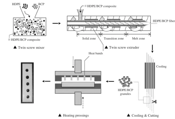
Fig. 1 shows the experimental schematic diagram for mak-ing a HDPE/BCP hybrid bone plate. In the first place, high-density polyethylene (HDPE) and biphasic calcium phosphate (BCP) powder were used as starting materials for fabrication of a HDPE/BCP hybrid bone plate. HDPE was used in the twin screw mixer and BCP powder was added with a parti-cle size of about 80~100 nm in diameter. Table 1 shows the typical properties of the constituents used for the experi-mental procedure. And then, HDPE and BCP mixtures were homogeneously made by the twin screw mixer (350/350 E, Brabender, Germany) for 1 h at a temperature of 150�C
depending on the volume ratio of HDPE/BCP. Secondly, the mixtures of HDPE/BCP were extruded homogeneously as a cylindrical rod shape using by a twin screw extruder (DSE 20, Brabender, Germany) at 120~150 rpm for 1 h at 150�C, which were about 3 mm in diameter. And to prepare of a HDPE/BCP granules, the HDPE/BCP fibers were used in the cooling (Typ.-844500.003, Brabender, Germany) for mor-phologic stabilization and cutting processing (Typ.-881207, Brabender, Germany). Compositions of the five composites are given in Table 1. Finally, the HDPE/BCP hybrid plates were fabricated using the granules of HDPE/BCP mixtures by a hot pressing method (2697, CARVER, USA) at 135~ 150�C under the pressure 3.0 MPa for 30 min.
Particularly, the BPC/HDPE hybrid plates were irradiated using γ-ray radiation (MDS Nordion, CA, IR221n wet storage Fig. 1. Schematic diagram showing the fabrication of the HDPE/BCP hybrid bone plate.
Table 1. Composition of the HDPE/BCP hybrid bone plate High density Biphasic calcium
Density Sample ID polyethylene phosphate
(HDPE, vol.%) (BCP, vol.%) (g cm-3)
100/0 100 0 0.964 90/10 90 10 1.137 80/20 80 20 1.355 70/30 70 30 1.572 60/40 60 40 1.743 HDPE HDPE/BCP composite HDPE/BCP composite HDPE/BCP fiber HDPE/BCP granules Solid zone Transition zone Melt zone
Twin screw mixer Twin screw extruder
Heat bands
Heating pressings
Cooling
Cooling & Cutting BCP
type C-188, ARTI, KAERI, Korea) to total doses of 25 kGy at a dose rate of 10 kGy h-1at room temperature in air for a
bridged bond and sterilization (Suarez et al. 2000; Kang and Nho 2001; Catano et al. 2005; Carmen et al. 2006).
3. Characterization of the HDPE/BCP hybrid plate
To evaluate a chemical analysis of materials, the hybrid plates were performed using a Fourier Transform Infrared Spectrometer (FT-IR, TENSOR-37, BRUKER, USA). The crystal structures and phases of the hybrid plates were identi-fied by X-ray Diffractometer (XRD, D/MAX-II, RIGAKU, Japan) operating at 40 kV and 150 mA. XRD with Cu Kα
source was employed for the range of 2θ==10~80�. The morphology and microstructures of the hybrid plates were investigated by a Scanning Electron Microscopy (SEM, JSM-6390, JEOL, Japan) with Au coating techniques (Automatic Magnetron Sputter Coater, 108A, Agar Scientific, UK). Densities, Vickers hardness and Young’s modulus of the HDPE/BCP hybrid plates were measured by using Gas Pycnometer (AccuPyc-1330, Micromeritics, USA), Micro Vickers Hardness Tester (HM-122 and VLPAK-2000, Aka-shi, Japan) and Ultrasonic Flaw Detector (5800-PR, Paname-trics, USA) techniques. Bending strength and tensile strength of the HDPE/BCP hybrid plates were evaluated using an Instron Universal Testing Machine (4443D2074, INSTRON, USA).
4. Evaluation of the biocompatibility
Biocompatibility of the hybrid bone plate on HaCaT (Hu-man Keratinocyte) cells was measured by a cell viability assay, with CCK-8 (Cell Counting Kit-8, Dojindo Lab., Japan) assay. CCK-8 assay is a dye and can be taken up by the mitochondria, and thus it was easily used to determine the cellular activity and to count the number of viable cells by spectrophotometrically measuring for the entire incuba-tion period (Kuhn et al. 2003; Maryama et al. 2004). A ready-for-use CCK-8 assay solution was used according to a pro-tocol suggested by the manufacturer. Briefly, the bone plates depending on the volume ratio of HDPE/BCP in FBS (Fetal Bovine Serum) free DMEM (Dulbecco’s Modified Eagle’s Medium) were incubated for 72 h at 37�C in a CO2incubator.
5×10³HaCaT cells in 100μl of the 10% FBS/DMEM
medi-um were seeded onto 96-well culture plate and allowed to adhere for 24 h before bone plate supernatant addition. After
24 h at 37�C in a CO2incubator, HaCaT cells in 96-well plate
were washed and the cells were exposed to bone plate super-natants at 10, 20, 40, 60, 80 and 100% concentrations for 24 h at 37�C in a CO2incubator and each concentration was
tested in triplicate. At the end of exposure, 10μl of CCK-8 solution was added to each well and the plate was incubated for 4 h at 37�C in a CO2incubator. After 4 h incubation, the
absorbance was measured by using an automated ELISA microplate reader (Bio-Tek Instruments, Winooski, VT, USA) at 450 nm.
5. Statistical analysis
Statistic analyses were performed using OriginPro ver. 7 software (OriginLab Inc). Each of the experimental points reported in this study are average values taken from five measurements.
RESULTS AND DISCUSSION
As can be seen in Fig. 2, the FT-IR analysis was perform-ed with attenuatperform-ed total reflection accessory (ATR, diamonds as an ATR element, the incident angle was 45�). Spectra show the infrared properties of HDPE/BCP (90/10 specimen). The positions of the FT-IR/ATR absorption peaks are summariz-ed in Table 2. It reveals absorption peaks of HDPE at 720, 1,470, 2,850 and 2,926 cm-1which are because of the -CH
3,
-CH2- and terminal vinyl groups in Fig. 2(a). The bands of
Fig. 2. FT-IR/ATR spectra of (a) HDPE, (b) BCP and (c) HDPE/ BCP (90/10) composites. 4000 3500 3000 2500 2000 1500 1000 Wavenumber (cm-1) C-H stretching of HDPE C-H bending PO stretching C-H rocking (a) (b) (c)
the -CH2- group of the asymmetrical stretching (2,926 cm-1) and symmetrical stretching (2,850 cm-1) are the most intense
in all the range of the assigned wavenumber. As shown in Fig. 2(b), the bands of the typical calcium phosphate system were absorbed strongly at 1,050 cm-1due to the PO
stretch-ing. Furthermore, the FT-IR/ATR spectrum of the HDPE/ BCP hybrid composites is shown in Fig. 2(c). The peaks of the biphasic calcium phosphate at 550, 613 and 1,050 cm-1
correspond to the PO43-bending mode. The C-H asymmetry bending (-CH3) in the spectra of the representative HDPE occurs at about 1,470 cm-1, and a CH
2rocking (-CH2-) app-ears near 720 cm-1, respectively. These results indicated that
HDPE has been combined with BCP.
Fig. 3 shows a comparison of the XRD patterns for the HDPE, BCP and HDPE/BCP composites (90/10). These XRD patterns are consistent with published HDPE in Fig. 3(a) (Han et al. 1999). Polyethylene is known to have a lamellar structure and this lamella structure usually has two phases, crystalline and amorphous. The bioceramics powder was also identified by a crystal analysis to be BCP, consist-ing of a 65~70/35~30 weight ratio of HAp/β-TCP in Fig. 3(b). As apparent in Fig. 3(c), the diffraction pattern obtained the presence of the HDPE/BCP (90/10) crystalline phase without other extra peaks for all the composites. It was con-firmed that BCP powders are uniformly dispersed in the HDPE matrix. When the calcium phosphate system was used as the implantation, homogenous distribution of the biphasic calcium phosphate system had an important effect on the bone formation behavior during the implanted periods.
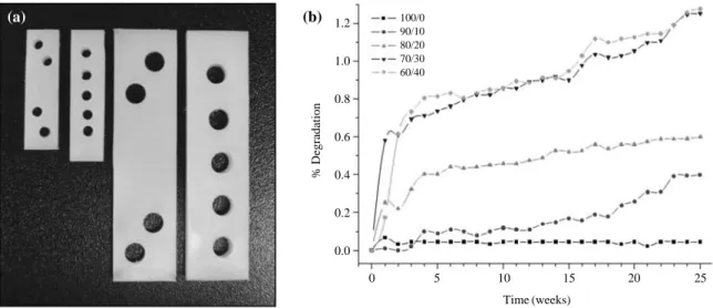
Fig. 4 shows the plates made of fabricated HDPE/BCP (80/20) composites, and the statistics of the biodegradability in the phosphate buffered saline (PBS) solution for 6 months. The dimensions of the HDPE/BCP hybrid plate were 800 mm in length, 20 mm in width and 2 mm in thickness in Fig. 4(a).
Table 2. Summarized FT-IR/ATR absorbance peak position of
HDPE
Group Vibrational mode Assigned wavenumber (cm-1)
-CH2- CH2rocking 720
-CH3 C-H asymmetry bending 1,470 -CH2- C-H symmetry stretching 2,850 -CH2- C-H asymmetry stretching 2,926
Fig. 3. XRD profiles of (a) HDPE, (b) BCP and (c) HDPE/BCP
(90/10) composites.
Fig. 4. Photograph (a) is showing the plates made of fabricated HDPE/BCP (80/20) composites, and biodegradability profiles (b) depending
on volume fractions of HDPE/BCP contents.
10 20 30 40 50 60 70 80 2θ degree Intensity (arb. unit) HDPE β-TCP HAp (a) (b) (c) 0 5 10 15 20 25 Time (weeks) 1.2 1.0 0.8 0.6 0.4 0.2 0.0 % Degradation 100/0 90/10 80/20 70/30 60/40 (a) (b)
The used hybrid plates consisted of an injection-molded polymer of HDPE and BCP bioceramics. The initial density was about 1.137 to 1.743 g cm-3in Table 1. The range of
these values was almost the same as human cortical bone and cancellous bone. These plates are also comfortable be-cause the HDPE/BCP have some flexibility. As shown in Fig. 4(b), biodegradation profiles of the fabricated HDPE/ BCP hybrid bone plate during 6 months (25 weeks) were investigated depending on the volume fractions of the HDPE/ BCP contents. As the BCP content increased, the degradabil-ity of the HDPE/BCP groups significantly increased because of the ionization of the BCP components such as Ca2++
and PO43-. Therefore, 100 vol.% HDPE contents (100/0) could not be degradated due to its high stability. The results of the biodegradability indicated an increasing degradability of the HDPE/BCP composites due to the high content of the BCP phase. The appreciable degradability was an important factor to improve the osteoconductivity and also enhance the in growth of the natural bone tissue.
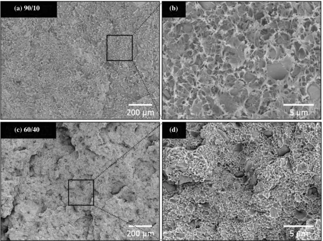
To identify the fracture characteristic of a HDPE/BCP
hybrid bone plate having different microstructures according to the existence of the BCP content, their fracture surfaces were observed in Fig. 5. In the low magnification image of the 90/10 specimen, the fracture surface was observed with a homogenous structure although the pulling-out phenome-non of the microstructure between the HDPE and BCP parti-cles were not revealed in Fig 5(a). In the magnified image (b) of the 90/10 specimen, the main fracture mode was the necking in the ductile fracture mode; many dimples were also revealed at local regions. However, as the BCP content increased, the main fracture mode was transformed to a brit-tle fracture type and a very roughness compared with the local regions of the 90/10 type, as shown in Fig. 5(c). From the enlarged images of the 60/10 specimen, especially, some fine BCP particles were founded on the fracture surface in Fig. 5(d), and this is typical evidence of a microcracking (crack interaction with residual strain field around dispersoid to create process zone ahead of crack tip) and a crack deflec-tion (surface roughening and crack tilting and a twisting dur-ing a propagation around dispersoid, cause by a thermal
ex-Fig. 5. SEM fracture surface of the HDPE/BCP hybrid bone plate: (a, b) 90/10 and (c, d) 60/40.
(a) 90/10 (b)
pansion mismatch and/or elastic modulus mismatch stress) in brittle fracture modes.
Fig. 6 shows the crack propagation made by Instron de-pending on the volume fractions of the HDPE/BCP contents. In the low magnification image (c) of the 60/40 composite, the crack deflection with the crack path was larger than that in the 90/10 composite. These images were taken from the crack tip regions marked with an arrow in Fig. 6(a, c). It was deduced that the HDPE/BCP hybrid microstructure has an effect on the crack deflection. As shown in Fig. 6(c), microcracking and crack bridging (pull out and crack bridg-ing by whisker and dispersoids) were observed more easily than in the 90/10 specimen due to the homogeneous addition of inorganic particles. From the enlarged magnification im-ages (b, d), it was inferred that the crack propagation was a typical ductile and brittle fracture pattern, respectively.
The dimpled marks made by a Vickers indentation depend-ing on the volume fractions of the HDPE/BCP contents are shown in Fig. 7. Spots that are not clear and battered were observed in the specimen of the HDPE/BCP containing 60 vol.% BCP in Fig. 7(a). However, as the volume fractions of the BCP powder increased, the dimpled spots were trans-formed dramatically. Spots at the corner and outer spots on
the corner are observed in the image (d) of the HDPE/BCP hybrid bone plate containing 40 vol.% BCP. It is due to the increase of the densification of the HDPE/BCP hybrid com-posites, because the BCP bioceramics powder as reinforcing agent is increasing.
Fig. 8. shows the Vickers hardness and Young’s modulus of the hybrid bone plate depending on the volume fractions of the HDPE/BCP contents. The values of the hardness and modulus of the HDPE/BCP plates which contained a 10 vol.% BCP content were about 7.0 Hv and 4.1 GPa, respec-tively. As the BCP content increased, the value of the Vickers hardness and Young’s modulus increased gradually because of the increased densification of the HDPE/BCP composites. Thus, their values increased with the reinforcing agent that contained a 60 vol.% BCP content which were approximately 11.1 Hv and 9.1 GPa because the BCP phase may be enhanc-ed by the mechanical stability of the HDPE phase. Especial-ly, from these evaluation, it was confirmed that the values of the Young’s modulus and densification of HDPE/BCP containing 60 vol.% BCP was the same as natural human bone.
The dependency of the bending strength and tensile streng-th on streng-the volume fraction of streng-the HDPE/BCP composites is
Fig. 6. Crack propagation of the HDPE/BCP hybrid bone plate: (a, b) 90/10 and (c, d) 60/40 composites.
(a) 90/10 (c) 60/40
shown in Fig. 9. The values of the bending strength in the 90/10 and 80/20 specimens were 61.19 and 46.45 MPa, while the tensile strengths were 22.9 and 21.7 MPa, respectively.
Furthermore, with increasing the BCP content up to 40 vol.%, the values of the bending and tensile strength also decreased because of the presence of a rigid filler (BCP). Generally, it
Fig. 9. SEM bending strength (MPa) and tensile strength (MPa) of
the hybrid bone plate depending on volume fractions of HDPE/BCP contents.
Fig. 7. The marks made by Vickers indentation depending on volume fractions of HDPE/BCP contents: (a) 90/10, (b) 80/20, (c) 70/30 and (d)
60/40.
(a) 90/10 (b) 80/20
(c) 70/30 (d) 60/40
90/10 80/20 70/30 60/40
HDPE/BCP content (vol.%)
Bending strength (MPa) Tensile strength (MPa) 65 60 55 50 45 40 35 30 24 22 20 18 16 14
Bending strength (MPa) Tensile strength (MPa)
Fig. 8. Vickers hardness (Hv) and Young’s modulus (GPa) of the
hybrid bone plate depending on volume fractions of HDPE/ BCP contents.
100/0 90/10 80/20 70/30 60/40 HDPE/BCP content (vol.%)
Vickers hardness (Hv) Young’s modulus (GPa) 12 11 10 9 8 7 6 10 8 6 4 2 Vickers hardness (Hv) Young’s modulus (GPa)
has been confirmed that hybrid organic/inorganic composites are necessary for a suitable modulus of a implantation. On the other hand, the hybrid composites are a necessary evil. As by increasing the inorganic content in the components without increasing the grain growth, the values of the bend-ing and tensile properties are decreased dramatically. These mechanical results indicated that HDPE/BCP composites are required with smaller inorganic articles or an acicular filer in the reverse direction of a stress due to an improvement of the microcracking and crack deflection for the reinforce-ment of the fracture mode.
The viability of the HaCaT cells was estimated after a exposure of the cells for 24 h to different concentrations of a bone plate supernatant. As observed in Fig. 10, the cells were exposed for 24 h with various concentrations (10, 20, 40 60, 80 and 100%) of different HDPE/BCP ratios (100/0, 90/10, 80/20 and 70/30 specimen). Viability was measured by the CCK-8 assay, as explained in detail in Section 2.4. The survival rate of the HaCaT cells was not altered by a treatment with the bone plate supernatants at a concentration of 10 to 100% for 24 h. In addition, all the bone plate super-natants at high concentrations (100%) for 72 h induced no decrease in the cell survival rate.
CONCLUSION
The hybrid bone plates were fabricated by a hot pressing
method using high-density polyethylene (HDPE) and differ-ent volume percdiffer-entages (10~40 vol.%) of biphasic calcium
phosphate (BCP) powder as a reinforcing agent, and the de-pendency of the morphological and mechanical properties on the BCP volume fractions was investigated. The BCP powders were homogeneously dispersed in the HDPE matrix by a twin screw mixer. According to the results, BCP bio-ceramics are also considered as deliberately biodegradable compounds, 90/10, 80/20, 70/30 and 60/40 specimens of HDPE/BCP hybrid composites were partially biodegraded, while no biodegradation was observed for the 100/0 groups. As the volume fractions of the BCP powder increased, it was confirmed that the fracture pattern changed from a duc-tile fracture mode to a brittle fracture mode. Particularly, the crack propagation of the microcracking and crack bridg-ing were observed more easily than the sbridg-ingle phase speci-men due to the homogeneous addition of inorganic particles. The morphologies of the matrix were dramatically changed, as the BCP bioceramics powder as a reinforcing agent was increased. The values of the density, Vickers hardness, Young’s modulus, bending strength and tensile strength of the 70/30 specimen were about 1.57%, 9.3 MPa, 7.9 Hv, 44.02 MPa and 20.4 MPa, respectively. It was confirmed that the density and Young’s modulus of the HDPE/BCP hybrid bone plates were almost like cancellous bones in human. In addition, the bone plates were not cytotoxic as indicated by the cell viability studies. The influence mechanism of the hybrid bone plate of various ratios on the cells is very com-plicated, which involves many factors, such as the chemical composition, pH values and concentration of extracts, and the incubation time. As a matter of fact, it is difficult to fully understand the influence mechanism in detail without a series control examination however the initial results of the cyto-toxicity in this study provide a good base for a further in vivo investigation of a bone tissue replacement.
ACKNOWLEDGMENT
This present work was supported by the Nuclear R & D Program from the Korea Ministry of Science and Technology.
REFERENCES
Aksakal B, Yildirim OS and Gul H. 2004. Metallurgical fail-Fig. 10. Cytotoxicity assays of HDPE/BCP hybrid bone plate
depending on the specimen extract and volume fractions.
100 80 60 40 20 10 Dilution of extraction Cell viability 120 100 80 60 40 20 0 100/0 90/10 80/20 70/30 60/40
ure analysis of various implant materials used in ortho-pedic applications. J. Failure. Analysis and Prevention
3(4):17-23.
Alhassan S and Goswami T. 2008. Wear rate model for UHM-WPE in total joint applications. Wear 265(1-2):8-13. Brown C, Williams S, Tipper JL, Fisher J and Ingham E. 2007.
Characterisation of wear particles produced by metal on metal and ceramic on metal hip prostheses under standard and microseparation simulation. J. Mater. Sci. Mater. Med.
18(5):819-827.
Carmen A, Arquimedes K, Rosestela P, Gema G, Nohemy D, Jeanette G and Yanixia S. 2006. HDPE/HA composites ob-tained in solution: Effect of the gamma radiation. Nucl. Instrum. Meth. Phys. Res. B 247(2):331-341.
Carmen A, Rosestela P, Arquimedes K, Gema G, Nohemy D, Yanixia S and Luis BJ. 2007. Characterization of hdpe/ha composites treated with titanate and zirconate coupling agents. Macromol. Symp. 247(1):190-198.
Catano L, Albano C, Karam A, Dominguez N, Sanchez Y and Gonzalez J. 2005. Effect of gamma irradiation on mechani-cal, thermal and rheological behavior of HDPE filled with seaweed residues. Nucl. Instrum. Meth. Phys. Res. B.
236(1-4): 348-353.
Chen Y, Zhang TH, Gan CH and Yu G. 2007. Wear studies of hydroxyapatite composite coating reinforced by carbon nanotubes. Carbon 45(5):998-1004.
Daculsi G. 1998. Biphasic calcium phosphate concept applied to artificial bone, implant coating and injectable bone sub-stitute. Biomaterials 19(16):1473-1478.
Daculsi G, Laboux O, Malard O and Weiss P. 2003. Current state of the art of biphasic calcium phosphate bioceramics. J. Mater. Sci. Mater. Med. 14(3):195-200.
Ferguson SJ, Wyss IP and Pichora DR. 1996. Finite element stress analysis of a hybrid fracture fixation plate. Med. Eng. Phys. 18(3):241-250.
Goodman SB. 2007. Wear particles, periprosthetic osteolysis and the immune system. Biomaterials 28(34):5044-5048. Goswami T and Alhassan S. 2008. Wear rate model for
UHM-WPE in total hip and knee arthroplasty. Mater Design
29(2):289-296.
Groot KD. 1980. Bioceramics consisting of calcium phosphate salts. Biomaterials 1(1):47-50.
Grubl A, Marker M, Brodner W, Giurea A, Heinze G, Meisin-ger V, Zehetgruber H and Kotz R. 2007. Long-term fol-low-up of metal-on-metal total hip replacement. J. Orthop. Res. 25(7):841-848.
Haaren EH and Heyligers IC. 2003. Implant wear and osteoly-sis with a hydroxylapatite-coated screw cup. Int. Orthop.
27(5):282-285.
Han JK, Song HY, Satio F and Lee BT. 2006. Synthesis of high
purity nano-sized hydroxyapaptite powder by microwave-hydrothermal method. Mater. Chem. Phys. 99(2-3):235-239.
Han SO, Lee DW and Han OH. 1999. Thermal degradation of crosslinked high density polyethylene. Polym. Degrad. Stabil. 63(2):237-243.
Homaeigohar SSH, Shokrgozar MA, Javadpour J, Khavandi A and Sadi AY. 2006. Effect of reinforcement particle size on in vitro behavior of β-tricalcium phosphate-reinforced high-density polyethylene: A novel orthopedic composite. J. Biomed. Mater. Res. A. 78(1):129-138.
Homaeigohar SSH, Shokrgozar MA, Khavandi A and Sadi AY. 2008. In vitro biological evaluation of -TCP/HDPE-A novel orthopedic composite: A survey using human osteo-blast and fibroosteo-blast bone cells. J. Biomed. Mater. Res. A.
84(2):491-499.
Homaeigohar SSH, Shokrgozar MA, Sadi AY, Khavandi A Javadpour J and Hosseinalipour M. 2005. In vitro evalua-tion of biocompatibility of beta-tricalcium phosphate-reinforced high-density polyethylene; an orthopedic com-posite. J. Biomed. Mater. Res. A. 75(1):14-22.
Huang S, Zhou K, Zhu W, Huang B and Li Z. 2006. Effects of in situ biomineralization on microstructural and mechani-cal properties of hydroxyapatite/polyethylene composites. J. Appl. Polym. Sci. 101(3):1842-1847.
Kalita SJ, Bhardwaj A and Bhatt HA. 2007. Nanocrystalline cal-cium phosphate ceramics in biomedical engineering. Mater. Sci. Eng. C. 27(3):441-449.
Kang PH and Nho YC. 2001. The effect of γ-irradiation on ultra-high molecular weight polyethylene recrystallized under different cooling conditions. Radiat. Phys. Chem.
60(1-2):79-87.
Khan M, Takahashi T, Kuiper JH, Sieniawska CE, Takagi K and Richardson JB. 2006. Current in vivo wear of metal-on-metal bearings assessed by exercise-related rise in plas-ma cobalt level. J. Orthop. Res. 24(11):2029-2035. Kuhn DM, Balkis M, Chandra J, Mukherjee PK and
Ghan-noum MA. 2003. Uses and limitations of the XTT assay in studies of candida growth and metabolism. J. Clin. Micro-biol. 41(1):506-508.
Lee BT, Youn MH, Paul RK, Lee KH and Song HY. 2007. In-situ sythesis of spherical BCP nano powders by microwave assisted process. Mater. Chem. Phys. 104(2-3):249-253. Legeros RZ, Lin S, Rohanizadeh R, Mijares D and Legeros JP.
2003. Biphasic calcium phosphate bioceramics: preparation, properties and applications. J. Mater. Sci. Mater. Med.
14(3):201-209.
Li Y, Weng W and Tam KC. 2007. Novel highly biodegrada-ble biphasic tricalcium phosphates compsoed of α-trical-cium phosphate and β-tricalcium phosphate. Acta
Bioma-terialia 3(2):251-254.
Manjubala I and Sivakumar M. 2001. In-situ synthesis of bipha-sic calcium phosphae ceramics using microwave irradia-tion. Mater. Chem. Physics. 71(3):272-278.
Maruyama K, Iwasaki F, Takizawa T, Yanagie H, Niidome T, Yamada E, Ito T and Koyama Y. 2004. Novel receptor-mediated gene delivery system comprising plasmid/prota-mine/sugar-containing polyanion ternary complex. Bioma-terials 25(11):3267-3273.
McKee GK. 1982. Total hip replacement-past, present and future. Biomaterials 3(3):130-135.
Nath S, Bodhak S and Basu B. 2007. Tribological investiga-tion of novel HDPE-HAp-Al2O3hybrid biocomposites
against steel under dry and simulated body fluid condition. J. Biomed. Mater. Res. A. 83(1):191-208.
Nie X, Leyland A and Matthews A. 2000. Deposition of layer-ed bioceramic hydroxyapatite/TiO2 coatings on titanium
alloys using a hybrid technique of micro-arc oxidation and electrophoresis. Sur. Coat. Tech. 125(1-3):407-414. Ozgur NE and Cuneyt TA. 1999. Manufacture of macroporous
calcium hydroxyapatite bioceramics. J. Euro. Ceram. Soc.
19(13-14):2569-2572.
Pandey A, Jan E and Aswath PB. 2006. Physical and mechani-cal behavior of hot rolled HDPE/HA composites. J. Mater. Sci. 41(11):3369-3376.
Poole CE, Patil SS, Lima DD and Colwell CW. 2005. Early fol-low-up for a hybrid total hip arthroplasty using a metal-backed acetabular component designed to reduce “Back-side” polyethylene wear. Hosp Special Surgery 1:31-34. Rahman A and Lau ACW. 1998. Singular stress fields in
inter-facial notches of hybrid metal matrix composites. Compos B. 29(6):763-768.
Roeder RK, Sproul MM and Turner CH. 2003. Hydroxyapatite whiskers provide improved mechanical properties in rein-forced polymer composites. J. Biomed. Mater. Res. A.
67(3):801-812.
Ryu HS, Youn HJ, Hong KS, Chang BS and Lee CK. 2002. An improvement in sintering property of β-tricalcium phos-phate by addition of calcium pyrophosphos-phate. Biomaterials
23(3):909-914.
Samuel S, Nag S, Scharf TW and Banerjee R. 2008. Wear resis-tance of laser-deposited boride reinforced Ti-Nb-Zr-Ta alloy composites for orthopedic implants. Mater. Sci. Eng. C. 28(3):414-420.
Sargeant A and Goswami T. 2006. Hip implants: Paper V.
Phy-siological effects. Mater Design 27(4):287-307.
Schmalzried TP. Why total hip resurfacing. J. Arthroplasty
22(7 suppl.):57-60.
Shao Z, Dyck LE, Wang H and Li XM. 2006. Antipsychotic drugs cause glial cell line-derived neurotrophic factor secre-tion from C6 glioma cells. J. Psychiatry Neurosci. 31(1): 32-37.
Slonaker M and Goswami T. 2004. Review of wear mechanisms in hip implants: Paper II-ceramics IG004712. Mater Design
25(5):395-405.
Sonoda M, Nishiyama T, Matsukawa Y and Moriyasu M. 2004. Cytotoxic activities of flavonoids from two Scutellaria plants in Chinese medicine. J. Ethnopharmacol 91(1):65-68. Spoerke ED, Murray NG, Li H, Brinson LC, Dunand DC and
Stupp SI. 2005. A bioactive titanium foam scaffold for bone repair. Acta Biomaterialia 1(5):523-533.
Suarez JCM, Mano EB and Pereira RA. 2000. Thermal beha-vior of gamma-irradiated recycled polyethylene blends. Polym. Degrad. Stabil. 69(2):217-222.
Xie J, Luan BL, Wang J, Liu XY, Rorabeck C and Bourne R. 2008. Novel hydroxyapatite coating on new porous titani-um and titanititani-um-HDPE composite for hip implant. Sur Coat Tech 202(13):2960-2968.
You C, Miyazaki T, Ishida E, Ashizuka M, Ohtsuki C and Tani-hara M. 2007. Fabrication of poly(vinyl alcohol)-apatite hybrids through biomimetic process. J. Euro. Ceram. Soc.
27(2-3):1585-1588.
Youn MH, Paul RK, Song HY and Lee BT. 2007. Fabrication of porous structure of BCP sintered bodies using microwave assisted synthesized HAp nano powder. Mater. Sci. For.
534-536(part1):49-52.
Yubao L, Klein CPAT, Xingdong Z and Groot KD. 1994. For-mation of a bone apatite-like layer on the surface of porous hydroxyapatite ceramics. Biomaterials 15(10):835-841. Zhang Y, Tanner KE, Gurav N and Silvio LD. 2007. In vitro
osteoblastic response to 30 vol% hydroxyapatitepolyethy-lene composite. J. Biomed. Mater. Res. A. 81(2):409-417. Zou C and Shen Z. 2007. An optimized in vitro assay for
screen-ing compounds that stimulate liver cell glucose utilization with low cytotoxicity. J. Pharmacol. Toxicol. Meth. 56(1): 58-62.
Manuscript Received: May 24, 2010 Revision Accepted: June 9, 2010