INTRODUCTION
Gallbladder cancer (GBC), though generally considered rare, is the most common malignancy of the biliary tract, accounting for 80–95% of biliary tract cancers. GBC is considered the most aggressive of biliary cancers with the shortest survival time.1
Complete surgical resection offers the only chance for a cure; however, only 10% of patients with GBC present with early-stage disease and are considered surgical candidates. Advanced GBC is characterized by early local invasion, extensive regional lymph node metastasis, vascular encasement, and distant
me-Significance of Metabolic Tumor Volume and
Total Lesion Glycolysis Measured Using
18
F-FDG PET/CT in Locally Advanced and
Metastatic Gallbladder Carcinoma
You Jin Chun
1, Hei-Cheul Jeung
2, Hyung Soon Park
3, Ji Soo Park
4,
Sun Young Rha
1, Hye Jin Choi
1, Jae-Hoon Lee
5, and Tae Joo Jeon
51Division of Medical Oncology, Department of Internal Medicine, Yonsei Cancer Center, Yonsei University College of Medicine, Seoul; 2Division of Medical Oncology, Department of Internal Medicine, Gangnam Severance Hospital, Yonsei University College of Medicine, Seoul; 3Division of Medical Oncology, Department of Internal Medicine, St. Vincent’s Hospital, The Catholic University of Korea, Suwon;
4Cancer Prevention Center, Yonsei Cancer Center, Yonsei University College of Medicine, Seoul;
5Department of Nuclear Medicine, Gangnam Severance Hospital, Yonsei University College of Medicine, Seoul, Korea.
Purpose: This study aimed to determine the prognostic value of new quantitative parameters of 18F-fluorodeoxyglucose positron
emission tomography/computed tomography (18F-FDG PET/CT), including metabolic tumor volume (MTV), in patients with
lo-cally advanced and metastatic gallbladder cancer (GBC).
Materials and Methods: In total, 83 patients initially diagnosed with locally advanced and metastatic GBC and who underwent
18F-FDG PET/CT at the time of initial diagnosis were retrospectively reviewed. The metabolic volume-based PET parameters of
pri-mary tumors and metastatic lesions were measured, including maximum and average standardized uptake values (SUV), MTV, and total lesion glycolysis. An overall survival (OS) analysis was performed using the Kaplan-Meier method with PET and clinical parameters. A Cox proportional hazards regression analysis was performed to determine independent prognostic factors.
Results: In univariate analysis, pathologic differentiation (p<0.001), performance status (PS; p=0.003), C-reactive protein (CRP) level (p=0.009), and PET-related SUVmt max (the highest SUV among the metastatic lesions) (p=0.040) and MTVtotal (the sum of the
MTVs of both the primary and metastatic lesions) (p=0.031), were significant for OS. In multivariate analysis, MTVtotal (hazard
ra-tio: 2.07; 95% confidence interval: 1.23–3.48; p=0.006) remained significant for the prediction of OS, as did differentiation (p=0.001), PS (p=0.001), and CRP (p=0.039).
Conclusion: In locally advanced and metastatic GBC, volume-based PET/CT parameters of the total tumor burden of malignan-cy, such as MTVtotal, were found to be useful for the identification of patients with poor prognosis.
Key Words: Gallbladder neoplasms, metastasis, 18F-FDG PET/CT, metabolic tumor volume, prognosis
pISSN: 0513-5796 · eISSN: 1976-2437
Received: November 26, 2018 Revised: April 20, 2019 Accepted: April 25, 2019
Corresponding author: Tae Joo Jeon, MD, PhD, Department of Nuclear Medicine,
Gangnam Severance Hospital, Yonsei University College of Medicine, 211 Eonju-ro, Gangnam-gu, Seoul 06273, Korea.
Tel: 82-2-2019-3740, Fax: 82-2-3462-5472, E-mail: [email protected] •The authors have no potential conflicts of interest to disclose. © Copyright: Yonsei University College of Medicine 2019
This is an Open Access article distributed under the terms of the Creative Com-mons Attribution Non-Commercial License (https://creativecomCom-mons.org/licenses/ by-nc/4.0) which permits unrestricted non-commercial use, distribution, and repro-duction in any medium, provided the original work is properly cited.
Yonsei Med J 2019 Jul;60(7):604-610 https://doi.org/10.3349/ymj.2019.60.7.604
tastasis, leading to a poor prognosis from unresectable or met-astatic disease.1,2
Accurate stratification for outcome prediction based only on anatomic stage is difficult. A more accurate and reliable prog-nostic system incorporating additional features of tumors, such as biological and molecular information, may be necessary to obtain a better prediction of prognosis and to choose the most appropriate treatment modality and follow-up plan for locally advanced and metastatic GBC. 18F-fluorodeoxyglucose
positron emission tomography/computed tomography (18
F-FDG PET/CT) is an increasingly available noninvasive test for malignancy based on glucose metabolism. It was recently dem-onstrated to be valuable for initial staging and for the detection of recurrent diseases in many kinds of tumors, including GBC.3 18F-FDG PET is useful not only for diagnosing and staging, but
also for evaluating the proliferative activity and malignancy grades of tumors reflecting prognosis. The standardized up-take value (SUV) of the primary tumor, a semi-quantitative pa-rameter derived from 18F-FDG PET, is a significant prognostic
factor for various types of cancer.4,5 Despite being a popular
landmark clinically, this parameter only has a single voxel value and cannot be used to indicate the metabolism of the whole tu-mor and metastatic lesions. In fact, many studies that have in-dicated SUV as a significant prognostic factor did not analyze parameters reflective of tumor volume. Recent studies have re-ported that volumetric PET parameters, such as metabolic tu-mor volume (MTV) and total lesion glycolysis (TLG), using a threshold-based automatic volume of interest (VOI) were bet-ter prognostic predictors for survival in patients with malignant pleural mesothelioma, esophageal cancer, and advanced head and neck squamous cell carcinomas.6-8 There is still little
evi-dence that volumetric PET parameters are significant prognos-tic predictor in patients with GBC. This study sought to investi-gate the prognostic value of SUV and volume-based metabolic parameters on used 18F-FDG PET/CT in comparison to other
clinical parameters in patients with advanced and metastatic GBC.
MATERIALS AND METHODS
PatientsA total of 83 patients at Gangnam Severance Hospital with bi-opsy-proven gallbladder adenocarcinoma who received 18
F-FDG PET/CT pretreatment between January 2007 and Decem-ber 2015 were included in this retrospective study. Exclusion criteria were patients with resectable early disease, double-pri-mary malignancy, previous cholecystectomy, and other histol-ogy types, such as cystic neoplasms, neuroendocrine tumors, or lymphomas. The Institutional Review Board of Severance Hospital, Yonsei University Health System approved this retro-spective study (IRB Number: 3-2015-0318) and waived the re-quirement to obtain informed consent.
The medical records of each patient were investigated for sex, age, histologic typing, performance status (PS), extrahepatic metastases, carcinomatosis, and treatment modality. Histologic typing was classified as well, moderately, or poorly differenti-ated, and PS was classified according to the Eastern Coopera-tive Oncology Group (ECOG) performance status. Computed tomography (CT) of the chest and abdominopelvic cavity, a ra-dionuclide bone scan, and 18F-FDG PET/CT were performed to
evaluate locally advanced disease or distant metastasis. Sur-vival status was retrieved from our medical records or from at-tempts to contact the patients or their referring physicians. All follow-up evaluations ended on December 30, 2015.
18F-FDG PET/CT imaging protocols
Imaging and data acquisition for metabolic parameters was conducted using the PET/CT system (Biograph TruePoint 40, Siemens Healthcare, Munich, Germany). PET/CT was per-formed before treatment. The fasting time before the adminis-tration of 18F-FDG (Nambuk Medical, Seoul, Korea) was at
least 6 hours, and serum glucose levels were not to have ex-ceeded 150 mg/dL. After the injection of 5.18 MBq/kg (0.14 mCi/kg) of 18F-FDG, each patient waited in a warm, quiet, dim
room for 60 minutes. An initial low-dose CT scan was followed by a PET scan from the skull base to the upper thigh level in the three-dimensional mode (1.5-min acquisition time per bed), and the scanned data were reconstructed using the iterative method, ordered subset expectation maximization, using two iterations and 21 subsets. Trans-axial spatial resolution of the PET system was 5 mm full-width at half maximum at the center of the field of view. The matrix size and thickness of the recon-structed PET image were 128×128 and 5 mm, respectively. Analysis of PET/CT data
The metabolic parameters from 18F-FDG PET/CT data were
evaluated by two experienced nuclear medicine physicians using dedicated software for the PET/CT workstation (Syngo VE32B, Siemens AG, Berlin, Germany). To define the contour-ing margins around the tumor, SUV >2.5 was used as previous-ly reported.9 The contour around the target lesions within the
boundaries was automatically generated and within the con-tour margin were combined to define the tumor volumes. MTV was defined as the sum of metabolic volumes of tumor tissues with increased FDG uptake. The SUV threshold value used in this study was 50% of the local maximum SUV intensity, iden-tified as a reasonable value in phantom studies.9,10 We selected
lesions with SUV >2.5, and selected regions within lesions with a SUV intensity greater than 50% for quantitative MTV measurement. TLG was representative of the metabolic activ-ity throughout the entire tumor and was calculated by multi-plying MTV and the mean SUV (SUVmean) of the MTV.
Appro-priately sized spherical VOIs, including each targeted locally advanced and metastatic lesion, were created by considering the tumor location in the trans-axial, sagittal, and coronal
planes. Physiologic activities in the adjacent liver, stomach, and bowel loops were avoided. These parameters, SUV, and MTV were automatically calculated by the Syngo software (Sie-mens, Erlangen, Germany).
Statistical analysis
The primary end point of this study was overall survival (OS), which was measured from the date of diagnosis of GBC to the date of death from any cause. The Kaplan-Meier method was used for survival analysis, and the difference in the rate was compared using a log-rank test. A prognostic model was es-tablished by finding all of the variables that significantly influ-enced OS (p<0.05) in univariate analysis. The clinical variables included in the univariate analysis were age, sex, pathologic differentiation, ECOG PS, extrahepatic metastases, carcinoma-tosis, C-reactive protein (CRP), and tumor markers [serum car-cinoembryonic antigen (CEA) and carbohydrate antigen 19-9 (CA19-9) levels]. Metabolic PET variables included the highest SUV of the locally advanced lesion (SUVLA max), the highest
SUV among the distant metastatic lesions (SUVmt max), the
MTV of the locally advanced lesion (MTVLA), the sum of the
MTVs of all metastatic lesions (MTVmt total), the highest MTV
among the metastatic lesions (MTVmt max), the sum of the MTVs
of both the locally advanced and metastatic lesions (MTVtotal),
the TLG of the locally advanced lesion (TLGLA), the sum of the
TLGs of all metastatic lesions (TLGmt total), the highest TLG
among the TLGmt (TLGmt max), and the sum of the TLGs of both
locally advanced and metastatic lesions (TLGtotal). For
metabol-ic parameters, the median value was used as the cut-off; for tu-mor markers, the normal range was used. A Cox proportional hazards regression analysis was performed to determine inde-pendent prognostic factors. Statistical analyses were per-formed using IBM SPSS Statistics for Windows, Version 23 (IBM Corp., Armonk, NY, USA). P values <0.05 were considered sig-nificant.
RESULTS
Patient characteristics
This study included 83 patients. The median clinical follow-up period was 9.9 months (range, 0.2–77.2 months). Baseline pa-tient and tumor characteristics, including age, sex, pathologic differentiation, ECOG PS, extrahepatic metastases, carcino-matosis, CRP, CEA, CA19-9, SUVLA max, SUVmt max, MTVLA,
MT-Vmt total, MTVmt max, MTVtotal, TLGLA, TLGmt total, TLGmt max, TLGtotal
levels, and treatment modality are presented in Table 1. Prognostic factors evaluated in univariate analysis The cut-off levels of serum CRP, CEA, and CA19-9 levels were set to 6 mg/L, 5 ng/mL, and 24 U/mL, respectively, based on the normal values at our institution. The median age of 67 years was used as a cut-off. The median values of the PET and
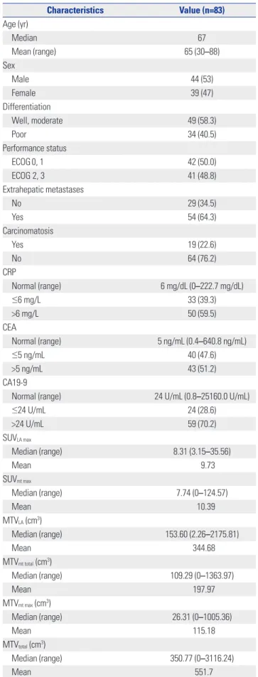
meta-Table 1. Baseline Patient Characteristics with Positron Emission To-mography Parameters of Primary and Metastatic Lesions
Characteristics Value (n=83) Age (yr) Median 67 Mean (range) 65 (30–88) Sex Male 44 (53) Female 39 (47) Differentiation Well, moderate 49 (58.3) Poor 34 (40.5) Performance status ECOG0, 1 42 (50.0) ECOG 2, 3 41 (48.8) Extrahepatic metastases No 29 (34.5) Yes 54 (64.3) Carcinomatosis Yes 19 (22.6) No 64 (76.2) CRP Normal (range) 6 mg/dL (0–222.7 mg/dL) ≤6 mg/L 33 (39.3) >6 mg/L 50 (59.5) CEA Normal (range) 5 ng/mL (0.4–640.8 ng/mL) ≤5 ng/mL 40 (47.6) >5 ng/mL 43 (51.2) CA19-9
Normal (range) 24 U/mL (0.8–25160.0 U/mL)
≤24 U/mL 24 (28.6) >24 U/mL 59 (70.2) SUVLA max Median (range) 8.31 (3.15–35.56) Mean 9.73 SUVmt max Median (range) 7.74 (0–124.57) Mean 10.39 MTVLA (cm3) Median (range) 153.60 (2.26–2175.81) Mean 344.68 MTVmt total (cm3) Median (range) 109.29 (0–1363.97) Mean 197.97 MTVmt max (cm3) Median (range) 26.31 (0–1005.36) Mean 115.18 MTVtotal (cm3) Median (range) 350.77 (0–3116.24) Mean 551.7
bolic parameters were as follows: SUVLA max, 8.31; SUVmt max,
7.74; MTVLA, 153.60 cm3; MTVmt total, 109.29 cm3; MTVmt max,
26.31 cm3; MTV
total, 350.77 cm3; TLGLA, 829.24 g; TLGmt total,
392.50 g; TLGmt max, 102.82 g; and TLGtotal, 2191.76 g. The
pa-tients were divided into two groups according to the median value of the parameters. Overall, 46 patients were younger than the median age of 67 years; 37 patients were older. Univariate analysis demonstrated that pathologic differentiation (p<0.001), PS (p=0.003), CRP level (p=0.009), SUVmt max (p=
0.040), and MTVtotal (p=0.031) were significantly prognostic. In
addition, as expected, chemotherapy with gemcitabine and cis-platin had a significant impact on prognosis (Table 2). Prognostic factors evaluated in multivariate analysis In multivariate analysis of adjusted treatment modalities, patho-logic differentiation [HR=2.42 (well differentiated and moder-ately differentiated vs. poorly differentiated); p=0.001], PS [HR= 2.28 (ECOG 0, 1 vs. 2, 3); p=0.001], CRP (HR=1.73; p=0.039), and MTVtotal [HR=2.07 (≤350.77 cm3 vs. >350.77 cm3); p=0.006]
were independent prognostic factors for the prediction of OS (Table 3, Fig. 1). In patients with locally advanced and
meta-static disease, MTVtotal, a volume-based metabolic PET
pa-rameter, was an important independent prognostic factor for OS, along with other PET parameters.
DISCUSSION
There have been various reports on the role of PET/CT in GBC Table 1. Baseline Patient Characteristics with Positron Emission
To-mography Parameters of Primary and Metastatic Lesions (Continued)
Characteristics Value (n=83) TLGLA (g) Median (range) 829.24 (5.62–16147.31) Mean 2546.76 TLGmt total (g) Median (range) 392.50 (0–28170.70) Mean 1762.47 TLGmt max (g) Median (range) 102.82 (0–28170.70) Mean 990.31 TLGtotal (g) Median (range) 2191.76 (0–28525.03) Mean 4386.3 Treatment modality Gemcitabine+cisplatin (n) 62 No treatment (n) 18 Others (n)* 3
ECOG, Eastern Cooperative Oncology Group; CRP, C-reactive protein; CEA, carcinoembryonic antigen; CA19-9, carbohydrate antigen 19-9; SUV, stan-dardized uptake value; SUVLA max, the highest SUV of the locally advanced le-sion; SUVmt max, the highest SUV among the distant metastatic lesions; MTV, metabolic tumor volume; MTVLA, the MTV of the locally advanced lesion; MTVmt total, the sum of the MTVs of all metastatic lesions; MTVmt max, the high-est MTV among the metastatic lesions; MTVtotal, the sum of the MTVs of both the locally advanced and metastatic lesions; TLG, total lesion glycolysis; TL-GLA, the TLG of the locally advanced lesion; TLGmt total, the sum of the TLGs of all metastatic lesions; TLGmt max, the highest TLG among the TLGmt; TLGtotal, the sum of the TLGs of both locally advanced and metastatic lesions.
Values are presented as number (percentage) unless otherwise noticed. *Others, one patient with concurrent chemotherapy (CCRT) using cisplatin, one patient with CCRT using fluorouracil, and one patient with radiotherapy alone.
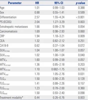
Table 2. Univariate Analysis of Prognostic Factors for Survival Outcomes
Parameter HR 95% CI p value Age 1.01 0.99–1.03 0.388 Sex 0.88 0.55–1.41 0.599 Differentiation 2.57 1.55–4.24 < 0.001 PS (ECOG) 2.04 1.27–3.29 0.003 Extrahepatic metastases 1.66 0.99–2.78 0.054 Carcinomatosis 1.69 0.98–2.93 0.060 CRP 1.94 1.18–3.21 0.009 CEA 1.32 0.82–2.12 0.251 CA19-9 0.62 0.37–1.04 0.072 SUVLA max 1.04 1.00–1.07 0.057 SUVmt max 1.02 1.00–1.04 0.040 MTVLA 1.60 0.99–2.59 0.057 MTVmt total 1.36 0.85–2.18 0.203 MTVmt max 1.10 0.682–1.75 0.716 MTVtotal 1.70 1.05–2.76 0.031 TLGLA 1.50 0.90–2.35 0.128 TLGmt total 1.41 0.88–2.26 0.157 TLGmt max 1.23 0.78–2.00 0.360 TLGtotal 1.50 0.93–2.40 0.098 Treatment modality* 0.44 0.26–0.76 0.003 HR, hazard ratio; CI, confidence interval; PS, performance status; ECOG, East-ern Cooperative Oncology Group; CRP, C-reactive protein; CEA, carcinoembry-onic antigen; CA19-9, carbohydrate antigen 19-9; SUV, standardized uptake value; SUVpri max, the highest SUV of the primary lesion; SUVmt max, the highest SUV among the metastatic lesions; MTV, metabolic tumor volume; MTVpri, the MTV of the primary lesion; MTVmt total, the sum of the MTVs of all meta-static lesions; MTVmt max, the highest MTV among the metastatic lesions; MT-Vtotal, the sum of the MTVs of both the primary and metastatic lesions; TLG, total lesion glycolysis; TLGpri, the TLG of the primary lesion; TLGmt total, the sum of the TLGs of all metastatic lesions; TLGmt max, the highest TLG among the TLGmt; TLGtotal, the sum of the TLGs of both primary and metastatic lesions. *Sixty-two patients received gemcitabine plus cisplatin chemotherapy, 18 pa-tients received no treatment, and 3 papa-tients received concurrent chemother-apy or radiotherchemother-apy alone.
Table 3. Multivariate Analysis of Prognostic Factors of Survival Outcomes
Parameter HR 95% CI p value Differentiation 2.42 1.43–4.09 0.001 PS 2.28 1.38–3.77 0.001 CRP 1.73 1.03–2.92 0.039 MTVtotal 2.07 1.23–3.48 0.006 Treatment modality 0.40 0.23–0.71 0.002 HR, hazard ratio; CI, confidence interval; PS, performance status; CRP, C-reac-tive protein; MTVtotal, the sum of the MTVs of both locally advanced and meta-static lesions.
diagnosis. In particular, 18F-FDG PET/CT seems to have a
po-tential role in staging, as these cancers are intensely FDG-avid. PET/CT has an overall diagnostic accuracy of 95.9% for the pri-mary disease and 85.7% and 95.9% for the detection of lymph nodes and metastatic lesions, respectively.11 Enhanced
utiliza-tion of glucose than normal tissues, more aggressive malignan-cies, and higher rates of glycolysis than less malignant or benign tumors have been observed in cancer cells.12-14 These glucose
metabolism differences can be measured quantitatively in vivo by PET after FDG administration.
We investigated the prognostic values of volume-based met-abolic parameters using 18F-FDG PET/CT in metastatic GBC,
compared with conventional clinical parameters. Many prog-nostic factors for advanced and metastatic GBC have been pro-posed: most are clinico-pathological parameters. Clinical or pathologic staging, including tumor extension and nodal in-volvement, and blood CEA levels have remained good prog-nostic values for GBC.15-17 However, these parameters cannot
be categorized in detail or presented as objective numbers in unresectable GBC. In other words, a significant prognostic mark-er that can quantify molecular and metabolic parametmark-ers is re-quired for the treatment decision of GBC patients.
Several recent studies have investigated the prognostic val-ue of 18F-FDG PET/CT parameters in GBC patients. Despite an
1.0 0.8 0.6 0.4 0.2 0.0 1.0 0.8 0.6 0.4 0.2 0.0 1.0 0.8 0.6 0.4 0.2 0.0 1.0 0.8 0.6 0.4 0.2 0.0 Survival probability
Survival probability Survival probability
Survival probability 0 10 20 30 40 50 60 0 10 20 30 40 50 60 0 10 20 30 40 50 60 0 10 20 30 40 50 60 Time (month) Time (month) Time (month) Time (month) MTVtotal ≤350.77 cm3 >350.77 cm3 CRP ≤6 mg/L >6 mg/L Performance status ECOG 1, 2 ECOG 2, 3 Differentiation Well, moderate Poor A C B D
Fig. 1. Kaplan-Meier curves for overall survival based on significant prognostic factors, including (A) pathologic differentiation [HR=2.42 (well differentiat-ed and moderately differentiatdifferentiat-ed vs. poorly differentiatdifferentiat-ed); p=0.001], (B) performance status [HR=2.28 (ECOG 0, 1 vs. 2, 3); p=0.001], (C) CRP [HR=1.73 (≤6 mg/dL vs. >6 mg/dL); p=0.039], and (D) MTVtotal [HR=2.07 (≤350.77 cm3 vs. >350.77 cm3); p=0.006] in gallbladder carcinoma. ECOG, Eastern Cooperative On-cology Group; CRP, C-reactive protein; MTVtotal, the sum of the MTVs of both the locally advanced and metastatic lesions; HR, hazard ratio.
absence of standardized cut-off values, poorer survival has consistently been correlated with a high SUVmax of the primary
lesion as measured on pretreatment 18F-FDG PET/CT scans of
biliary tract carcinoma.18 Only one previous study investigated
the utility of 18F-FDG PET/CT volumetric parameters to predict
clinical outcomes in GBC. Yoo, et al.19 analyzed the various
metabolic volume-based PET parameters of primary tumors, including maximum and average SUV, MTV, and TLG, mea-sured on 18F-FDG PET/CT scans of 44 patients with GBC. They
showed that those with an MTV cut-off of 135 cm3 (p=0.001) and
a TLG cut-off of 7090 g (p<0.050) had significantly longer OS than those with lower MTV and TLG values. In the present study, pathologic differentiation, PS, and serum CRP levels were significant factors according to univariate analysis, while SUV and MTV were significant independent prognostic fac-tors. MTVtotal was determined to be a meaningful independent
prognostic factor in multivariate analysis with adjustment for treatment modality.
Histologically, the gallbladder does not have submucosa, and the cancer infiltrates directly into the muscularis propria. The gallbladder wall is thin, and the cancer is able to easily infil-trate to adjacent organs, such as the liver, duodenum, and pan-creas. As GBC is associated with a high rate of local invasion and distant metastases, resulting in poor survival, we hypoth-esized that the metabolic activity of all primary and metastatic lesions on 18F-FDG PET/CT scans might be more helpful to
guide treatment decisions than the primary lesion only. Thus, our study included locally advanced and metastatic GBC pa-tients and measured the MTV and TLG of both metastatic and locally advanced primary lesions. In doing so, we deduced through multivariate analysis that the total MTV, including metastatic lesions, was the most significant prognostic factor.20
Quantified metabolic activity can provide valuable informa-tion to help prognosticate and assess treatment response in clinical oncology.21 While CT scan and MRI readily reveal the
anatomic distribution of tumors, they do not permit the quanti-fication of the metabolic activity of a tumor. Anatomically large tumors in CT scan can have low metabolic activity, and small le-sions of metastases can be highly active. Therefore, it is thought that MTV measurement with PET/CT is an important factor for prediction of survival prognosis.22,23 While SUV
mt max (the
highest SUV among the metastatic lesions) was significant in univariate analysis, it was excluded from multivariate analysis because SUVmt max, interpreted as a single voxel value, may not
reflect the tumor’s general metabolism due to tumor hetero-geneity. In addition, TLG was not significant for survival pre-diction: it is calculated by multiplying the tumor volume by the SUVmean. Metastatic GBC can show diverse SUVs in both a
local-ly advanced primary lesion and in multiple metastatic lesions. Therefore, as the TLG is calculated as the SUVmean, its
impor-tance in survival prediction may be weakened. MTV appeared to be more important for prediction of survival prognosis than SUV due to the variety of SUVs obtained.
Our present study had several limitations. First, it was de-signed as a retrospective study and included a relatively small number of patients. Therefore, our results may not be applica-ble to all patients with locally advanced and metastatic GBC. Second, it was difficult to clarify the boundary between the primary lesion and liver infiltrative lesion, since GBC readily invade the liver. Hence, we defined locally advanced GBC in-cluding liver invasion and obtained MTV according to locally advanced primary lesion. Third, the cut-off value of each PET parameter was set to a median value due to a small number of samples and a wide range of parameter values. It is necessary to find an accurate cut-off value with a larger number of pa-tients.
Despite these limitations, our study is the first to demon-strate the clinical value of volume-based PET parameters of GBC in a metastatic clinical setting, and our results support a more detailed follow-up or stratification of aggressive therapy in high MTVtotal patients due to their poor prognosis.
Addi-tional larger-scale prospective studies using 18F-FDG PET/CT
are required to validate our results.
AUTHOR CONTRIBUTIONS
Conceptualization: You Jin Chun, Hei-Cheul Jeung, Tae Joo Jeon. Data curation: You Jin Chun, Hei-Cheul Jeung, Tae Joo Jeon. Formal analysis: You Jin Chun. Funding acquisition: Hei-Cheul Jeung, Tae Joo Jeon. Investigation: You Jin Chun, Hei-Cheul Jeung, Tae Joo Jeon. Methodology: You Jin Chun, Tae Joo Jeon. Project administration: You Jin Chun. Resources: You Jin Chun, Hyung Soon Park, Ji Soo Park, Sun Young Rha, Hye Jin Choi. Software: Jae-Hoon Lee, Tae Joo Jeon. Su-pervision: Hei-Cheul Jeung, Tae Joo Jeon. Validation: Tae Joo Jeon. Vi-sualization: You Jin Chun. Writing—original draft: You Jin Chun. Writ-ing—review & editing: You Jin Chun, Hei-Cheul Jeung, Tae Joo Jeon.
ORCID iDs
You Jin Chun https://orcid.org/0000-0003-2667-6570 Hei-Cheul Jeung https://orcid.org/0000-0003-0952-3679 Hyung Soon Park https://orcid.org/0000-0003-3879-3109 Ji Soo Park https://orcid.org/0000-0002-0023-7740 Sun Young Rha https://orcid.org/0000-0002-2512-4531 Hye Jin Choi https://orcid.org/0000-0001-5917-1400 Jae-Hoon Lee https://orcid.org/0000-0002-9898-9886 Tae Joo Jeon https://orcid.org/0000-0002-7574-6734
REFERENCES
1. Strom BL, Soloway RD, Rios-Dalenz JL, Rodriguez-Martinez HA, West SL, Kinman JL, et al. Risk factors for gallbladder cancer. An international collaborative case-control study. Cancer 1995;76: 1747-56.
2. Randi G, Franceschi S, La Vecchia C. Gallbladder cancer world-wide: geographical distribution and risk factors. Int J Cancer 2006; 118:1591-602.
3. Corvera CU, Blumgart LH, Akhurst T, DeMatteo RP, D’Angelica M, Fong Y, et al. 18F-fluorodeoxyglucose positron emission tomog-raphy influences management decisions in patients with biliary
cancer. J Am Coll Surg 2008;206:57-65.
4. Allal AS, Dulguerov P, Allaoua M, Haenggeli CA, El-Ghazi el A, Lehmann W, et al. Standardized uptake value of 2-[(18)F] fluoro-2-deoxy-D-glucose in predicting outcome in head and neck carci-nomas treated by radiotherapy with or without chemotherapy. J Clin Oncol 2002;20:1398-404.
5. Davies A, Tan C, Paschalides C, Barrington SF, O’Doherty M, Utley M, et al. FDG-PET maximum standardised uptake value is associ-ated with variation in survival: analysis of 498 lung cancer patients. Lung Cancer 2007;55:75-8.
6. Hyun SH, Choi JY, Shim YM, Kim K, Lee SJ, Cho YS, et al. Prognos-tic value of metabolic tumor volume measured by 18F-fluorode-oxyglucose positron emission tomography in patients with esoph-ageal carcinoma. Ann Surg Oncol 2010;17:115-22.
7. Lee HY, Hyun SH, Lee KS, Kim BT, Kim J, Shim YM, et al. Volume-based parameter of 18F-FDG PET/CT in malignant pleural
meso-thelioma: prediction of therapeutic response and prognostic im-plications. Ann Surg Oncol 2010;17:2787-94.
8. Moon SH, Choi JY, Lee HJ, Son YI, Baek CH, Ahn YC, et al. Prog-nostic value of 18F-FDG PET/CT in patients with squamous cell carcinoma of the tonsil: comparisons of volume-based metabolic parameters. Head Neck 2013;35:15-22.
9. Lee P, Weerasuriya DK, Lavori PW, Quon A, Hara W, Maxim PG, et al. Metabolic tumor burden predicts for disease progression and death in lung cancer. Int J Radiat Oncol Biol Phys 2007;69:328-33. 10. Ciernik IF, Dizendorf E, Baumert BG, Reiner B, Burger C, Davis
JB, et al. Radiation treatment planning with an integrated posi-tron emission and computer tomography (PET/CT): a feasibility study. Int J Radiat Oncol Biol Phys 2003;57:853-63.
11. Ramos-Font C, Gómez-Rio M, Rodríguez-Fernández A, Jiménez-Heffernan A, Sánchez Sánchez R, Llamas-Elvira JM. Ability of FDG-PET/CT in the detection of gallbladder cancer. J Surg Oncol 2014;109:218-24.
12. Bos R, van Der Hoeven JJ, van Der Wall E, van Der Groep P, van Di-est PJ, Comans EF, et al. Biologic correlates of (18)fluorodeoxyglu-cose uptake in human breast cancer measured by positron emis-sion tomography. J Clin Oncol 2002;20:379-87.
13. Kurokawa T, Yoshida Y, Kawahara K, Tsuchida T, Okazawa H,
Fu-jibayashi Y, et al. Expression of GLUT-1 glucose transfer, cellular proliferation activity and grade of tumor correlate with [F-18]-fluo-rodeoxyglucose uptake by positron emission tomography in epi-thelial tumors of the ovary. Int J Cancer 2004;109:926-32. 14. Chung JK, Lee YJ, Kim SK, Jeong JM, Lee DS, Lee MC. Comparison
of [18F]fluorodeoxyglucose uptake with glucose transporter-1 ex-pression and proliferation rate in human glioma and non-small-cell lung cancer. Nucl Med Commun 2004;25:11-7.
15. Donohue JH. Present status of the diagnosis and treatment of gall-bladder carcinoma. J Hepatobiliary Pancreat Surg 2001;8:530-4. 16. Donohue JH, Stewart AK, Menck HR. The National Cancer Data
Base report on carcinoma of the gallbladder, 1989-1995. Cancer 1998;83:2618-28.
17. Manfredi S, Benhamiche AM, Isambert N, Prost P, Jouve JL, Faivre J. Trends in incidence and management of gallbladder carcinoma: a population-based study in France. Cancer 2000;89:757-62. 18. Furukawa H, Ikuma H, Asakura K, Uesaka K. Prognostic
impor-tance of standardized uptake value on F-18 fluorodeoxyglucose-positron emission tomography in biliary tract carcinoma. J Surg Oncol 2009;100:494-9.
19. Yoo J, Choi JY, Lee KT, Heo JS, Park SB, Moon SH, et al. Prognostic significance of volume-based metabolic parameters by (18)F-FDG PET/CT in gallbladder carcinoma. Nucl Med Mol Imaging 2012; 46:201-6.
20. Maldonado A, González-Alenda FJ, Alonso M, Sierra JM. PET-CT in clinical oncology. Clin Transl Oncol 2007;9:494-505.
21. Fendler WP, Philippe Tiega DB, Ilhan H, Paprottka PM, Heinemann V, Jakobs TF, et al. Validation of several SUV-based parameters de-rived from 18F-FDG PET for prediction of survival after SIRT of he-patic metastases from colorectal cancer. J Nucl Med 2013;54:1202-8. 22. Oh JR, Seo JH, Chong A, Min JJ, Song HC, Kim YC, et al. Whole-body metabolic tumour volume of 18F-FDG PET/CT improves the prediction of prognosis in small cell lung cancer. Eur J Nucl Med Mol Imaging 2012;39:925-35.
23. Ryu IS, Kim JS, Roh JL, Lee JH, Cho KJ, Choi SH, et al. Prognostic value of preoperative metabolic tumor volume and total lesion gly-colysis measured by 18F-FDG PET/CT in salivary gland carcino-mas. J Nucl Med 2013;54:1032-8.