서 론 Clostridium difficile은 혐기성 아포형성 그람양성 간균
Clostridium difficile 분리주에서의 중합효소 연쇄반응법을
이용한 B 독소 유전자의 검출
이혁민・김영아・박광일・이경원・정윤섭 연세대학교 의과대학 임상병리과학교실, 세균내성연구소Detection of Toxin B Gene of Clostridium difficile by Polymerase
Chain Reaction from Clinical Isolates
Hyukmin Lee, M.D., Young Ah Kim, M.D., Kwang Il Park, M.D., Kyungwon Lee, M.D. and Yunsop Chong, Ph.D.
Department of Clinical Pathology and Research Institute of Bacterial Resistance, Yonsei University College of Medicine, Seoul, Korea
원 본 접 수:1999년 1 월 19일 접수번호:CM99-1 4 수정본접수:1999년 2 월 23일 교 신 저 자:이 경 원 (120-752) 서울 서대문구 신촌동 134번지 연세대학교 의과대학 임상병리과학교실・세균내성연구소 전화:02-361-5866 Fax:02 -313 -0956
Background:Clostridium difficile causes antibiotic-associated diarrhea or pseudomembranous
colitis by producing of toxins in patients treated with antimicrobial agents. Stool cultures for C.
difficile and tests for the presence of its toxin are the most widely used methods for the diagnosis of
infection. The aim of this study was to determine the usefulness of polymerase chain reaction for the detection of toxin B gene from C. difficile isolates.
Methods:In this study, 85 strains of C. difficile were used, which were isolated from stool
specimens of patients with suspected antibiotic-associated diarrhea or pseudomembranous colitis from 1987 to 1994 using cefoxitin-cycloserine-fructose agar. DNA of the C. difficile isolates was extracted by boiling and by conventional methods. The primers used for toxin B gene amplification were YT-17, GGTGGAGCTTCAATTGGAGAG-3' and YT-18, 5'-GTGTAACCTACTTTCATAACACCAG-3'. Amplification products were electrophoresed in a 1% agarose gel containing ethidium bromide and the presence of the 399 bp band was examined under ultraviolet light. The results were compared with those of toxin A detection by PCR and with the results of quantitative cultures.
Results:Toxin B gene was detected in 74% (63/85) of the C. difficile isolates. Toxin B gene was
detected in all strains with toxin A gene, but not in the strains without toxin A gene. DNA extraction by boiling and by conventional methods gave the same detection rate. The positive rate of toxin B gene was slightly higher in the strains which were isolated with a higher colony count from stool than nontoxigenic ones.
Conclusions:The PCR detection of toxin B gene is a useful method for identifying the toxigenic C. difficile strain in the clinical laboratory, and the boiling method is simple for DNA extraction.
The use of a toxin test can reduce false positive diagnosis due to the presence of nontoxigenic strains among the isolates.
(Korean J Clin Microbiol 1999;2:77~81)
Key words :Clostridium difficile, Toxin A, Toxin B, Enterotoxin, Cytotoxin, Polymerase chain reaction
으로 A 독소 (enterotoxin)와 B 독소 (cytotoxin)를 생성 하여 위막성 대장염이나 항생제 유발 설사를 일으킨다 [1]. 이 감염은 clindamycin, ampicillin, cephalosporin 등의 항균제 사용으로 흔히 유발되는데, 근래 면역억제 환 자, 고령환자의 증가 등으로 인하여 그 발생 빈도는 점차 증가하고 있다[2, 3]. 세브란스 병원에서도 C. difficile 배양 양성율은 1989년에는 6% (8/142)에 불과하 였으나 1997년에는 29% (176/609)로 급격히 증가하였다 [4]. 이 세균은 일부 건강인도 보균하고 있고, 감염환자 는 경한 설사로부터 위막성 대장염 등의 다양한 임상 양상을 보이며[3, 5-7], 적절한 치료를 하지 않을 경우 는 고령이거나 만성쇠약 감염환자 중의 10-20%가 사망 할 수 있어서 이 감염증의 신속하고 정확한 진단이 필 요하다[3, 5-7]. C. difficile 감염을 진단하는 방법에는 세균 배양과 독소 생성시험이 있다. 세균 배양은 감도가 높으나, 분 리 세균 중에는 독소 비생성 균주가 있어서 위양성 결 과를 피할 수 없고, 시간이 오래 걸리는 단점이 있다[9]. 독소 생성시험에는 라텍스 응집시험, 세포 배양법 등 이 있는데, 앞의 방법은 감도와 특이도가 비교적 높지 않으며, 세포 배양방법은 특이도는 높으나 기술적으로 어려운 단점이 있다[9]. C. difficile 독소의 유전자 염기서열이 밝혀짐에 따라 [10] 중합효소 연쇄반응법(polymerase chain reaction, PCR) 을 이용한 독소 유전자의 검출방법이 근래 소개되었다 [11-13]. 이 방법은 감도와 특이도가 우수하고, 비교적 신속하게 결과를 알 수 있는 장점이 있다고 보고된 바 있으나 우리나라에서는 아직 이에 대한 보고가 없다. 이 연구에서 저자들은 순배양된 C. difficile 균주에서 PCR 법으로 B 독소 유전자를 검출하는 방법의 유용성 을 알아보고자 하였다. 대상 및 방법 C. difficile 배양은 1987년부터 1994년 사이에 항생제 유발 설사와 위막성 대장염이 의심되어 의뢰된 세브란 스병원 환자의 변을 대상으로 하였다. C. difficile 정량 배양을 위해서는 연변 혹은 수양변 0.1 mL를 thioglycollate 배지로 10배수 단계 희석하여 3개의 희석 액을 만들고, 그 0.1 mL씩을 미리 환원시킨 cycloserine-cefoxitin-fructose agar (CCFA)에 접종하였다. 이들 과정은 혐기성 상자 안에서 시행하였으며 24-48 시간 35℃로 혐기성 배양 후 의심스러운 집락이 있으면, 전통적인 재래식 방법과 ATB 32A Kit (bioMerieux SA, Marcy l'Etoile, France)를 사용하여 동정하였다. 변 1 mL 중의 세균 수는 희석배수를 곱하여 계산하였다.
독소 유전자 시험은 냉동 보관하였던 85주의 C. difficile을 대상으로 하였고 음성 대조균으로 C. difficile
이외의 Clostridium spp., 즉 C. clostridioforme, C. perfringens, C. septicum, C. tertium 각 2주와 C. butyricum, C. histolyticum, C. innocuum, C. sordellii, C. sporogenes, C. tetani 각 1주를 사용하였고, 또한 호기성 그람음성 간 균인 Escherichia coli, Klebsiella pneumoniae 및 Pseudomonas aeruginosa 각 1주를 사용하였다.
A 독소 유전자 검출을 위한 PCR은 Kato 등[12]에 의해 일본 Gifu Anaerobe Institute에서 시행되었다. B 독 소 유전자 검출을 위한 PCR은 Gumerlock 등의 방법 [13]으로 본원에서 시행하였다. C. difficile에서의 DNA 추출은, 21주에서는 통상적인 방법[14]으로, 64주에서는 가열법[15, 16]으로 하였다. 통상적인 추출 방법은 순배 양된 C. difficile 집락을 미리 환원시킨 50 mL의 brain heart infusion (BHI) broth에 접종하여 37℃에서 48시간 배양한 후 6,000 x g에서 20분간 원심분리하고 50 mM Tris (pH 7.4)로 세척하였다. 침전물은 2 mL Tris-borate- EDTA (TBE) buffer (pH 8)에 부유시켰다. Lysozyme을 최 종농도 0.5 mg/mL가 되게 첨가하고, 37℃에서 15분간 반응시킨 후 sodium dodecyl sulfate (SDS)를 최종 농도 0.5%가 되게 첨가하여 60℃에서 10분간 처리하였다. 1,000 x g에서 10분간 원심분리하여 얻은 상층액을 phenol-chloroform-isoamyl alcohol (24:25:1)로 처리하여 단백을 제거하고 상층액에 95% cold ethanol을 첨가하여 -20℃에서 하룻밤 혹은 -70℃에서 1 시간 두어서 DNA를 침전시킨 후 침사를 Tris-EDTA buffer (pH 7.4)에 부유시 켜서 PCR에 사용하였다. 가열법은 순배양된 집락 2-3 개를 100 μL의 멸균증류수에 넣고 잘 혼합하여 10분 간 끓인 후, 13,000 x g에서 1분 30초간 원심분리하여 상층액을 PCR에 사용하였다. B 독소 유전자 시발체는 YT-17: 5'-GGTGGAGCTTCAATTGGAGAG-3'와 YT-18: 5'-GTGTAACCTACTTTCATAACACCAG-3'를 사용하였다 [12]. 반응혼합액은 PreMix- Top (Bioneer, 충북 청원)에 DNA 추출액 1μL와 각 시발체를 1μL (20 pmole)씩 및 멸균증류수 17μL를 혼합하여 시험에 사용하였다. 먼
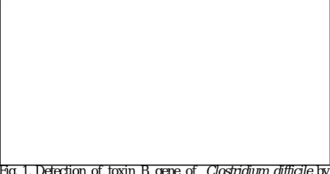
Fig. 1. Detection of toxin B gene of Clostridium difficile by PCR. Lane 1, negative control (D.W.); lanes 2 and 9, size marker (123 bp); lane 3, positive control strain; lanes 4 to 8, toxin B-positive strains; lane 10, toxin B-negative strain; lanes 11 to 17, toxin B-positive strains.
3 저 95℃에서 5분간 처리한 후, 95℃ 30초, 58℃ 30초, 72℃ 30초의 조건으로 50회 반응시켰다. 반응액 10μL 를 0.5μg/mL의 ethidium bromide가 포함된 1% agarose gel에서 전기영동 후, 399 bp 의 밴드가 있는 지를 관찰 하였다(Fig. 1). 결 과 독소 유전자가 검출된 균주는 시험된 85주 중 63주 (74%)이었다. A 독소 유전자가 검출된 균주 모두에서 B 독소 유전자가 검출되었으나, A 독소 유전자가 검출 되지 않은 나머지 22주(26%)에서는 B 독소 유전자도 검출되지 않았다. 음성 대조균으로 사용된 C. difficile 이외의 다른 Clostridium 균종 들과 호기성 그람음성 간균에서는 B 독소 유전자가 검출되지 않았다. A 독소 유전자 검출 결과와 비교할 때 DNA 추출방법에 따른 차이는 없었다(Table 1). 정량 배양 결과를 확인할 수 있었던 59주의 변 1 g 중의 C. difficile 수와 독소 생성여부를 비교해 본 결과, C. difficile 분리수가 많았던 검체에서 분리된 균주에서 독소 생성 양성율이 높았다(p=0.037, Mann-Whitney's one- sided method, Table 2).
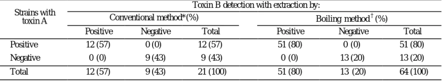
고 찰 C. difficile은 A 독소나 B 독소를 생성하는 균주만이 대장 점막에 손상을 주고 장내에 체액을 축적시켜 위 막성 대장염이나 항균제 유발 설사를 일으킨다[17]. 한 편, A 독소가 B 독소보다 임상 증상과 관련이 많다는 보고가 있으나 이에 대해서는 아직 논란이 많다[18]. C. difficile에 의한 감염을 진단하기 위해서는 변에서 C. difficile을 검출하거나 그 독소를 검출해야 한다. C. difficile을 검출하는 방법에는 CCFA 배지 등을 이용한 혐기성 배양방법과 16S rRNA를 PCR로 검출하는 방법 이 있다[19]. 배양방법은 감도는 높으나 검사 소요 시 간이 길고 독소 비생성 주로 인한 위양성 결과를 보이 는 것이 단점이다. 한편 16S rRNA를 이용하여 C. difficile을 검출하는 방법은 배양 방법에 비하여 소요시 간이 4-5시간 정도로 짧은 장점이 있으나 변에서 PCR 을 직접 시행하기가 기술적으로 어려운 단점이 있다 [19]. 독소 검출 방법에는 세포 배양법, 라텍스 응집시험, PCR 법 등이 있다[9]. 세포 배양법은 특이도는 높으나 시험 에 소요되는 시간이 길고, 특수한 장비와 기술이 필요 한 것이 단점이다. 또한 변 검체 내에 억제 물질이 있 어서 감도가 낮아질 수 있다. 라텍스 응집시험은 소요 시간이 1시간 정도로 짧으나 감도와 특이도가 비교적 낮은 것으로 알려져 있다[9]. PCR 방법은 소요 시간이 4-6 시간 정도로 비교적 짧고 A 독소 또는 B 독소 유전자를 검출하므로 특이 도가 높은 유용한 검사로 보고되었다[13]. 초기에는 A 독소에 대한 PCR 법이 주로 이용되었으나 A 독소가 C. sordellii의 세포독소와 구조가 유사하여 교차 반응을 일으키는 것으로 알려져 있다[20, 21]. 본 연구에서는 B 독소에 대한 PCR 법으로 시험하였는데, 음성 대조균으 로 사용한 C. difficile 이 외의 Clostridium spp. 뿐 아니라 변에서 흔히 분리될 수 있는 E. coli, K. pneumoniae에서 도 위양성 결과가 없어 특이도가 매우 높았다. 총 85 주의 C. difficile에 대하여 B 독소 유전자를 시험한 결 과, 74% (63/85)가 양성으로, 약 2/3만이 독소를 생성한 Table 1. Comparison of two DNA extraction methods for toxin B gene detection by PCR
Toxin B detection with extraction by:
Conventional method*(%) Boiling method†(%) Strains with
toxin A
Positive Negative Total Positive Negative Total Positive 12 (57) 0 (0) 12 (57) 51 (80) 0 (0) 51 (80) Negative 0 (0) 9 (43) 9 (43) 0 (0) 13 (20) 13 (20) Total 12 (57) 9 (43) 21 (100) 51 (80) 13 (20) 64 (100) *DNA extraction by Gumerlock et al.[15, 16]. Concordance rate of toxin A and B gene detection by PCR was 100%.
†
DNA extraction by Tang et al.[14]. Concordance rate of toxin A and B gene detection by PCR was 100%.
Table 2. Comparison of quantitative culture results with toxin B gene detection rate by PCR
Toxin B* C. difficile in
stool (CFU/mL)
No. of strain
tested Positive Negative
≥106 39 33 (85%) 6 (15%)
102 – <106 20 12 (60%) 8 (40%)
Total 59 45 14
다는 다른 보고와 비슷하였다[3]. 따라서 C. difficile 배 양 결과만으로 환자를 진단할 경우 약 30%의 환자에 게 불필요하게 항생제를 투여하게 된다고 하겠다. 한편, 독소 생성 C. difficile은 대부분이 두 가지 독소 를 모두 생성하는 것으로 알려져 있으나[3], B 독소 만 을 생성하는 균주가 있고[22-24] 이러한 균주에 의해서 도 항균제 유발설사를 일으킬 수 있음이 증명되었다 [18]. 특히, 인도네시아 등의 일부 국가에서는 이 균주 의 비율이 31.8%까지 보고되었으나[23] 본 연구에서는 시험균주 모두 A 독소와 B 독소 유전자 두 가지를 생 성하거나 한 가지도 생성하지 않아서 두 독소 중 한가 지만을 검출해도 충분할 것으로 생각되었다. 본 연구에서는 PCR 시험을 위한 DNA 추출 방법으 로 통상적인 방법과 가열법을 비교하였는데 두 방법 모두의 B 독소 유전자 검출 결과가 A 독소 유전자 검 출 결과와 일치하였다. 통상적인 DNA 추출 방법은 소 요시간이 길고, 여러 단계의 조작을 필요로 하나[14], 가열법은 간단하고, 빠르므로 검사실에서 사용하기에 편리한 방법으로 생각되었다[15, 16]. Poirier 등[25]은 C. difficile의 정량적인 세균 배양을 실시했을 경우 변에서 배양되는 세균 수와 독소의 생 성사이에는 유의한 관계가 있다고 하였다. 본 연구에 서도 변 1 g당 C. difficile 수가 많았던 균주의 독소 유 전자 양성율이 약간 높았다. 한편, 정상 신생아나 C. difficile 감염 증상이 없는 입원환자에서 분리되는 C. difficile 중에서도 독소 생성 균주가 있음이 보고되어 [9] 독소 생성 시험만으로는 위양성 결과를 얻을 수 있 다. 따라서 이 세균 감염의 정확한 진단을 위해서는 정성적인 세균 배양보다는 정량적인 세균 배양을 독소 생성 시험과 함께 시행하는 것이 바람직하다고 하겠 다. 배양된 세균으로 PCR을 시행하기 위해서는 2-4일의 배양시간이 소요된다. 따라서 독소 유전자를 직접 변 에서 PCR로 검출하는 방법이 이용될 수 있다. 그러나 변에는 heme, bilirubin, urobilinogen, bile salt 등 DNA의 증폭을 억제하는 물질이 존재하므로[26] 변을 immuno- magnetic enrichment[27], phenol-chloroform technique[28] 등으로 전 처리한 후에 PCR을 시행하는 방법이 소개 된 바 있는데, 앞으로 이에 대한 연구도 필요할 것으 로 생각된다. 결론적으로 순배양된 C. difficile 균주에서 B 독소 유 전자를 PCR 법으로 검출하는 것은 검사실에서 이용하 기 쉬운 방법이며 배양 결과의 위양성을 줄일 수 있으 므로 항생제 유발 설사와 위막성 대장염의 정확한 진 단에 매우 유용하며 이를 위한 DNA 추출은 간편한 가 열법으로도 충분하다는 결론을 얻었다. 요 약 목 적:Clostridium difficile은 A 독소와 B 독소를 생성 하여 항생제를 사용하는 환자에게 항생제 유발 설사나 위막성 대장염을 일으킬 수 있다. 이 세균 감염을 정 확히 진단하기 위해서는 세균배양과 독소시험을 함께 사용하는 것이 바람직하다. 본 연구에서는 순배양된 C. difficile 균주에서 중합효소 연쇄반응법(PCR)을 이용한 B 독소 유전자 검출 방법의 유용성을 검토하였다. 방 법:세브란스병원에서 1987-1994년 사이에 분리된 C. difficile 85 주를 대상으로 하였다. 순배양된 균주에 서 통상적인 방법과 가열법으로 DNA를 추출하였고, PCR은 시발체 YT-17, 5'-GGTGGAGCTTCAATTGGAGAG-3'와 YT-18, 5'-GTGTAACCTACTTTCATAACACCAG-3'을 사용하여 95℃ 30초, 58℃ 30초, 72℃ 30초의 조건으로 50회의 반 응 후, 1% agarose gel에 전기영동하여 399 bp의 증폭산 물을 관찰하였다. 이 결과를 PCR에 의한 A 독소 유전 자 검출 결과 및 정량적 세균배양 결과와 비교하였다. 결 과:총 85주 중 63주(74%)에서 B 독소 유전자가 검출되었다. PCR에 의한 A 독소 유전자가 검출된 모든 균주에서 B 독소 유전자가 검출되었으나, A 독소 유전 자가 검출되지 않은 나머지 22주(26%)에서는 B 독소 유전자도 검출되지 않았다. 독소 유전자 검출율은 DNA 추출 방법에 따른 차이는 없었으며, 변 1 g 당 C. difficile 수가 많았던 검체에서 분리된 균주에서 높았다. 결 론:순배양된 C. difficile 균주에서 B 독소 유전자 를 PCR 법으로 검출하는 것은 검사실에서 이용하기 쉬운 방법이며 배양 결과의 위양성을 줄일 수 있으므 로 항생제유발 설사와 위막성 대장염의 정확한 진단에 매우 유용하며 이를 위한 DNA 추출은 간편한 가열법 으로도 충분할 것으로 판단되었다. 참 고 문 헌
1. Thamlikitkul V, Danpakdi K, Chokloikaew S. Incidence of diarrhea and Clostridium difficile toxin in stools from hospitalized patients receiving clindamycin, beta-lactams, or nonantibiotic medications. J Clin Gastroenterol 1996;22:161-3.
2. Ho M, Yang D, Wyle FA, Mulligan ME. Increased incidence of Clostridium difficile-associated diarrhea following decreased restriction of antibiotic use. Clin Infect Dis 1996;23(S1):S102-6.
3. Fekety R. Antibiotic-associated colitis. In:Mandelll GL, Bennett JE, Dolin R. ed. Mandell, Douglas and Bennett's Principles and Practice of Infectious Diseases. 4th ed. New York:Chuchill Livingstone, 1995:978-87.
4. 김영아, 박광일, 박남재, 이경원, 정윤섭. Clostridium difficile 순배양에서 PCR을 이용한 toxin B gene의 검출. 제 33차 대한임상병리학회 추계학술대회 초 록 M-56, 1997.
5 Clindamycin-associated colitis due to a toxin-producing species of Clostridium in hamsters. J Infect Dis 1977; 136:701-5.
6. Bartlett JG, Chang TW, Gurwith M, Gorbach SL, Onderdonk AB. Antibiotic-associated pseudomembranous colitis due to toxin-producing clostridia. N Eng J Med 1978;298:531-4.
7. 이희주 및 정윤섭. 항균제유발 설사가 의심되는 환 자에서 분리된 Clostridium difficile 균주의 독소생성 에 관한 검토. 대한임상병리학회지 1992;12:479-85. 8. Fekety R and Shah AB. Diagnosis and treatment of
Clostridium difficile colitis. JAMA 1993;269:71-5. 9. Bartlett JG. Clostridium difficile: history of its role as an
enteric pathogen and the current state of knowledge about the organism. Clin Infect Dis 1994;18(S4):S265- 72. 10. Braun V, Hundsberger T, Leukel P, Sauerborn M, von
Eichel-Streiber C. Definition of the single integration site of the pathogenicity locus in Clostridium difficile. Gene 1996;181:29-38.
11. Kato N and Kato H. Molecular detection and identification of anaerobic bacteria. J Infect Chemother 1997;3:5-14. 12. Kato N, Ou CY, Kato H, Bartely SL, Brown VK, Dowell
VR Jr, et al. Identification of toxigenic Clostridium difficile by the polymerase chain reaction. J Clin Microbiol 1991;29:33-7.
13. Gumerlock PH, Tang YJ, Weiss JB, Silva J Jr. Specific detection of toxigenic strains of Clostridium difficile in stool specimens. J Clin Microbiol 1993;31: 507-11. 14. Gumerlock PH, Tang YJ, Meyers FJ, Silva J Jr. Use of the
polymerase chain reaction for the specific and direct detection of Clostridium difficile in human feces. Rev Infect Dis 1991;13:1053-60.
15. Silva J Jr, Tang YJ, Gumerlock PH. Genotyping of Clostridium difficile isolates. J Infect Dis 1994;169: 661-4.
16. Tang YJ, Houston ST, Gumerlock PH, Mulligan ME, Gerding DN, Johnson S, et al. Comparison of arbitrarily primed PCR with restriction endonuclease and immunoblot anaylses for typing Clostridium difficile isolates. J Clin Microbiol 1995;33:3169-73.
17. Lyerly DM, Krivan HC, Wilkins TD. Clostridium difficile: its disease and toxins. Clin Microbiol Rev 1988;1:1-18. 18. Borriello SP. Pathogenesis of Clostridium difficile
infection. J Antimicrob Chemother 1998;41(SC):S13-9. 19. Kuhl SJ, Tang YJ, Navarro L, Gumerlock PH, Silva J. Jr.
Diagnosis and monitoring of Clostridium difficile infections with the polymerase chain reaction. Clin Infect Dis 1993;16(S4):S234-8.
20. Brazier JS. The diagnosis of Clostridium difficile-
associated disease. J Antimicrob Chemother 1998;41 (SC):S29-40.
21. Wren BW, Heard SR, al-Saleh AI, Tabaqchali S. Characterisation of Clostridium difficile strains by polymerase chain reaction with toxin A- and B-specific primers. J Med Microbiol 1993;38:109-13.
22. Borriello SP, Wren BW, Hyde S, Seddon SV, Sibbons P, Krishna, et al. Molecular, immunological, and biological characterization of a toxin A-negative, toxin B-positive strain of Clostridium difficile. Infect Immun 1992;60:4192-9.
23. Kato H, Kato N, Watanabe K, Iwai N, Nakamura H, Yamamoto T, et al. Prevalence of toxin A-negative, toxin B-positive Clostridium difficile among childern and adults. 7th International Cong Infect Dis, abstract no. 21.008, 1996.
24. Kato H, Kato N, Watanabe K, Iwai N, Nakamura H, Yamamoto T, et al. Identification of toxin A-negative, toxin B-positive Clostridium difficile by PCR. J Clin Microbiol 1998;36:2178-82.
25. Poirier L, Lamothe F, Vincelette J, Bourgault AM. Usefulness of semi-quantitative cultures in the diagnosis of Clostridium difficile associated disease. Eur J Clin Microbiol Infect Dis 1991;10:770-2.
26. Higuchi R. Simple and rapid preparation of samples for PCR. In: Erlich HA. PCR technology: Principles & application for DNA amplification. New York: Stockton, 1989.
27. Wolfhagen MJHM, Fluit AC, Torensma R, Poppelier MJJG, Verhoef J. Rapid detection of toxigenic Clostridium difficile in fecal samples by magnetic immuno PCR assay. J Clin Microbiol 1994;32:1629-33.
28. Kato N, Ou CY, Kato H, Bartley SL, Luo CC, Killgore GE, et al. Detection of toxigenic Clostridium difficile in stool specimens by the polymerase chain reaction. J Infect Dis 1993;167:455-8.