저작자표시 2.0 대한민국 이용자는 아래의 조건을 따르는 경우에 한하여 자유롭게 l 이 저작물을 복제, 배포, 전송, 전시, 공연 및 방송할 수 있습니다. l 이차적 저작물을 작성할 수 있습니다. l 이 저작물을 영리 목적으로 이용할 수 있습니다. 다음과 같은 조건을 따라야 합니다: l 귀하는, 이 저작물의 재이용이나 배포의 경우, 이 저작물에 적용된 이용허락조건 을 명확하게 나타내어야 합니다. l 저작권자로부터 별도의 허가를 받으면 이러한 조건들은 적용되지 않습니다. 저작권법에 따른 이용자의 권리는 위의 내용에 의하여 영향을 받지 않습니다. 이것은 이용허락규약(Legal Code)을 이해하기 쉽게 요약한 것입니다. Disclaimer 저작자표시. 귀하는 원저작자를 표시하여야 합니다.
A THESIS FOR THE DEGREE OF MASTER OF SCIENCE
Anti-BACE-1 activity of Three Constituents
Identified in the Adults of Blattella germanica
바퀴 성충에서 동정된 3 종 화합물의
항 BACE-1 활성
By
XIAOHAN LI
Major in WCU Biomudulation
Department of Agricultural Biotechnology
Seoul National University
A THESIS FOR THE DEGREE OF MASTER OF SCIENCE
Anti-BACE-1 activity of Three Constituents
Identified in the Adults of Blattella germanica
UNDER THE DIRECTION ADVISER JEONG-YONG SUH SUBMITTED TO THE FACULTY OF THE GRADUATE SCHOOL
OF SEOUL NATIONAL UNIVERSITY
By Xiao Han Li
Major in WCU Biomodulation Department of Agricultural Biotechnology
Seoul National University August, 2018
APPROVED AS A QUALIFIED DISSERTATION OF Xiao Han Li FOR THE DEGREE OF MASTER OF SCIENCE
BY THE COMMITTEE MEMBERS
Chairman Dr. Jae-Yong Han
Vice chairman Dr. Jeong-Yong Suh
Anti-BACE-1 activity of Three Constituents Identified in
the Adults of Blattella germanica
WCU Biomudulation Seoul National University
Xiaohan Li
ABSTRACT
Alzheimer's disease (AD), acknowledged as progressive multifarious neurodegerative disorder, is the leading cause of presenile and senile dementia in both developed and developing countries. There are two hallmarks of AD, amyloid plaques and neurofibrillary tangles (NFTs). The human β-amyloid cleaving enzyme (BACE-1) is a key enzyme responsible for amyloid plaque production, which implicates the progress and symptoms of AD. AD is currently treated using acetylcholinesterase inhibitors and N-methyl-D-aspartate receptor antagonists. Until now, there is no cure for AD, and no treatments can stop or reverse its symptoms. Therefore, there is a pressing need to develop new, safe, improved naturally occurring anti-AD agents.
In this study, a fluorescence resonance energy transfer-based enzyme assay was used to identify the BACE-1 inhibitory constituents from methanol extracts from the
constituents were determined to be the polyunsaturated omega-6 fatty acid linoleic acid, the steroid cholest-5-en-3-ol, and the flavanonol fustin. Based on IC50 values, cholest-5-en-3-ol (21.13 μM) and linoleic acid (23.02 μM) were the most potent BACE-1 inhibitory constituents. The IC50 of fustin was 34.88 μM. Overall, these compounds were significantly less potent inhibitors of BACE-1 than either Inhibitor IV (IC50, 1.51 μM) or epigallocatechin gallate (IC50, 13.45 μM). German cockroach whole-body constituents containing cholest-5-en-3-ol, linoleic acid, and fustin are potential therapeutics or lead molecules for the prevention or treatment of AD.
Key words: Natural products, Alzheimer’s disease, Blattella germanica, BACE-1,
cholest-5-en-3-ol, fustin, linoleic acid
CONTENTS
ABSTRACT... IV LIST OF TABLES... IX LIST OF FIGURES... XI INTRODUCTION... 1 LITERATURE REVIEW... 5 1. ALZHEIMER’S DISEASE... 51.2.1 Early stage... 7
1.2.2 Moderate stage... 8
1.2.3 Advanced stage... 9
1.3. Pathological hallmarks of Alzheimer’s disease... 9
1.4. Worldwide death rate and Epidemiology of Alzheimer’s disease... 10
1.5. Risk factors and causes for Alzheimer’s disease... 11
1.5.1 Age... 11
1.5.2 Genetic factors... 12
1.5.3Nongenetic factors... 12
1.6. Market and cost of Alzheimer’s disease... 13
1.7. Treatments for Alzheimer’s disease... 14
1.8. Pathogenesis hypothesis of Alzheimer’s disease... 15
1.8.1. Cholinergic hypothesis of Alzheimer’s disease... 15
1.8.2 Amyloid cascade hypothesis of Alzheimer’s disease... 16
1.8.3. Tau hypothesis of Alzheimer’s disease... 18
2. AMYLOID PRECURSOR PROTEIN... 19
3. PRESENILIN 1AND PRESENILIN 2... 21
5. BACE-1(Β-SECRETASE 1)... 23
5.1. Basic knowledge of BACE-1... 24
5.2. BACE-1 structure and catalytic mechanism... 26
5.4. In vitro BACE-1 FRET assay principle... 31
MATERIALS AND METHODS... 33
1. INSTRUMENTAL ANALYSIS... 33
2. MATERIALS... 34
3. INSECT MATERIAL... 35
4. FLUORESCENCE RESONANCE ENERGY TRANSFER ENZYME ASSAY... 35
5. BIOASSAY-GUIDED FRACTIONATION AND ISOLATION... 36
6. DATA ANALYSIS... 46
RESULTS... 48
1. FRETASSAY-GUIDED FRACTIONATION AND IDENTIFICATION... 48
2. IN VITRO BACE-1INHIBITORY ACTIVITY OF THE ISOLATED COMPOUNDS... 72
DISCUSSION... 74
LITERATURE CITED... 77
LIST OF TABLES
Table 1. In vitro human BACE-1 inhibitory activity of each fraction obtained from the solvent partitioning of the methanol extract of the adults of Blattella germanica using a FRET-based enzyme assay
44
Table 2. In vitro BACE-1 inhibitory activity of each subfraction from the hexane-soluble fraction derived form adults of Blattella germanica using a FRET bioassay
Table 3. In vitro BACE-1 inhibitory activity of each subfraction from the Ethyl acetate-soluble fraction derived form adults of Blattella germanica using a FRET-based enzyme assay
47
Table 4. 1H and 13C NMR spectral data for compound1.
51
Table 5. 1H and 13C NMR spectral data for compound2
57
Table 6. 1H and 13C NMR spectral data for compound3.
62
Table 7. In vitro BACE-1 inhibitory activity of three compounds obtained from the solvent partitioning of the methanol extract of Blattella germanica adults using a FRET-based enzyme assay
LIST OF FIGURES
Figure. 1. Neuropathological hallmarks of Alzheimer’s disease. 9
Figure. 2. Cholinergic hypothesis of Alzheimer’s disease. 15
Figure 3. Schematic to show the hydrolysis of APP. 19
Figure 4. Evolutionary tree showing the relationship between BACE-1, BACE-2, and other aspartic protease.
23 Figure 5 Structure of the BACE1 protein. Based on PyMOL rendering of PDB 1fkn. 25
Figure 6. Schematic representation of BACE-1 catalytic mechanism 26
Figure 7. Principle of FRET based BACE-1 activity assay 30
Figure 8. Solvent partition of Blattella germanice adults methanol extract 34
Figure 9. Procedures to isolate Bace-1 inhibitory compounds from hexane-soluble fraction of Blattella germanica adults methanol extract.
37
Figure 10. HPLC chromatogram of compound1 38
Figure 11. HPLC chromatogram of compound2 38
Figure 12. Procedures to isolate Bace-1 inhibitory compounds from ethyl acetate-soluble fraction of Blattella germanica adults methanol extract.
40
Figure 13. HPLC chromatogram of compound3 41
Figure 14. EM-MS spectrum of compound1 49
Figure 15. 1H NMR spectrum of compound1 49
Figure 16. 13C NMR spectrum of compound1 50
Figure 17. DEPT spectrum of compound1 50
Figure 18. Structure of Cholest-5-en-3-ol 53
Figure 19. EI-MS spectrum of compound2 55
Figure 20. 1H NMR spectrum of compound2 55
Figure 21. 13C NMR spectrum of compound2 56
Figure 23. Structure of Linoleic Acid 58
Figure 24. EI-MS spectrum of compound3 60
Figure 25. 1H NMR spectrum of compound3 60
Figure 26. 13C NMR spectrum of compound3 61
Figure 27. DEPT spectrum of compound3 61
INTRODUCTION
Alzheimer's disease (AD) is the most universal cause of presenile and senile dementia in both developed and developing countries (Kalaria et al., 2008). The World Health Organization (WHO) predicts that neurodegenerative diseases will replace cancer as the second most common cause of death in 2040. (Winklhofer et al., 2008) AD is one of the most typical neurodegenerative diseases. It was first discovered in 1906 by German psychiatrist and pathologist Arosheimer Alzheimer. It is mainly divided into familial Alzheimer's disease and Alzheimer's dementia. Among them, the latter is more common. (Berchtold NC, Cotman CW, 1998) Its main clinical manifestations are progressive neurological disorders such as memory impairment, cognitive impairment, personality changes, and language disorders. The main pathological features of Alzheimer's disease are diffused atrophy of the entire brain and significant tissue changes - age spots and neurofibrillary tangles, as well as neuronal dysfunction, enlarged volume, reduced numbers, and synaptic loss. The patient’s initial symptoms are forgetfulness, which in turn deteriorates as the decline in comprehension, orientation, memory, and judgment. The patient enters a complete state of decline in the
late stages of illness, the intelligence is completely lost, language and movement disorders become more and more apparent, and stays in bed all day long. And life can not take care of themselves, eventually the patient died of multiple failures and secondary infections. (Xu W et al., 2009) As the world’s population ages, Alzheimer’s disease has become a serious problem for the medical community.
However, the etiology and pathogenesis of AD have not yet been thoroughly studied. Senile plaques, neurofibrillary tangles and loss of neurons are the three main pathological features of AD. Excessive production of β-amyloid (Aβ) produces neurotoxicity and produces a series of neurological impairments that are the β-secretase at the N-terminus of the amyloid precursor (APP) and the sequential cleavage of the C-terminal γ- secretase produced. Amyloid-β (Aβ) fibrosis, aggregation and deposition in the brain and the formation of senile plaques (SPs) are important pathological factors leading to the pathogenesis of AD.( Asai M et al., 2006) APP is a transmembrane protein that is processed by a series of enzymes. Most of the normal conditions are first cleaved by α-secretase in the domain of Aβ to produce secretory s APPα and C83. C83 is produced by cleavage of γ-secretase. Segments P3 and C fragment CTF and competitively inhibit APP cleavage by BACE-1. In rare cases, BACE-1 cleaves at the Asp1 position to produce 99 amino acids of C99 or Glu11 to cleave 89 amino acids of
C89. C99 or C89 produces amyloid Aβ under the cleavage of γ-secretase, and Aβ over-aggregates to form senile plaques. The production and imbalance of Aβ is considered to be an important factor in the pathogenesis. Therefore, if it can reduce the production of Aβ in the early stage, it is a method for intervention in the treatment of AD.
Natural compounds are extracted from natural products such as plants , insects and microorganism have been suggested as alternative sources for anti-AD products. The natural products constitute a potential source of bioactive secondary substances that have been perceived by the general public as relatively safe and often act at multiple and novel target sites (Raskin et al., 2002; Jassim and Naji, 2003). In higher organisms, insects are the most diverse species and the largest biomass group, accounting for about 80% of all animal species found in the world. They are the largest biological resources on the planet that have not yet been fully recognized and used. Most insects contain abundant nutrients such as essential amino acids, proteins, fats, trace elements, inorganic salts, vitamins, and carbohydrates. Moreover, insects have the characteristics of short breeding cycle, easy breeding, strong disease resistance and adaptability, and high food conversion rate. These advantages make insect resources become a research and development hotspot in recent years. (Hai-Long Jiang et al., 2012)
In this study, is using German Cockroach (Blattella germanica). Although it is controlled as a global vector insect, its medicinal value has long attracted attention. It is rich in compounds such as proteins, lipids, and peptides. And it has many functions, such as antibacterial and antiviral, and resistance. Oxidation, anti-tumor effect. Possible pharmacological effects of German Cockroach and their role in inhibiting BACE-1 activity. Detailed tests are needed to understand how to improve the anti-AD potency and stability of the compounds isolated from Blattella germanica for eventual commercial development.
LITERATURE REVIEW
1.1. The discovery of Alzheimer’s disease
At 1901, Auguste Deter, a woman in Frankfurt, Germany Memory impairments occurred, directions were confusing, often lost and nonsense. They were then sent to a mental hospital. Her first doctor, Alois Alzheimer, recorded the first visit to her, and Auguste became the first patient with a detailed record of Alzheimer's disease.
Dr. Alois has been engaged in neuropathological studies. As early as 1898, he discovered that in patients with dementia, partly because of the deterioration of the primary ganglia of their cerebral cortex, and later Auguste died of illness in 1906. Alois performed an autopsy to study her brain neuropathy. During the autopsy process, Alois noted that Auguste's brain was reduced in size, weight was reduced, her sulci was deepened and widened, her cerebral cortex was atrophied, and the temporal lobe, especially in the hippocampus, shrank. Later, he conducted a histopathological study and observed the silver-stained brain cortex sections with a microscope and found significant pathological changes in the different stages of nerve fibers: in early-stage diseased nerve cells, some of the nerve fibers showed normal morphology. Some of the nerve fibers are thickened and stiff; in the mid-stage, the nerve fibers are gradually approaching, forming a thick nerve bundle, and gradually approaching the nerve cell body; in the end stage, the cell body and nuclei of the nerve cell disintegrate, and only
the entangled nerve bundles. About one-quarter to one-third of the brain's nerve cells have the above-mentioned pathological changes. At the same time, Alois observes that almost all neurons in the cerebral cortex contain tiny, millet-like particles with special material deposited. This particular material was later named the "amyloid plaque." In the same year, Alois reported on Auguste's case at a scientific meeting and presented his findings, which was the first description of a new subtype of Alzheimer's disease by researchers. (Shampo M A et al., 2013, Engelhardt E al., 2015)
In 1910, a German psychiatrist Emil Kraepelin who worked with Dr. Alzheimer, first named “Alzheimer’s Disease” in the eighth edition of his book Psychiatrie (Hippius, 2003).
1.2.The signs and symptoms of Alzheimer’s disease
1.2.1 Early stage
In people with AD, at the early stage, the increasing impairment of learning and memory but not obviously. In some small part, the difficulties at executive functions, language, perception, or execution of movements are more obviously than memory problems. (Carlesimo GA, Oscar-Berman M al., 1992) AD will not affect all memory capacity equally. The affect of person's older episodic memory, implicit memory, and
semantic memory are lesser degree than new memories. ( Marko Jelicic et al., 1995) Shrinking vocabulary and decreased word fluency are the mainly characterized of language problems. That will leading oral and written language have some difficulties. (Vanessa Taler et al., 2008) But in this stage, patients usually capable of interacting basic ideas adequately. While doing something such as dressing, may have some certain movement coordination or planning difficulties, but usually unnoticed. As the disease progresses, although patients can usually continue many tasks by themselves, but may need other people assistance or supervision.( Frank EM, 1994)
1.2.2 Moderate stage
Patients gradually being unable of most common activities on daily living because of the progressive deterioration and eventually hinders independence. Speaking difficulties become more obviously due to an inability to recall words, which leads to incorrect word substitutions sometime. Writing and reading skills are also progressively lost. Complex behaviors become less coordinated as AD progresses. During this stage, the problem about memory become worse, and the person may sometimes fail to recognise close relatives. The impaired of long-term memory is gradually appear.( H. Förstl and A. Kurz. 1999)
The changes about neuropsychiatric and behaviour become more prevalent. Wandering, irritability and labile affect are common manifestations, which leading to crying, resistance to caregiving, or unpremeditated aggression.( H. Förstl and A. Kurz. 1999) Approximately 30% of patients will develop illusionary misidentifications or other delusional symptoms. Urinary incontinence will also appear.
1.2.3 Advanced stage
During this stage, patients are completely dependent on caregivers. At begining, language is reduced to very simple phrases, even single words, then eventually complete loss of speech. People can often understand by return emotional signals. Not only aggressiveness still be present, exhaustion and extreme apathy are much more common symptoms. ( H. Förstl and A. Kurz. 1999) Patients will not be able to perform ultimately, even the simplest tasks. The cause of death usually is not the disease itself but an external factor, such as pneumonia or infection of pressure ulcers.
1.3. Pathological hallmarks of Alzheimer’s disease
Amyloid plaques and neurofibrillary tangles (NFTs) (Fig. 1) are two hallmarks of AD. Amyloid plaques are abnormal cluster of peptide fragments (Aβ), NFTs are
abnormal collections of protein tau and are found inside the neurons (Silbert, 2007). Normal tau proteins can make microtubules stable. However, abnormal hyperphosphorylated tau protein will separate from the microtubules and then collection to generate NFTs.
Fig. 1. Neuropathological hallmarks of Alzheimer’s disease.
1.4. Worldwide death rate and Epidemiology of Alzheimer’s disease
In 2015, around 29.8 million people worldwide are living with AD. AD and other dementia resulted in about 1.9 million deaths in 2015. The World Health Organization (WHO) predicts that neurodegenerative diseases will replace cancer as the second most
in the people with the age over the age of 60, 15–20% in the people over 75 years old and 25–50% in the people over 85 years old (Duthey, 2013). The dementia prevalence rate in individuals over the age of 60 are 6.4% in North America, Western Europe 5.4%, 4.6% in Latin America, 4.0% in China and Western Pacific regions, and 1.6% in Africa (Qui et al., 2009). The developed countries prevalence of dementia is higher than developing countries. Maybe because of the cerebrovascular risk factor such as obesity, smoking, diabetes and hypertension (Ferri et al., 2005; Rizzi et al., 2014). The incidence of AD is also related to both gender and age. Alzheimer's Association described in 2014, compared with men, more women are suffering AD or other dementias.
1.5. Risk factors and causes for Alzheimer’s disease
1.5.1 Age
Advancing age is the most well-known risk factor causing AD. Most people get AD after 65 years old, the rate of AD is almost doubles every 5 years form 65 years old, which means the people with age over 90 prevalence are higher than 25% (Qiu et al., 2009). The age-dependent hypothesis has been established. In this hypothesis, normal synaptic dysfunction of brain is leaded by advancing age, and this dysfunction induces
the decline of normal cognitive. However, three factors including chronic neuroinflammatory responses, altered brain, and initiating injury cell physiology divert the normal brain decline into the AD pathophysiology, which leading to the major neuronal loss and brain dysfunction, finally causing the onset of AD (Herrup, 2010).
1.5.2 Genetic factors
AD can be mainly divided into early-onset familial AD (FAD) and sporadic late-onset AD(Finder, 2010). Most cases of Alzheimer's disease do not exhibit autosomal-dominant inheritance and are termed sporadic AD, in which environmental and genetic differences may act as risk factors.Based on research of twin and family, the genetic heritability ranges from 49% to 79%. Around 0.1% of the cases of Alzheimer's disease are familial forms of autosomal dominant inheritance, which have an onset before 65 years old. Most of FAD can be attributed to mutations in one of these three genes: those encoding amyloid precursor protein (APP) and presenilins 1 and 2. This AD generally affects people with the age below 65, and the symptoms can appear between people around the age of 30 and 40 (Duthey, 2013; Binetti, 2009).
Blood pressure, Cerebrovascular disease, body weight, plasma lipid levels, type 2 diabetes, metabolic syndrome, traumatic brain injury and smoking all can be nongenetic risk for AD (Reitza and Mayeux, 2014). However, some nongenetic factors can protective people away form AD. Including physical activity, intellectual activity and diet. Physical exercise can improve body and brain healthy, by increasing oxygen extraction, glucose utilization and cerebral blood flow(Reitza and Mayeux, 2014). Intellectual activities are benefited to cognitive function, such as reading, writing, and game playing(Reitza and Mayeux, 2014). Diets high in vegetable, fish, fruit, low in red meat, and sometimes intake of wine can also reduce the risk of AD (Scarmeas et al., 2009). That may because in this diet the antioxidants are high, that may suppressing of neuronal damage. In addition, fruits and vegetables' high content vitamin E can reduce Aβ-associated lipid peroxidations and cell apoptosi(Butterfield et al., 2002).
1.6. Market and cost of Alzheimer’s disease
AD cost including direct costs and indirect costs. Direct costs like hospital resources, family payments to caregivers, drugs, medical and social service. Indirect costs such as the loss of income by patients or family members' reduction(Castro et al., 2010). The worldwide costs for AD were sharply increased form 315 billion USD in
2005 to 422 billion USD in 2009 and 604 billion USD in 2010 (Wimo et al., 2013). In these worldwide costs, over 70% are contributed to developed countries, such as Europe and North America.
The other lower than 30% are costed by low-income countries, and these costs accounts for around 1% of the worldwide gross domestic product, respectively (Wimo
et al., 2010). The total costs of one people who with dementia, higher-income countries
are 38 times higher than low-income countries, and the social service direct costs are 120 times higher.
1.7. Treatments for Alzheimer’s disease
Until now, treatment can only delay the deterioration of the disease and there is no cure that can be cured. There are two kinds of treatments for AD approved by U. S. Food and Drug Administration (FDA). They are four inhibitors of AChE, such as donepezil, tacrine, rivastigmine, and galanthamine. And the uncompetitive NMDA receptor antagonist, such as memantine (Parsons et al., 2013). Tacrine was in 1993, is the first approved AChE inhibitor, however, taday is rarely used because of the associated side effects, like possible liver damage (Schneider, 2013). Donepezil is approved to use at all stage of the treatment. Rivastigmine and galantamine are
approved to use mild to moderate stage of treatment. Increasing levels of acetylcholine is a chemical message involved in memory, judgment and thinking, these made AChE inhibitors approved to treat AD. However, they will have some side effects, including vomiting, nausea, loss of appetite and anorexia, (Schneider, 2013). The NMDA receptor antagonist used in treat AD by regulating the activity of glutamate. However, side effects including headache, fatigue, confusion, constipation, and dizziness (Burock and Naqvi, 2014).
1.8. Pathogenesis hypothesis of Alzheimer’s disease
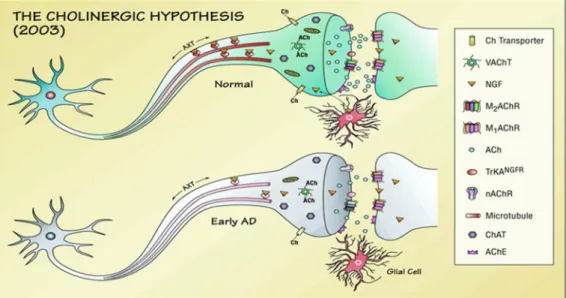
1.8.1. Cholinergic hypothesis of Alzheimer’s disease
The cholinergic hypothesis is the first emerged try to explain AD over decades. Most currently available drug therapies are based on this hypothesis. This hypothesis states that the cognitive decline in the central nervous system which caused by the dysfunction of acetylcholine induces is associated with AD. The differences in cholinergic neurons occurred in the AD patients brain are compared with the healthy young neuron (Fig. 2). Recent studies shows that choline acetyltransferase and/or AChE activity were not affected in the brain of AD patients who had the mild cognitive impairment (MCI) or on the early stage of AD, that make some challenges to this
hypothesis (Terry andBuccafusco, 2003). The cholinergic hypothesis nowadays has not
maintained widely support, mainly because medications intended to treat the acetylcholine deficiency ont only have some big side effect have not been very effective.
Fig. 2. Cholinergic hypothesis of Alzheimer’s disease. Schematic representation
of the known and proposed changes in cholinergic neurons that occur in the aged and
early Alzheimer’s disease (AD) brain compared with healthy young neurons.
1.8.2 Amyloid cascade hypothesis of Alzheimer’s disease
In 1991, the amyloid hypothesis postulated that extracellular amyloid beta (Aβ) deposits are the fundamental cause of the disease. The most obvious pathological
features of Alzheimer's disease are caused by a series of processes of deposition and accumulation of Aβ in the brain regions of related memory, and Aβ aggregation is analyzed and considered to be neurotoxic Aβ in the production(John Hardy, David Allsop, 1991).And the degradation caused by the imbalance in the removal. This view is the "amyloid cascade hypothesis," which has dominated the field for decades. Although there is no clear evidence that there is a direct relationship between Aβ and memory impairment and neurological function, the important role of Aβ in the neuropathology of AD is beyond any doubt ( Tuomo Polvikoski et al., 1995). In recent years, studies have shown that soluble Aβ oligomers may be the cause of early, preclinical pathological changes, and that subsequent cascade reactions lead to synaptic function impairment that results in severe neuronal atrophy or even loss of neurons. AD is a polypeptide having different amino acid amounts through cleavage of amyloid precursor protein (APP) by β-secretase and γ-secretase. Among them, Aβ1-40 and Aβ1-42 are two major types associated with AD. Both Aβ1-40 and Aβ1-42 are neurotoxic and participate in the formation of amyloid plaques and oligomers in the brain. Aβ1-42 is the main component of amyloid plaques and oligomers in the brain, and oligomers are currently considered to be the most direct neurotoxic factors. Aβ1-40 is a normal soluble product in the brain and cerebrospinal fluid. It is the main form of
Aβ in the blood and is the main component of Aβ deposition in the cerebral blood vessel wall. It also has a certain damage to the cerebral blood vessels. Because the balance between the production and clearance of Aβ in AD patients gradually changes, the concentration of Aβ in the brain increases, accelerating the series of complex reactions triggered by the accumulation of Aβ deposition and accumulation of aggregated Aβ, including the phosphorylation of Tau protein. Changes in synapses, synaptic changes, gliosis, loss of transmitters, and inflammatory responses eventually lead to a series of pathological phenomena such as neuronal dysfunction, death, neurofibrillary tangles, and plaque formation (Holmes C et al., 2008).
1.8.3. Tau hypothesis of Alzheimer’s disease
Studies find that changes in tau protein will lead to the disintegration of microtubules in paitents’ brain cells. The tau hypothesis shows that tau protein abnormalities initiate the disease cascade. Microtubules are mainly composed of α-β-microtubule heterodimers (Goedert M et al., 1991). Tau protein is a microtubule-associated protein that can be regulated by protein kinases and protein phosphatases. Under normal physiological conditions, binding of Tau protein to tubulin promotes the synthesis of microtubules by tubulin and eventually induces bundles of
microtubules. The non-phosphorylated Tau protein moves to the nucleus under stress and acts as a protective agent when bound to DNA. However, after Tau protein hyperphosphorylation, the binding rate of tubulin to tubulin is significantly reduced, and it binds microtubules to a certain extent, thereby degrading and deleting the function of nerve fibers and ultimately producing neurofibrillary tangles (Iqbal K, et al., 2005). The appearance of neurofibrillary tangles affects the normal physiological functions of neurotransmitters and blocks the transmission of signals between nerve cells, eventually leading to the formation of AD (Chun W, Johnson GV. 2007).
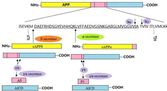
2. Amyloid precursor protein
The amyloid precursor protein (APP) is a transmembrane glycoprotein expressed widely in the brain and central nervous system. There are two types of cleavage that are important for the production of Aβ(Fig.3). One is The outer region of the cell cleaves β-secretase and the other is γ-secretase cleaves in the transmembrane region(Zhang et
al., 2011; Evin and Li, 2012). APP is first cleaved by α- or β-secretase, leaving almost
all of the extracellular domain detached, producing a - Or the β-C terminal fragment. The α- and β-C terminal fragments are then cut by the γ-secretase in the transmembrane region, and then release P3 and Aβ to the cell's external environment. (Caille et al.,
2004). In addition, γ-secretase cleavage generates A cosmid polypeptide AICD.α-secretase normally produces non-amyloidogenic proteins when it is cleaved by APP. After cleaving α-secretase, it produces APP and α-C-terminal fragments of soluble n-terminal products, which are then Γ-secretase cleaves the cleavage site. (Zhang et al., 2011) Because the fragment produced in Aβ molecule does not contain intact Aβ, it does not have the conditions for forming amyloid deposits, and APPsα has a neurotrophic effect and can protect nerve cells (Nikolaev et al., 2009). The cleavage of β-secretase can lead to the production of Aβ. After cleavage, β-secretase can produce APPsβ and β-C-terminal fragments, and then cut through γ-secretase to form different lengths of Aβ. Typically, γ-secretase cleaves Aβ40, but sometimes produces more neurotoxic Aβ42. In addition, the production of Aβ can be blocked by α-secretase
cleavage, because α-secretase competes with β-secretase, and the effect of these two secretases on APP can be said to be shifted.
Figure 3. Schematic to show the hydrolysis of APP. APP is hydrolyzed into
peptide fragments (with Aβ marked pink), including soluble sAPPβ, Aβ, AICD or sAPPα, P3 and AICD. The formation of Aβ 40 (Aβ 42 ) is accomplished by hydrolysis of APP with β-secretase through γ 40 (γ 42 ).
3. Presenilin 1 and Presenilin 2
Presenilin (PS) 1 and 2 are closely related to the onset of AD, they mutations can indirectly increase the deposition of Aβ. The full-length PS is composed of nine transmembrane domains on the built-in omentum. There are two homologous genes,
PS1 and PS2. They are mainly expressed on neurons and widely expressed in the whole brain. With regard to all the exact functions of the PS protein, it is not yet clear that PS1 is an important component of the formation and survival of neurons and progenitor cells in a specific brain region, and knocking out of the PS1 gene results in death of the mouse embryo. In the experiment, the forebrain PS gene of mice was knocked out, and thus AD-like progressive neurodegenerative changes without Aβ deposition occurred in the mice, and changes included synaptic plasticity, forebrain regression, and spatial memory impairment. The PS mutation is also closely related to AD, a variety of PS1 gene mutations will lead to increased neurotoxicity of Aβ42, and the strong hydrophobicity of Aβ42 makes it easier to aggregate, causing a cascade reaction, resulting in AD neurodegeneration change (Holmes C et al., 2008). Studies have shown that mutations in PS1 significantly accelerate the rate of Aβ deposition in mutant transgenic mice, but increased expression of Aβ42 was detected in knockout mice if they were transfected with human PS1 gene.
4. Apolipoprotein E
Apolipoprotein E (ApoE), a plasma protein involved in the transport of cholesterol substances, plays an important role in the pathophysiology of Aβ regulation. ApoE is an
important apolipoprotein in the central nervous system and is mainly secreted by astrocytes and microglia. It is a key lipid transporting carrier in the central nervous system and plays a role in repairing damaged neurons, maintaining lipid balance, removing toxins, and maintaining synaptic connections (Polvikoski T et al., 1995). Studies have shown that ApoE as a molecular chaperone of Aβ can bind to different types of Aβ, leading to changes in Aβ in terms of toxicity, deposition, and structure. Studies have shown that retinoic acid receptor agonists cause increased expression of ApoE, which can increase the scavenging capacity of soluble Aβ in the AD mouse model, reduce Aβ plaques and reverse cognitive impairment, and at the same time improve synaptic function. Experiments have shown that ApoE2 has a neuroprotective effect and can reduce the probability of AD onset. However, ApoE4 is associated with the onset probability of delayed-onset and sporadic AD. The proportion of patients carrying ApoE4 alleles is a normal cognitively-diagnosed group. 2-3 times. It has been shown in the literature that ApoE4 can accelerate the progression of aging AD in the brain and is associated with cognitive impairment. The cortical and hippocampus of ApoE4 transgenic mice exhibit neurodegenerative changes. In summary, ApoE is also important in the metabolism of Aβ.
5.1. Basic knowledge of BACE-1
β-secretase (BACE1), also named beta-site amyloid precursor protein cleaving enzyme 1, membrane-associated aspartic protease 2, beta-site APP cleaving enzyme 1, aspartyl protease 2, memapsin-2, and ASP2, is a humans enzyme encoded by the BACE1 gene, for product 42-residue amyloid beta peptide (Aβ42), which involved in APP amyloidogenic processing pathway (Mancini et al., 2011). APP cleavage by β-secretase and γ-secretase to generate Aβ, and then Aβ aggregation inducing amyloid plaque, which is one of the hallmark of AD and is toxicity to neurons in the brain of AD patients. Five groups reported the molecular cloning of the β-secretase independently, and although they use different approaches try to identify the β-secretase, they all strongly supporting the conclusion that the cloned protein was indeed β-secretase (Hussain et al., 1999; Lin et al., 2000; Sinha et al., 1999; Vassar et al., 1999; Yan et al., 1999). Bace-1 is a 501 aminoacid type 1 transmembrance aspartic protease related to the pepsin family. And BACE-2 is a 518 amino acids single transmembrane aspartyl protease (Sun et al., 2005). BACE-1 gene is on chromosome 11, whereas BACE-2 gene maps on chromosome 21 (Cheon et al., 2008).
with β-secretase, BACE-2 doesn't have high neuronal expression(Bennett et al., 2000); Laird et al., 2005). Whatsmore, although BACE-2 enzyme can generate Aβ in vitro, the preferred BACE-2 enzyme cleavage site in APP is within Aβ(Basi et al., 2003; Farzan et al., 2000; Fluhrer et al., 2002; Yan et al., 2001), which made BACE-2 precluding the formation of Aβ. These results show that, BACE-2 unlikely to be as a major of β-secretase in the brain, but at the same time, concerns have raised that BACE-1 inhibitors might also will inhibit BACE-2 and that will cause BACE-2-related mechanism-based side-effects.
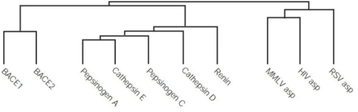
BACE-1 and BACE-2 are build a new family of transmembrane aspartic proteases. This family are most closed to pepsin family, that family expressed few in humans. They are more assosiated with the retroviral aspartic protease, which included the mouse Moloney leukaemia virus, HIV protease, and Rous sarcoma virus (Fig. 4) (Citron, 2004a).
Fig. 4. Evolutionary tree showing the relationship between BACE-1, BACE-2, and other aspartic protease
5.2. BACE-1 structure and catalytic mechanism
BACE-1 is very important for AD treatment and AD drug development. Some study already reported numerous BACE-1 complexes structures(Hong et al., 2000; Patel et al., 2004). BACE-1 is a class I transmembrane protein that consisting of a connecting strand, an NH2-terminal protease domain, and a cytosolic domain and a transmembrane region. Sequence homology with the other aspartic proteases shows that BACE-1 has a pro sequence which about 48 residues at the NH2-terminal region of its. The bilobal structure of BACE-1 has the conserved general folding of aspartic proteases (Fig.5). BACE-1 active site is characterized by the presence of hydrophilic and small
This is considered to be the regulate substrate approach to the BACE-1 active site and be considered the right geometry of the substrate in for the catalytic process (Shimizu et
al., 2008; Mancini et al., 2011). In addition, in acidic pH 4.0–4.5 BACE-1 shows high
activity, and this pH is usually used in vitro assay (Mancini et al., 2011). BACE-1 by a general acid-base mechanism, which is common to aspartyl protease, shows enzymatic activity. Two aspartic acids were characterized in aspartyl protease, thus for BACE-1 enzyme, they are Asp32 and Asp228 (Mancini et al., 2011). The BACE-1 catalytic mechanism is present in Fig. 6. The protonated Asp32 form a hydrogen bond with the carbonyl oxygen of the cleavage bond, and the non-protonated Asp228 is related to lytic
water. Two peptidic products release by the tetrahedral intermediate collapses induction, and the enzyme will restored for another catalysis cycle.
Fig. 5 Structure of the BACE1 protein. Based on PyMOL rendering of PDB 1fkn.
Fig. 6. Schematic representation of BACE-1 catalytic mechanism 5.3. BACE-1 inhibitors for Alzheimer’s disease
BACE-1 is a required enzyme in the amyloidogenic pathway for product Aβ. Thus it is considered that develop BACE-1 inhibitors is the therapeutic agents. To develop therapeutic potential would require of Bace-1 inhibitors, not only need good potency and pharmacokinetic properties, also need low molecular weight (700 daltons) and high lipophilicity purpose to penetrate the blood-brain barrier. BACE-1 inhibitors can be divided to peptidic inhibitors, peptidomimetic inhibitors and non-peptide compounds, and have synthetic inhibitors and natural compounds. Peptidomimetics are the first generation of BACE-1 inhibitors. Mainly due to the BACE-1 enzyme large open active site having high affinity to binding polypeptide substrate, they are extremely potent inhibitors in vitro(Vassar, 2014). However, because of the blood-brain-barrier (BBB) and penetration poor oral bioavailability, so in vivo, these peptides are not good potent inhibitor against BACE-1(Silvestri, 2009). And small-molecular inhibitors, high oral bioavailability and good penetrability to BBB, have already been developed and expressed improved pharmacological characteristics. As the second generation BACE-1 inhibitors, they possess improved pharmacological characteristic. But, they can't achieve enough concentration in the brain because they were the substrates of P-glycoprotein, which is the ATP-dependent drug efflux pump for xenobiotics in the BBB(Probst et al., 2012).Recently, the third generation BACE-1 inhibitors, which have
small MW and great potent inhibitory activity have already been developed. And they were reported to have good brain penetration that can reduce cerebral Aβ in the preclinical animal models. There are several of potent BACE-1 inhibitor drugs that have been developed in the human clinical trials(Vassar, 2014). Several companies are in the early stages of development and testing of this potential class of treatment. Inc was reported that phase I results for its candidate verubecestat (MK-8931) in April 2012 Merck & Co.,. Merck already began a Phase II/III trial for this medicine in December 2012 and plan to completed in July 2019. However, Merck halted its late-stage trial of verubecestat for mild to moderate Alzheimer's disease in February 2017, after it was reported that having "virtually no chance" of working according to an independent panel of experts. This occurred just three months after Eli Lilly & Co. announced that its own setback with solanezumab. The results of Merck's trial of verubecestat on AD patients with early stage Alzheimer's are still expected in February 2019. Eli Lilly and AstraZeneca Company announced an agreement to codevelop AZD3293 in September 2014. A pivotal Phase II/III clinical trial of AZD3293 was started in late 2014 and that is planned to recruit 1,500 patients and is planned end in May 2019. Eli Lilly’s inhibitor LY2886721 is another BACE-1 inhibitor that has reached phase II trials. The data on phase I trial were the first presented at the Alzheimer’s Association International
conference in 2012. Daily dosing during two weeks, reduced BACE-1 activity by 50– 75% and CSF Aβ42 by 72% (Willis et al., 2012; Bowman Rogers and Strobel, 2013). Recently, Lilly reported the phase II trial of LY2886721 was terminated due to the liver abnormalities that were found in 4 out of 45 patients (Rogers, 2013). This toxicity, however, does not have to be related to the working mechanism of the inhibitor, but can represent off-target effects as the livers of BACE-1 knockout mice are normal.
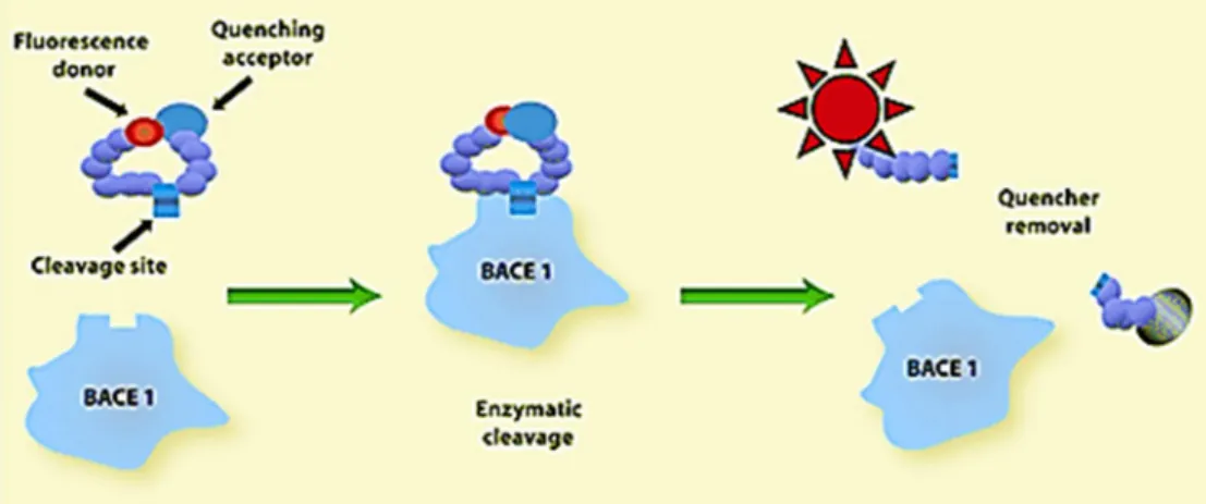
5.4. In vitro BACE-1 FRET assay principle
In most common assay method utilized to study BACE-1 inhibitory activity in vitro is the FRET assay. FRET is sensitive and is easily applied in high-throughput screening (HTS) for BACE-1 inhibitory compounds. FRET assay principle is shown in Fig. 7. The synthetic peptide with a fluorophore (donor group) and a quencher (acceptor group) was used as the substrates. In the uncleaved substrate, the fluorescence is quenched due to intramolecular resonance energy transfer from the donor group to quenching group. When the substrate is cleaved by BACE-1, the fluorescence is not quenched due to the disturbance of the energy transfer, and the fluorescent signal can be measured. The increase fluorescent signal is linearly related to the rate of proteolysis. While BACE-1 is inhibited by an inhibitor, this fluorescent signal is be reduced.
According to this, HTS of BACE-1 inhibitory compounds was performed.
MATERIALS AND METHODS
1. Instrumental analysis
The1H and 13C nuclear magnetic resonance (NMR) spectra were recorded in MeOD on an AVANCE 600 spectrometer (Bruker, Rheinspettem, Germany) at 600 and 150 MHz, respectively, using tetramethylsilane as an internal standard. The chemical shifts are given in δ parts per million (ppm). Distortionless enhancement polarization
transfer (DEPT) spectra were acquired using Bruker software to differentiate among 13C signals for CH3, CH2, CH, and quaternary carbon. The ultraviolet (UV) spectra were obtained in acetonitrile or methanol on a Kontron UVICON 933/934 spectrophotometer (Milan, Italy), mass spectra on a Jeol JMS-DX 303 spectrometer (Tokyo, Japan). Silica gel 60 (0.063–0.2 mm) (Merck, Darmstadt, Germany) was used for column chromatography. Merck precoated silica gel plates (Kieselgel 60 F254, 0.20 mm) were used for analytical thin layer chromatography (TLC). An Isolera one medium-pressure liquid chromatograph (Biotage, Uppsala, Sweden) and an Agilent 1200 high-performance liquid chromatograph with binary solvent pump (Agilent, Santa Clara, CA, USA) were used for isolation of active principles.
2. Materials
Three constituents, cholest-5-en-3-ol, linoleic acid and fustin, were identified in B.
germanica, in this study, and their pure organic compounds were purchased from S-A,
Sigma-Aldrich (St. Louis, MO, USA); Ext, Extrasynthese(Lyon area, France). BACE-1 Inhibitor IV, curcumin, and epigallocatechin gallate (EGCG) were purchased from Merck (Darmstadt, Germany) and Sigma-Aldrich (St. Louis, MO, USA), respectively. Recombinant human β-secretase (BACE-1) and fluorogenic peptide substrate
7-methoxycumarin-4-acetyl-[Asn670, Lue671]-amyloid β/A4 precursor protein 770 fragment 667-676-(2,4 dinitrophenyl) Lys-Arg-Arg amide trifluoroacetate salt were purchased from Sigma-Aldrich. All of the other chemicals and reagents which used in this study were of analytical grade quality and are available commercially.
3. Insect material
Cultures of B. germanica were maintained in the laboratory for nine years without exposure to any known insecticide. They were reared with calf chow pellets (Samyang, Seoul) in glass jars (30 cm diameter × 30 cm) at 27 ± 1°C and 55 ± 5% relative humidity under a 12:12 h light:dark cycle.
4. Fluorescence resonance energy transfer enzyme assay
The methods of Wang et al. (2014) and Lv et al. (2008) were used with a slight modification to assess the BACE-1 inhibitory activity of the test compounds. The assay mixtures containing 2 μL of 0.3 units/μL recombinant human BACE-1, 20 μL of 0.1 mg/mL fluorogenic peptide substrate, 76 μL of 50 mM sodium acetate (pH 4.5), and the
isolated compounds (1–1,000 μg/mL) in 2% dimethyl sulfoxide were preincubated for 2 h at 37°C in darkness, followed by adding 20 μL of 2.5 M sodium acetate to terminate the reaction. Inhibitor IV, curcumin, and EGCG served as standard references and were similarly formulated. The fluorescence intensity was measured using a SpectraMAX Gemini XS plate reader (Molecular Devices, Sunnyvale, CA) at 320 nm excitation and 405 nm emission at room temperature. The inhibition percentage was determined with the following equation: % inhibition = 100 – [(FS – FS0)/(FC – FC0)] ´ 100, where FS and FS0are the fluorescence of samples at 120 min and 0 time, and FCand FC0 are the fluorescence of control at 120 min and 0 time, respectively (Lv et al., 2008). Results were expressed as mean ± standard error (SE) of triplicate samples of three independent experiments.
5. Bioassay-guided fractionation and isolation
The air-dried B. germanica adults (3 kg) was pulverized, extracted with methanol (3 L, three times) at room temperature for 3 days, and filtered. The combined filtrate was concentrated by rotary evaporation at 40°C to yield approximately 148 g of a
tawny tar. The extract (20 g) was sequentially partitioned into hexane- (8.26 g), chloroform- (1.01 g), ethyl acetate- (0.36 g), butanol- (4.82 g), and water-soluble (5.55 g) portions for subsequent bioassay (Fig. 8). This fractionation procedure was repeated two times. The organic solvent-soluble portion were concentrated under vacuum at 40°C, and the water-soluble portion was concentrated at 50°C. To isolate the active constituents, 0.1–0.5 mg/mL of each B. germanica adult-derived fraction was tested in a FRET enzyme assay as described by Lv et al. (2008) and Wang et al. (2015).
Fig. 8. Solvent partition of Blattella germanica adult methanol extract.
The hexane-soluble fraction (10 g) was the most biologically active fraction (Table 2) and was chromatographed on a 5.5 ´ 70 cm silica gel (600g) column by elution with a gradient of chloroform and methanol [100:0 (1 L), 99:1 (2 L), 97:3 (2 L), 95:5 (2 L), 90:10 (1 L), 80:20 (1 L), 60:40 (1 L), and 0:100 (1 mL) by volume ], and then, elution with methanol (3 L) was performed to afford 54 fractions (each approximately 200 mL) (Fig. 9). The column fractions were monitored by TLC on silica gel plates developed with a chloroform and methanol (97:3 by volume) mobile phase. Fractions with similar
Rfvalues on the TLC plates were pooled. The spots were detected by spraying the plate with 2% H2SO4and then heating the samples on a hot plate. Active fractions 9–10 (H3)
were obtained. Fraction H2 was separated by MPLC with a UV detector at 254 nm and 365 nm and column cartridge (340 g silica gel) with 510 mL column volume through elution with a gradient of chloroform and methanol [100:0 (1530 mL), 99: 1 (1530 mL), 98:2 (1020 mL), 97:3 (1020 mL), 95:5 (1020 mL), 90:10 (1020 mL), 80:20 (510 mL), and 0:100 (1530 mL) by volume] at a flow rate of 50 mL/min to provide 54 fractions (each approximately 200 mL). The column fractions were monitored by TLC on silica gel plates as described previously. Active fractions 21–24 (H33) were pooled and separated by MPLC with a UV detector at 254 nm and 365 nm and column cartridge (100 g silica gel) with a column volume of 132 mL by elution with a gradient of chloroform and methanol [100:0 (396 mL), 99: 1 (396 mL), 98:2 (264 mL), 97:3 (396 mL), 95:5 (396 mL), 90:10 (264 mL), and 0:100 (396 mL) by volume] at a flow rate of 25 mL/min to provide 181 fractions (each approximately 20 mL). The column fractions were monitored by TLC on silica gel plates as described previously. Fractions 38–73 (H333) were pooled and separated by MPLC with a UV detector at 254 nm and 365 nm and column cartridge (25 g silica gel) with a column volume of 33 mL by elution with a gradient of chloroform and methanol [100:0 (110 mL), 99: 1(100 mL), 98:2 (66 mL), 97:3 (99 mL), 95:5 (66 mL), 90:10 (66 mL), and 0:100 (100 mL) by volume] at a flow rate of 15 mL/min to provide 82 fractions (each approximately 20 mL). The column
fractions were monitored as described previously. Fractions 17–58 (H3332) and 59– 82(H3333) were obtained. Fraction H3332 was purified by preparative TLC with chloroform and methanol (97:3 by volume). Preparative high-performance liquid chromatography (HPLC) was performed to separate the constituents from active fraction with a 7.8 mm i.d. × 300 mm µBondapak C18 column (Waters, Milford, MA, USA) and mobile phase of methanol and water (80:20 by volume) at a flow rate of 1
mL/min. Chromatographic separation was monitored using a UV detector at 206 nm. Finally, active compound 1 (30.47 mg) was isolated at a retention time of 18.36 min (Fig. 10). Another active fraction, H3333, was purified by preparative TLC with chloroform and methanol (97:3 by volume) (Fig. 11). A preparative HPLC was performed to separate the constituents from active fraction with a µBondapak C18 column and mobile phase of methanol and water (85:15 by volume) at a flow rate of 1
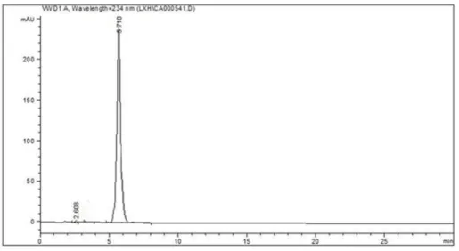
mL/min. Chromatographic separation was monitored using a UV detector at 234 nm. Finally, active compound 2 (5.26 mg) was isolated at a retention time of 5.18 min (Fig. 10).
Fig. 9. Procedures to isolate BACE-1 inhibitory compounds from the hexane-soluble fraction of Blattella germanica adults methanol extract.
Fig. 10. HPLC chromatogram of compound 1.
The active ethyl acetate-soluble fraction (2 g) was separated by MPLC with a UV detector at 254 nm and 365 nm and SNAP column cartridge (100 g silica gel) with a volume of 132 mL column by elution with a gradient of chloroform and methanol [100:0 (264 mL), 99: 1(132 mL), 98:2 (264 mL), 97:3 (396 mL), 95:5 (264 mL), 90:10 (264 mL), 80:20 (264 ml), and 0:100 (396 mL) by volume] at a flow rate of 25 mL/min to provide 128 fractions (each approximately 20 mL) (Fig. 12). The column fractions were monitored by TLC on silica gel plates as described previously. Active fractions 37–64 (E4) was obtained. Fractions E4 was separated by preparative TLC [chloroform : methanol (96:4) by volume]. Of the five fractions, active fraction E44 (145.54 mg) was obtained. Fraction E44 was purified by preparative TLC with chloroform and methanol (96:4 by volume). A preparative HPLC was performed to separate the constituents from active fraction with a µBondapak C18 column and mobile phase of methanol and water (85:15 by volume) at a flow rate of 1 mL/min. Chromatographic separation was monitored using a UV detector at 210 nm. Finally, active compound 3 (11.53 mg) was isolated at a retention time of 4.45 min (Fig. 13).
Fig. 12. Procedures to isolate BACE-1 inhibitory compounds from the ethyl acetate-soluble fraction of Blattella germanica adult methanol extract.
Fig. 13. HPLC chromatogram of compound 3.
6. Data analysis
The fifty percent inhibitory concentration (IC50) was defined as the concentration
of the compound that resulted in a 50% loss of BACE-1 activity. The IC50values were
determined using GraphPad Prism 5.1 software (GraphPad Software, La Jolla, CA,
USA). The IC50 values for the treatments were considered significantly different from
one another when their 95% confidence limits (CLs) did not overlap. The results are expressed as the means± standard errors (SEs) of triplicate samples from three independent experiments.
RESULTS
1. FRET assay-guided fractionation and identification
The fractions obtained from the solvent partitioning of the methanol extract from the adults of B. germanica were tested for inhibitory activity against human BACE-1
using a FRET enzyme assay (Table 1). Significant differences in inhibitory activity were observed among the fractions and were used to identify the peak activity fractions for the next step of purification. As judge by the 24 h IC50 values, the hexane-soluble and ethyl acetate-soluble fractions showed the most potent inhibitory activity. Moderate and weak inhibition was obtained using the chloroform-soluble fraction and water-soluble and butanol-soluble fractions, respectively.
Table 1. In vitro human BACE-1 inhibitory activity of each fraction obtained from the solvent partitioning of the methanol extract from the adults of Blattella germanica using a fluorescence resonance energy transfer enzyme assay
Methanol extract 19.42 (17.07–22.09) 1.1 ± 0.08 4.88 0.961 Hexane-soluble fr.c 19.39 (16.68–22.55) 1.0 ± 0.08 5.46 0.946 Chloroform-soluble fr. 50.71 (47.84–53.75) 1.3 ± 0.05 2.35 0.991 Ethyl acetate-soluble fr. 19.66 (17.29–22.36) 1.0 ± 0.07 4.59 0.962 Butanol-soluble fr. 96.46 (86.35–107.8) 1.3 ± 0.08 3.15 0.973 Water-soluble fr. 89.64 (84.55–95.04) 1.4 ± 0.06 2.00 0.990
a CL denotes confidence limit.
b Pearson’s chi-square goodness-of-fit test.
c Fraction.
The BACE-1 inhibitory activity of each subfraction derived from the hexane-soluble fraction is given in Table 2.
Table 2. In vitro human BACE-1 inhibitory activity of each subfraction from the hexane-soluble fraction derived from the adults of Blattella germanica using a fluorescence resonance energy transfer enzyme assay
Fraction
% inhibition at test concentration (mg/mL)
0.5 0.1 H1 83.7 ± 0.27 78.7 ± 0.69 H2 91.0 ± 1.17 85.3 ± 1.53 H3 92.1 ± 1.43 89.1 ± 1.31 H4 88.1 ± 2.47 84.5 ± 1.03 H5 77.6 ± 0.49 63.4 ± 0.72 H6 83.5 ± 0.86 77.5 ± 0.89 H7 81.2 ± 1.28 66.4 ± 0.14 H8 69.8 ± 1.52 43.5 ± 0.67 H31 85.1 ± 0.45 72.6 ± 0.37 H32 90.6 ± 0.87 82.5 ± 1.28 H33 94.1 ± 1.62 93.1 ± 0.38 H34 90.6 ± 0.94 88.2 ± 0.76 H35 83.3 ± 0.36 72.7 ± 2.43 H36 78.4 ± 2.12 53.5 ± 1.11 H331 53.1 ± 2.16 36.4 ± 2.73 H332 96.8 ± 1.34 85.7 ± 1.13
H333 96.2 ± 1.23 81.3 ± 1.56 H334 93.0 ± 0.58 66.5 ± 1.75 H335 92.7 ± 1.51 72.4 ± 0.64 H3331 82.3 ± 0.25 64.2 ± 0.77 H3332 93.8 ± 1.18 80.1 ± 1.29 H3333 90.3 ±1.27 72.6 ± 2.63 H3333 90.3 ± 1.27 72.6 ± 2.63
The BACE-1 inhibitory activity of each subfraction derived from the ethyl acetate-soluble fraction is shown in Table 3.
Table 3. In vitro human BACE-1 inhibitory activity of each subfraction from the ethyl acetate-soluble fraction derived from the adults of Blattella germanica using a fluorescence resonance energy transfer enzyme assay
Fraction
% inhibition at test concentration (mg/mL)
0.5 0.1 E1 82.0 ± 0.27 72.1 ± 1.36 E2 82.9 ± 1.78 63.9 ± 1.73 E3 83.3 ± 0.48 72.4 ± 2.26 E4 91.1 ± 0.23 82.2 ± 1.29 E5 71.1 ± 1.12 52.2 ± 2.36 E6 40.4 ± 2.75 33.7 ± 2.84 E41 79.2 ± 1.17 60.7 ± 3.23 E42 72.7 ± 0.84 48.9 ± 2.74 E43 80.5 ± 0.74 69.8 ± 2.57 E44 84.6 ± 0.92 73.1 ± 1.81 E45 69.1 ± 2.44 38.6 ± 2.93
FRET assay-guided fractionation of the adults of B. germanica led to the identification of three active compounds through spectroscopic analyses, including EI-MS and NMR spectroscopy. Compound 1 was obtained as white powder. The mass
spectrum of the isolate exhibited a molecular ion at m/z 386 [M]+ (Fig. 14) and 1H NMR spectra (Fig. 15) showed 46 protons. Its 13C NMR spectra (Fig. 16) showed 27 carbons in the molecule comprising methoxy groups and ethyl groups as indicated in
DEPT (Fig. 17), suggesting the molecular formula C27H46O. Compound 1: EI-MS (70
eV), m/z (% relative intensity): 386 [M]+ (100), 57 (20), 95 (21), 107 (22), 133 (15), 145 (22) , 159 (17), 199 (7) , 213 (23), 231 (14), 255 (23), 275 (51), 301 (31), 326 (5),
353 (29), 368 (37), 412 (1). 1H NMR (MeOD, 600 MHz) and 13C NMR (MeOD, 150
MHz); See Table 4. This compound was characterized as cholest-5-en-3-ol (or epicholesterol)
[(1S,2R,10S,11S,14R,15R)-2,15-dimethyl-14-[(2R)-6-methylheptan-2-yl]tetracyclo[8.7. 0.02,7.011,15]heptadec-7-en-5-ol] (Fig. 18). The interpretations of proton and carbon signals were largely consistent with the findings of Xu et al. (2015).
Fig. 16. 13C NMR spectrum of compound 1.
Fig. 17. DEPT spectrum of compound 1. Table 4. 1H and 13C NMR spectral data for compound 1
Position Partial structure δC, ppm (MeOD, 150 MHz) δH, ppm (MeOD, 600 MHz) δC, ppm (Xu et al., 2015) δH, ppm (Xu et al., 2015) 1 CH2 38.67 1.38m, 1.13m 38.2 1.36m, 1.12m 2 CH2 32.42 1.56m, 1.31m 32.4 1.56m, 1.31m 3 CH 72.57 3.49m 71.6 3.52m
4 CH2 43.62 2.23 m, 1.98m 43.3 2.22m, 1.98m 5 C 142.12 142.3 6 CH 122.57 5.33 (J = 4.8 Hz) 121.5 5.35d (J = 5.5 Hz) 7 CH2 33.15 2.04 m 32.5 2.04m, 1.77m 8 CH 33.39 1.45 m 32.5 1.46m 9 CH 51.86 1.44 m 51.8 1.45m 10 C 37.80 37.2 11 CH2 22.31 1.52 m, 1.27m 21.3 1.50m, 1.26m 12 CH2 41.29 1.56 m, 1,31m 40.5 1.55m, 1.31m 13 C 43.14 43.3 14 CH 57.70 1.40 m 57.0 1.41m 15 CH2 25.43 1.60 m, 1.35m 24.9 1.62m, 1.34m 16 CH2 29.44 1.62 m, 1.36m 28.9 1.61m 1.35m 17 CH 58.31 1.47 m 57.6 1.47m 18 19 20 CH3 CH3 CH 12.42 19.97 37.49 0.71s 1.01s 1.64m 12.2 19.8 36.6 0.68s 1.00s 1.65m
21 22 23 24 25 26 27 CH3 CH2 CH2 CH2 CH CH3 CH3 19.34 37.24 25.05 40.81 29.28 23.29 23.05 0.94 (J = 6.5 Hz) 1.25m 1.26m 1.27m 1.62m 0.86d (J = 6.5 Hz) 0.87d (J = 6.5 Hz) 19.1 36.2 24.5 40.2 28.6 23.0 22.8 0.95d (J = 6.5 Hz) 1.26m 1.27m 1.27m 1.63m 0.86d (J = 6.5 Hz) 0.87d (J = 6.5 Hz)
Fig. 18. Structure of cholest-5-en-3-ol. The chemical formula of the steroid compound
Compound 2 was obtained as colorless oil. The mass spectrum of the isolate exhibited a molecular ion at m/z 280 [M]+ (Fig. 19) and 1H NMR spectra (Fig. 20)
showed 32 protons. Its 13C NMR spectra (Fig. 21) showed 18 carbons in the molecule
comprising methoxy groups and ethyl groups as indicated in DEPT (Fig. 22), suggesting the molecular formula C18H32O2. Compound 2: EI-MS (70 eV), m/z (% relative intensity): 280 [M]+(100), 55 (51), 67 (100), 81 (91), 95 (65), 109 (30) , 123 (14), 137 (8) , 150 (6), 168 (4), 182 (5), 196 (4), 209 (2), 262 (29), 263 (1), 279 (15). 1H
NMR (MeOD, 600 MHz) and 13C NMR (MeOD, 150 MHz): See Table 5. This
compound was characterized as linoleic acid [(9Z,12Z)-octadeca-9,12-dienoic acid] (C18:2) (Fig. 23). The interpretations of proton and carbon signals were largely consistent with those of previous studies (Miao et al., 2010).
Fig. 19. EI-MS spectrum of compound 2.
Fig. 21. 13C NMR spectrum of compound 2.
Table 5. 1H and 13C NMR spectral data for compound 2 Position Partial structure δC, ppm (MeOD, 150 MHz) δH, ppm (MeOD, 600 MHz) δC, ppm (Miao et al., 2010) δH, ppm (Miao et al., 2010) 1 C=O 180.58 179.6 2 CH2 34.07 2.35t (J = 7.5Hz) 34.0 2.35t (J = 7.2 Hz) 3 CH2 24.59 1.64m 24.6 1.64m 4 CH2 29.03 1.27~1.39m 29.1 1.27m 5 CH2 28.98 1.27~1.39m 29.0 1.27m 6 CH2 31.49 1.27~1.39m 31.9 1.27m 7 CH2 29.31 1.27~1.39m 29.3 1.27m 8 CH2 27.15 2.06m (J = 7.0 Hz) 27.0 2.04m (J = 7.0 Hz) 9 CH 127.84 5.35m 127.9 5.35m 10 CH 130.09 5.35m 130.1 5.35m 11 CH2 25.57 2.78t (J = 7.0Hz) 24.7 2.77t (J = 6.4Hz) 12 CH 128.01 5.35m 128.1 5.35m 13 CH 129.90 5.35m 129.7 5.35m 14 CH2 27.12 2.06t (J = 7.0Hz) 27.1 2.06m 15 CH2 29.10 1.27~1.39m 29.1 1.27~1.39m 16 17 18 CH2 CH2 CH3 29.54 22.53 13.99 1.27~1.39m 1.27~1.39m 0.90t (J = 7.0Hz) 29.5 22.7 14.0 1.27~1.39m 1.27~1.39m 0.89t (J = 6.9Hz)
Fig. 23. Structure of linoleic acid. The chemical formula of the unsaturated fatty acid
Compound 3 was obtained as yellow powder. The mass spectrum of the isolate exhibited a molecular ion at m/z 288 [M]+ (Fig. 24) and 1H NMR spectra (Fig. 25)
showed 15 protons. Its 13C NMR spectra (Fig. 26) showed 12 carbons in the molecule
comprising methoxy groups and ethyl groups as indicated in DEPT (Fig. 27), suggesting the molecular formula C15H12O6. Compound 3: EI-MS (70 eV), m/z (% relative intensity): 288 [M]+(100), 51 (4), 77 (6), 81 (7), 121 (3), 123 (48) , 137 (100),
152 (10) , 179 (1), 197 (1), 213 (2), 231 (2), 244 (1), 259 (30).1H NMR (MeOD, 600
MHz) and 13C NMR (MeOD, 150 MHz); See Table 6. This compound was
characterized as fustin (or dihydrofisetin)
[(2R,3R)-2-(3,4-dhydroxyphenyl)-3,7-dihydroxy-2,3-dihydrochromen-4-one] (Fig. 28). The interpretations of proton and carbon signals were largely consistent with those of previous studies (Shrestha et al., 2013).
Fig. 24. EI-MS spectrum of compound 3.
Fig. 26. 13C NMR spectrum of compound 3.
Table 6. 1H and 13C NMR spectral data for compound 3 Position Partial structure δC, ppm (MeOD, 150 MHz) δH, ppm (MeOD, 600 MHz) δC, ppm (Shrestha et al., 2013) δH, ppm (Shrestha et al., 2013) 1 2 CH 85.80 4.93d (J = 11.8 Hz) 85.55 4.93d (J = 11.6 Hz) 3 CH 74.73 4.47d (J = 11.8 Hz) 74.50 4.47d (J = 11.6 Hz) 4 C=O 194.61 194.25 5 CH 130.24 7.72d (J = 8.7 Hz) 129.91 7.71 (J = 8.6 Hz) 6 CH 112.25 6.52dd (J = 8.7, 2.2 Hz) 111.98 5.35d (J = 8.7, 2.2 Hz) 7 C 167.04 166.63
8 CH 103.85 6.32d (J = 2.2 Hz) 103.59 6.30d (J = 2.2 Hz) 9 C 165.25 164.86 10 C 113.59 113.29 1’ C 130.24 129.96 2’ CH 116.05 6.97d (J = 2.0 Hz) 115.77 6.97d (J = 2.0 Hz) 3’ C 147.25 146.92 4’ C 146.46 146.86 5’ CH 116.20 6.79d (J = 8.1 Hz) 24.9 6.79d (J =8.0 Hz) 6’ CH 121.07 6.85dd (J = 8.2, 2.0 Hz) 28.9 6.85dd (J = 8.0, 2.0 Hz)
Fig.28. Structure of fustin. The chemical formula of the flavonoid fustin is C15H12O6, with a molar mass of 288.26 g/mol.