저작자표시-비영리-변경금지 2.0 대한민국 이용자는 아래의 조건을 따르는 경우에 한하여 자유롭게 l 이 저작물을 복제, 배포, 전송, 전시, 공연 및 방송할 수 있습니다. 다음과 같은 조건을 따라야 합니다: l 귀하는, 이 저작물의 재이용이나 배포의 경우, 이 저작물에 적용된 이용허락조건 을 명확하게 나타내어야 합니다. l 저작권자로부터 별도의 허가를 받으면 이러한 조건들은 적용되지 않습니다. 저작권법에 따른 이용자의 권리는 위의 내용에 의하여 영향을 받지 않습니다. 이것은 이용허락규약(Legal Code)을 이해하기 쉽게 요약한 것입니다. Disclaimer 저작자표시. 귀하는 원저작자를 표시하여야 합니다. 비영리. 귀하는 이 저작물을 영리 목적으로 이용할 수 없습니다. 변경금지. 귀하는 이 저작물을 개작, 변형 또는 가공할 수 없습니다.
A DISSERTATION FOR THE DEGREE OF DOCTOR OF PHILOSOPHY
Modular Configuration of DEMETER DNA
Demethylase and Its Application to
Epigenome Editing
DEMETER DNA 탈메틸 효소의 모듈형 구성
개발과 후성유전체 편집 기술을 위한 응용 연구
AUGUST 2019
HOSUNG JANG
MAJOR IN HORTICULTURAL SCIENCE AND BIOTECHNOLOGY DEPARTMENT OF PLANT SCIENCE
COLLEGE OF AGRICULTURE AND LIFE SCIENCES THE GRADUATE SCHOOL OF SEOUL NATIONAL UNIVERSITY
i
Modular Configuration of DEMETER DNA
Demethylase and Its Application to
Epigenome Editing
HOSUNG JANG
Department of Plant Science, Seoul National University
ABSTRACT
DNA methylation is a key epigenetic modification which regulates gene expression and chromatin structure in higher eukaryotes. DNA methylation is often mitotically heritable but can be removed by active mechanisms in an enzyme-dependent manner. In Arabidopsis, the DEMETER (DME) DNA glycosylase specifically excises 5-methylcytosine (5mC), generating harmful 3′ blocking intermediates, which should be immediately processed by subsequent base excision repair machineries. DME and three other family members, REPRESSOR OF SILENCING 1 (ROS1), DEMETER-LIKE 2 (DML2) and DML3 share similar domain structures, but display distinct catalytic efficiencies. The DME family proteins are large, multi-domain DNA glycosylases with variable sequences
ii
connecting conserved domains, but the contribution of these structures to the regulation of catalytic activity and the enzymes required for downstream demethylation pathway after 5mC excision are largely unknown. In this study, I extensively manipulated DME protein by reducing in size to identify the minimal regions necessary for 5mC excision activity. Domain swapping experiments revealed that the glycosylase domains of DME family members are functionally equivalent, and compatibility between conserved domains is critical for DNA demethylation in vitro. In addition, I demonstrated that ABASIC ENDONUCLEASE 1-LIKE (APE1L) and ABASIC ENDONUCLEASE-REDOX PROTEIN (ARP) are responsible for trimming unusual 3′ end structures after 5mC excision, which proposes more complete DNA demethylation pathway in plants. Finally, I applied DME to epigenome editing by fusion with a transcription activator-like effector (TALE) DNA binding module. TALE-DME fusion protein showed delicate modulation of DNA methylation at specified genomic loci. Taken together, these studies will broaden our understanding of the fundamental regulatory mechanism of DNA methylation and transcription, and provide a promising avenue to produce various epigenetic traits by targeted DNA demethylation.
Keywords: DEMETER, DNA demethylation, domain structure, AP endonuclease, epigenome editing, TALE DNA binding module
iii
CONTENTS
ABSTRACT………i CONTENTS………..……iii LIST OF TABLES...………vii LIST OF FIGURES………..……...viii LIST OF ABBREVIATIONS………...xi GENERAL INTRODUCTION ………...…1CHAPTER 1. Biochemical study of the domain structure of DNA demethylase ABSTRACT.………18
INTRODUCTION……….………...20
MATERIALS AND METHODS………...………..………23 Construction of the minimized DME with catalytic core
Cloning of the glycosylase domain swapping constructs between DME family proteins
Cloning of domain swapping constructs between DME and DML2 Protein expression and purification
iv
In vitro DNA glycosylase assay Single turnover kinetics of DME Electrophoretic mobility shift assay
Sequence alignment and phylogenetic analysis
RESULTS………...…………..…35
Three conserved domains of DME comprise the minimal entity for 5mC excision in vitro..………...…………..…35
Conserved domains of DME family proteins are catalytically compatible in vitro………...…………..…40
Functional motifs in the conserved domains of DME..………..…49
DISCUSSION……….………..52
REFERENCES……….58
CHAPTER 2.Biochemical study of the DNA demethylation pathway ABSTRACT.………64
INTRODUCTION……….………...65
MATERIALS AND METHODS………...………..………69 Expression and purification of the proteins
In vitro 5mC excision assay In vitro 3′ phosphodiesterase assay
v
In vitro nucleotide incorporation assay In vitro 3′ phosphatase assay
In vitro AP endonuclease assay on AP sites Electrophoretic mobility shift assay
RESULTS………...…………..…78
DME and ROS1 5mC DNA glycosylases generate a 3′-PUA as a primary blocking intermediate in early 5mC excision………78
Enzyme-dependent δ-elimination process following 5mC excision..…80
Arabidopsis encodes three putative AP endonucleases APE1L, APE2 and ARP..………85
APE1L and ARP process major 5mC excision intermediate 3′-PUA to generate 3′-OH………86
3′ phosphatase activity of ARP...………90
Biochemical activities of MBP-free AP endonucleases.………94
ARP has AP site incision activity...………94
DNA binding activity of AP endonucleases...………98
DISCUSSION……….………100
vi
CHAPTER 3. Application to epigenome editing by targeted DNA demethylation
ABSTRACT...………115 INTRODUCTION……….……….117 MATERIALS AND METHODS...………121
Cloning of the TALE-DME fusion constructs Plant materials and growth conditions
Agrobacterium-mediated Arabidopsis transformation Genotyping of the transgenic plants
RNA isolation and gene expression analysis Quantitative real-time PCR (qRT-PCR) analysis Locus specific bisulfite sequencing
RESULTS..………...…………..130 Generation of transgenic plants expressing TALE-DME fusion proteins.……….…130 DNA demethylation activity of TALE-DME in transgenic plants...…132 DISCUSSION……….………140 REFERENCES...………143 ABSTRACT IN KOREAN...……….……….148
vii
LIST OF TABLES
CHAPTER 1
Table 1-1. Oligonucleotides used in this study...……….…31 Table 1-2. Catalytic rate constants (kcat-st) of purified proteins calculated
from single turnover kinetics..………39
CHAPTER 2
Table 2-1. List of oligonucleotides for biochemical assays.………75
CHAPTER 3
Table 3-1. Oligonucleotides used in this study...………127 Table 3-2. TALE-DME targeting sequences..………128
viii
LIST OF FIGURES
CHAPTER 1
Figure 1-1. Structures of manipulated DME fragments and chimeric proteins between DME and other family members..………..……….…….32 Figure 1-2. Sequences of the synthetic linkers and structures of chimeric fragments generated by domain swapping between DME and DML2 ………..…..33 Figure 1-3. SDS-PAGE analysis of purified proteins used for in vitro 5mC
excision assay.………34 Figure 1-4. The in vitro 5mC excision activity of minimal DME catalytic
core with three conserved domains.………37 Figure 1-5. The conserved motifs of the DME family proteins in Arabidopsis.………43 Figure 1-6. The in vitro 5mC excision activity of chimeric proteins generated by swapping glycosylase domains between DME family members.………44 Figure 1-7. The in vitro 5mC excision activity of DME-DML2 chimeric
proteins...………46 Figure 1-8. Electrophoretic mobility shift assay of DME-DML2 chimeric
ix
Figure 1-9. Sequence alignment of the conserved domains of DME family proteins...………51
CHAPTER 2
Figure 2-1. Schematic diagrams of manipulated DME and ROS1...………76 Figure 2-2. SDS-PAGE analysis of purified proteins...………77 Figure 2-3. The 5mC excision products generated by DME and ROS1 DNA glycosylases………82 Figure 2-4. Enzyme-dependent δ-elimination process during 5mC excision...………84 Figure 2-5. In vitro reconstitution of DNA demethylation with Arabidopsis AP endonucleases...………88
Figure 2-6. The 3′ phosphatase activity of Arabidopsis AP
endonucleases.………92 Figure 2-7. In vitro AP endonuclease activities of Arabidopsis MBP-free AP
endonucleases….………96 Figure 2-8. Electrophoretic mobility shift assay of Arabidopsis AP
endonucleases….………99 Figure 2-9. Model of active DNA demethylation pathway in
x
CHAPTER 3
Figure 3-1. Diagrams of the manipulated vectors for cloning………129 Figure 3-2. Schematic diagrams of TALE binding sites and TALE-DME constructs..………131 Figure 3-3. Bisulfite conversion of genomic DNA extracted from each T1 transgenic plant.………134 Figure 3-4. DNA demethylation activity of TD3 and TD5 TALE-DME fusion proteins in T1 transgenic plants.………136 Figure 3-5. Expression level of FWA in TALE-DME T1 transgenic plants.………138 Figure 3-6. Flowering time of TALE-DME T1 transgenic plants..………139
xi
LIST OF ABBREVIATION
3′-PUA 3′- phosphor-α, β-unsaturated aldehyde
5caC 5-carboxylcytosine
5fC 5-formylcytosine
5hmC 5-hydroxymethylcytosine
5mC 5-methylcytosine
AP Apurinic/apyrimidinic
APE1L Abasic endonuclease 1-like
APE2 Abasic endonuclease 2
ARP Abasic endonuclease-redox protein
BER Base excision repair
CGI CpG island
CMT3 CHROMOMETHYLASE 3
CRISPR Clustered, regularly interspaced, short palindromic repeats
DME DEMETER
DML2 DME-LIKE 2
DML3 DME-LIKE 3
DNMT DNA methyltransferase
DRM2 DOMAINS REARRANGED METHYLTRANSFERASE 2
EEP Endonuclease/exonuclease/phosphatase
Endo III Endonuclease III
Endo IV Endonuclease IV
FACT Facilitates chromatin transaction
FCL 4Fe-4S cluster loop
xii
FWA FLOWERING WAGENINGEN
GMO Genetically modified organisms
GPD Glycine/proline-rich loop with a conserved aspartic acid
hAPE1 human AP endonuclease 1
HhH Helix-hairpin-helix
hOGG1 human 8-oxoguanin DNA glycosylase
IDR Interdomain region
LIG1 DNA ligase I
MEA MEDEA
MET1 DNA METHYLTRANSFERASE 1
ncRNA noncoding RNA
ROS1 REPRESSOR OF SILENCING 1
RRM RNA recognition motif
SSB Single-strand break
TALE Transcription activator-like effector
TDG Thymine-DNA glycosylase
TET Ten-eleven translocation
UHRF1 Ubiquitin-like PHD and RING finger domains 1
ZDP Zinc finger DNA 3′ phosphoesterase
1
GENERAL INTRODUCTION
DNA methylation
DNA methylation is a stable epigenetic modification crucial for gene imprinting, transposon silencing, and many developmental processes in higher eukaryotes (Huh et al., 2008; Smith and Meissner, 2013). DNA methylation is achieved by adding a methyl group on C5 position of cytosine (5-methylcytosine, 5mC), and usually occurs in the symmetric context of CG dinucleotides in mammals. The initial DNA methylation patterns are established by DNA methyltransferase 3 (DNMT3), and maintained by maintenance methyltransferase DNMT1 (Law and Jacobsen, 2010; Wu and Zhang, 2010). During cell division, DNMT1 interacts with ubiquitin-like PHD and RING finger domains 1 (UHRF1) which has strong preference for hemimethylated DNA, and methylates the cytosine on the complementary strand (Arita et al., 2008; Law and Jacobsen, 2010; Wu and Zhang, 2010; Lyco, 2018).
In plants, DNA methylation occurs at cytosines in all sequence contexts including the symmetric CG and CHG (H represents A, T or C), and the asymmetric CHH sites. Three different enzymes are responsible for catalyzing these DNA methylation patterns: CG and CHG methylation are
2
maintained by DNA METHYLTRANSFERASE 1 (MET1) and
CHROMOMETHYLASE 3 (CMT3), respectively, whereas CHH
methylation is achieved through de novo methylation by DOMAINS REARRANGED METHYLTRANSFERASE 2 (DRM2) (Law and Jacobsen, 2010; Wu and Zhang, 2010).
DNA demethylation in mammals
DNA demethylation is a reverse process of DNA methylation, which can be achieved through either passive or active mechanisms. Passive DNA demethylation occurs in a replication-dependent manner when DNA methyltransferases such as DNMT1 and MET1 are down-regulated or absent. During the early embryogenesis, both maternal and paternal genomes of the mammalian zygote undergo global DNA demethylation which is referred to as epigenetic reprogramming (Wu and Zhang, 2010; Iurlaro et al, 2017). As cell division progresses, the maternal genome experiences gradual and passive DNA demethylation with exclusion of DNMT1 from the nucleus (Carlson et al., 1992). However, paternal genome undergoes rapid genome-wide DNA demethylation immediately after fertilization, implying that this process requires an enzyme-dependent mechanism (Wu and Zhang, 2010; Guo et al., 2014).
3
Ten-eleven translocation (TET) family proteins are mammalian DNA demethylases which mediate the oxidation of 5mC to 5-hydroxymethylcytosine (5hmC) (Tahiliani et al., 2009). In the mammalian zygote, 5hmC accumulates in the paternal genome along with dramatic decrease of 5mC, which indicates that TET-mediated active DNA demethylation occurs in mammals (Wossidlo et al., 2011). TET proteins further catalyze the oxidation of 5hmC to formylcytosine (5fC) and 5-carboxylcytosine (5caC), albeit TET proteins prefer 5mC to 5hmC or 5fC as a substrate (He et al., 2011; Ito et al., 2011; Hu et al., 2015; Wu and Zhang, 2017). The 5fC and 5caC bases can be removed by thymine-DNA glycosylase (TDG), and subsequent base excision repair (BER) enzymes such as AP endonuclease complete DNA demethylation (He et al., 2011; Maiti and Drohat, 2011; Kohli and Zhang, 2013; Wu and Zhang, 2017).
DNA demethylation in plants
Although plants and mammals have highly conserved DNA methylation systems, they have evolved distinct DNA demethylation machineries. In plants, the DEMETER (DME) DNA glycosylase family proteins catalyze direct excision of 5mC from DNA and initiate active DNA demethylation to permit transcription of target genes (Choi et al., 2002;
4
Gong et al., 2002; Agius et al., 2006; Gehring et al., 2006). As a bifunctional 5mC DNA glycosylase/lyase, DME catalyzes the cleavage of a sugar-phosphate backbone after 5mC excision via β- and δ-elimination reactions, producing harmful 3′-blocking intermediates such as 3′- phosphor-α, β-unsaturated aldehyde (3′-PUA) and 3′-phosphate (Gehring et al., 2006; Lee et al., 2014). These 5mC excision intermediates are processed to provide 3′-OH by BER enzymes such as AP endonuclease and 3′ phosphatase for subsequent polymerization. Biochemical studies have revealed that Arabidopis ABASIC ENDONUCLEASE 1-LIKE (APE1L) and ABASIC ENDONUCLEASE-REDOX PROTEIN (ARP) are capable of processing a major intermediate 3′-PUA, whereas ZINC FINGER DNA 3′ PHOSPHOESTERASE (ZDP) and ARP are involved in processing 3′-phosphate (Martinez-Macias et al., 2012; Lee et al., 2014). Genetic studies showed that the ape1l zdp double mutant is lethal, suggesting their roles in DME-initiated DNA demethylation during seed development (Li et al., 2015b). Although the specific DNA polymerase involved in subsequent polymerization is not well-characterized, DNA polymerase λ presumably performs the incorporation of unmethylated cytosines to abasic sites (Uchiyama et al., 2004; Uchiyama et al., 2009), and DNA ligase I completes the final ligation step of BER (Córdoba-Cañero et al., 2011; Li et al., 2015a).
5
Function and structure of the plant DNA demethylase
Recent genome-wide DNA methylome analysis revealed that DME removes DNA methylation at numerous target loci in the Arabidopsis central cell, and the resulting changes in DNA methylation patterns are inherited by the fertilized endosperm (Park et al., 2016). During early endosperm development, DME establishes maternal-specific expression of MEDEA (MEA), FERTILIZATION-INDEPENDENT SEED2 (FIS2) and FLOWERING WAGENINGEN (FWA), while the paternal alleles remain methylated and silenced (Choi et al., 2002; Kinoshita et al., 2004; Gehring et al., 2006; Jullien et al., 2006). In particular, DME specifically removes 5mC of maternal MEA allele for gene activation, which is required for endosperm and seed development. Consequently, homozygous dme mutants are embryonic lethal, whereas the DME/dme heterozygote produces 50% of viable seeds (Choi et al., 2002). On the other hand, three additional DME family members are reported in Arabidopsis - REPRESSOR OF SILENCING 1 (ROS1), DME-LIKE 2 (DML2) and DML3. ROS1 is required for regulation of transgene expression and production of stomatal lineage cells, whereas both DML2 and DML3 appear to facilitate DNA demethylation to regulate proper development in somatic tissues (Gong et al., 2002; Penterman et al., 2007; Yamamuro et al., 2014).
6
The DME family proteins have a modular structure, and share three conserved C-terminal domains (the glycosylase domain and flanking domains A and B), albeit they show distinct 5mC excision efficiencies in vitro (Penterman et al., 2007; Mok et al., 2010; Ponferrada-Marin et al., 2011). The DME family belongs to the helix-hairpin-helix (HhH) superfamily of DNA glycosylase, which contains an HhH motif and a glycine/proline-rich loop with a conserved aspartic acid (GPD) in the central glycosylase domain. The catalytic aspartic acid and lysine residues in the HhH-GPD motif are essential for DME activity (Choi et al., 2004; Gehring et al., 2006; Mok et al., 2010). Homology modeling of DME suggests that domain A and the glycosylase domain are involved in composition of the 5mC recognition pocket (Ponferrada-Marin et al., 2011; Brooks et al., 2014), and domain A is also required for non-specific DNA binding activity through its mixed charge cluster motif (Mok et al., 2010). Domain B contains a permuted CXXC and a divergent RNA recognition motif (RRM), but its function remains largely unknown (Iyer et al., 2011).
Targeting mechanism of the plant DNA demethylase
In contrast to the global DNA demethylation in mammals, no strong evidence exists for genome-wide demethylation in plants (Zhu, 2009). It is
7
plausible that certain guiding molecules are required for DME targeting. It is still largely unknown how DME family proteins are guided to target loci to initiate DNA demethylation and transcriptional activation, but several studies have provided some clues about targeting mechanism of DME. DME family proteins might be guided to target sequences by interacting with other chromatin binding modules. Indeed, there are numerous cases in which histone modifying enzymes such as histone acetyltransferases and histone methyltransferases are recruited to the target sites via the interaction with bromodomain or chromodomain proteins that bind to acetylated histones and methylated DNA, respectively (Marmorstein and Zhou, 2014; Patel, 2016). In Arabidopsis, histone H1.2 was identified as a DME-interacting protein via yeast two-hybrid and in vitro pull-down assays (Rea et al., 2012), suggesting that DME can also be guided by chromatin-associated proteins. In addition, DME family proteins can be recruited to target genomic regions by recognizing specific chromatin signatures unique to DNA demethylation targets. These signatures may include open chromatin structure with low histone density, or novel histone modifications required for active demethylation (Qian et al., 2012).
Alternatively, DME family proteins may utilize some transcription-associated patterns such as mRNA transcripts or noncoding RNAs
8
(ncRNAs). It is noteworthy that some Polycomb group complexes are guided to their targets using ncRNA as an adaptor molecule in animals and plants (Fitzpatrick et al., 2002; Mak et al., 2002; Rinn et al., 2007; Heo and Sung, 2011). Although the involvement of RNA molecules in DME targeting is largely hypothetical, the presence of CXXC and RRM motifs in domain B suggests their potential role as RNA binding modules for the guidance. In addition, RNA binding proteins may guide DME family proteins to target loci, as exemplified by ROS3 which is a small RNA binding protein that guides ROS1 for sequence-specific DNA demethylation (Zheng et al., 2008), albeit the direct interaction between ROS1 and ROS3 is still elusive.
DME is reported to possess two classes of target sites including GC-rich heterochromatin regions and accessible euchromatin regions (Frost et al., 2018). First, DME is known to target intergenic and heterochromatin regions for DNA demethylation (Ibarra et al., 2012). The Arabidopsis Facilitates chromatin transaction (FACT) histone chaperone complex is required for guidance of DME to heterochromatic targets and specific imprinted genes (Ikeda et al., 2011; Frost et al., 2018). The FACT complex is conserved in diverse organisms including yeast, mammals and plants, and is required for transcription of RNA polymerase II through nucleosomal
9
templates (Hondele and Ladurner, 2013; Hsieh et al., 2013). This suggests that FACT complex might also assist DME demethylation through nucleosome-rich heterochromatin regions (Frost et al., 2018). Second, DME appears to be preferentially guided to less compact euchromatin regions, and many of DME targets in the central cell are found near the promoters of imprinted genes such as MEA, FWA and FIS2 (Choi et al, 2002; Kinoshita et al., 2004; Gehring et al., 2006; Jullien et al., 2006), and even euchromatic transposable elements (Ibarra et al., 2012). These studies reveal the function of DNA demethylase and enhance our understanding of DNA methylation dynamics in plants.
The thesis work focused on the biochemical study of the plant DNA demethylase and downstream events of the DNA demethylation pathway. This study also includes the application of plant DNA demethylase to epigenome editing and addresses the following three topics:
Chapter 1: Biochemical study of the domain structure of DNA demethylase
Chapter 2: Biochemical study of the DNA demethylation pathway Chapter 3: Application to epigenome editing by targeted DNA demethylation
10
REFERENCES
Agius, F., Kapoor, A., and Zhu, J.K. (2006). Role of the Arabidopsis DNA glycosylase/lyase ROS1 in active DNA demethylation. Proc. Natl. Acad. Sci. USA 103: 11796-11801.
Arita, K., Ariyoshi, M., Tochio, H., Nakamura, Y., and Shirakawa, M. (2008). Recognition of hemi-methylated DNA by the SRA protein UHRF1 by a base-flipping mechanism. Nature 455: 818-821.
Brooks, S.C., Fischer, R.L., Huh, J.H., and Eichman, B.F. (2014).
5-methylcytosine recognition by Arabidopsis thaliana DNA
glycosylases DEMETER and DML3. Biochemistry 53: 2525-2532. Carlson, L.L., Page, A.W., and Bestor, T.H. (1992). Properties and
localization of DNA methyltransferase in preimplantation mouse embryos: implications for genomic imprinting. Genes Dev. 6: 2536-2541.
Choi, Y., Gehring, M., Johnson, L., Hannon, M., Harada, J.J., Goldberg, R.B., Jacobsen, S.E., and Fischer, R.L. (2002). DEMETER, a DNA glycosylase domain protein, is required for endosperm gene imprinting and seed viability in Arabidopsis. Cell 110: 33-42.
Cordoba-Canero, D., Roldan-Arjona, T., and Ariza, R.R. (2011). Arabidopsis ARP endonuclease functions in a branched base excision DNA repair pathway completed by LIG1. Plant J. 68: 693-702.
Fitzpatrick, G.V., Soloway, P.D., and Higgins, M.J. (2002). Regional loss of imprinting and growth deficiency in mice with a targeted deletion of KvDMR1. Nat. Genet. 32: 426-431.
11
Frost, J.M., Kim, M.Y., Park, G.T., Hsieh, P.H., Nakamura, M., Lin, S.J.H., Yoo, H., Choi, J., Ikeda, Y., Kinoshita, T., Choi, Y., Zilberman, D., and Fischer, R.L. (2018). FACT complex is required for DNA demethylation at heterochromatin during reproduction in Arabidopsis. Proc. Natl. Acad. Sci. USA 115: E4720-E4729.
Gehring, M., Huh, J.H., Hsieh, T.F., Penterman, J., Choi, Y., Harada, J.J., Goldberg, R.B., and Fischer, R.L. (2006). DEMETER DNA glycosylase establishes MEDEA polycomb gene self-imprinting by allele-specific demethylation. Cell 124: 495-506.
Gong, Z., Morales-Ruiz, T., Ariza, R.R., Roldan-Arjona, T., David, L., and Zhu, J.-K. (2002). ROS1, a repressor of transcriptional gene silencing in Arabidopsis, encodes a DNA glycosylase/lyase. Cell 111: 803-814.
Guo, F., Li, X., Liang, D., Li, T., Zhu, P., Guo, H., Wu, X., Wen, L., Gu, T.P., Hu, B., Walsh, C.P., Li, J., Tang, F., and Xu, G.L. (2014). Active and passive demethylation of male and female pronuclear DNA in the mammalian zygote. Cell Stem Cell 15: 447-459.
He, Y.F., Li, B.Z., Li, Z., Liu, P., Wang, Y., Tang, Q., Ding, J., Jia, Y., Chen, Z., Li, L., Sun, Y., Li, X., Dai, Q., Song, C.X., Zhang, K., He, C., and Xu, G.L. (2011). Tet-mediated formation of 5-carboxylcytosine and its excision by TDG in mammalian DNA. Science 333: 1303-1307.
Heo, J.B., and Sung, S. (2011). Vernalization-mediated epigenetic silencing by a long intronic noncoding RNA. Science 331: 76-79.
Hondele, M., Stuwe, T., Hassler, M., Halbach, F., Bowman, A., Zhang, E.T., Nijmeijer, B., Kotthoff, C., Rybin, V., Amlacher, S., Hurt, E.,
12
and Ladurner, A.G. (2013). Structural basis of histone H2A-H2B recognition by the essential chaperone FACT. Nature 499: 111-114. Hsieh, F.K., Kulaeva, O.I., Patel, S.S., Dyer, P.N., Luger, K., Reinberg,
D., and Studitsky, V.M. (2013). Histone chaperone FACT action during transcription through chromatin by RNA polymerase II. Proc. Natl. Acad. Sci. USA 110: 7654-7659.
Hu, L., Lu, J., Cheng, J., Rao, Q., Li, Z., Hou, H., Lou, Z., Zhang, L., Li, W., Gong, W., Liu, M., Sun, C., Yin, X., Li, J., Tan, X., Wang, P., Wang, Y., Fang, D., Cui, Q., Yang, P., He, C., Jiang, H., Luo, C., and Xu, Y. (2015). Structural insight into substrate preference for TET-mediated oxidation. Nature 527: 118-122.
Huh, J.H., Bauer, M.J., Hsieh, T.F., and Fischer, R.L. (2008). Cellular programming of plant gene imprinting. Cell 132: 735-744.
Ibarra, C.A., Feng, X., Schoft, V.K., Hsieh, T.F., Uzawa, R., Rodrigues, J.A., Zemach, A., Chumak, N., Machlicova, A., Nishimura, T., Rojas, D., Fischer, R.L., Tamaru, H., and Zilberman, D. (2012). Active DNA demethylation in plant companion cells reinforces transposon methylation in gametes. Science 337: 1360-1364.
Ikeda, Y., Kinoshita, Y., Susaki, D., Ikeda, Y., Iwano, M., Takayama, S., Higashiyama, T., Kakutani, T., and Kinoshita, T. (2011). HMG domain containing SSRP1 is required for DNA demethylation and genomic imprinting in Arabidopsis. Dev. Cell 21: 589-596.
Ito, S., Shen, L., Dai, Q., Wu, S.C., Collins, L.B., Swenberg, J.A., He, C., and Zhang, Y. (2011). Tet proteins can convert 5-methylcytosine to 5-formylcytosine and 5-carboxylcytosine. Science 333: 1300-1303. Iurlaro, M., von Meyenn, F., and Reik, W. (2017). DNA methylation
13
Dev. 43: 101-109.
Iyer, L.M., Abhiman, S., and Aravind, L. (2011). Natural history of eukaryotic DNA methylation systems. Prog. Mol. Biol. Transl. Sci. 101: 25-104.
Jullien, P.E., Kinoshita, T., Ohad, N., and Berger, F. (2006). Maintenance of DNA methylation during the Arabidopsis life cycle is essential for parental imprinting. Plant Cell 18: 1360-1372.
Kinoshita, T., Miura, A., Choi, Y., Kinoshita, Y., Cao, X., Jacobsen, S.E., Fischer, R.L., and Kakutani, T. (2004). One-way control of FWA imprinting in Arabidopsis endosperm by DNA methylation. Science 303: 521-523.
Kohli, R.M., and Zhang, Y. (2013). TET enzymes, TDG and the dynamics of DNA demethylation. Nature 502: 472-479.
Law, J.A., and Jacobsen, S.E. (2010). Establishing, maintaining and modifying DNA methylation patterns in plants and animals. Nat. Rev. Genet. 11: 204-220.
Lee, J., Jang, H., Shin, H., Choi, W.L., Mok, Y.G., and Huh, J.H. (2014). AP endonucleases process 5-methylcytosine excision intermediates during active DNA demethylation in Arabidopsis. Nucleic Acids Res. 42: 11408-11418.
Li, Y., Duan, C.G., Zhu, X., Qian, W., and Zhu, J.K. (2015a). A DNA ligase required for active DNA demethylation and genomic imprinting in Arabidopsis. Cell Res. 25: 757-760.
Li, Y., Cordoba-Canero, D., Qian, W., Zhu, X., Tang, K., Zhang, H., Ariza, R.R., Roldan-Arjona, T., and Zhu, J.K. (2015b). An AP endonuclease functions in active DNA demethylation and gene imprinting in Arabidopsis. PLoS Genet. 11: e1004905.
14
Lyko, F. (2018). The DNA methyltransferase family: a versatile toolkit for epigenetic regulation. Nat. Rev. Genet. 19: 81-92.
Maiti, A., and Drohat, A.C. (2011). Thymine DNA glycosylase can rapidly excise 5-formylcytosine and 5-carboxylcytosine: potential implications for active demethylation of CpG sites. J. Biol. Chem. 286: 35334-35338.
Mak, W., Baxter, J., Silva, J., Newall, A.E., Otte, A.P., and Brockdorff, N. (2002). Mitotically stable association of polycomb group proteins eed and enx1 with the inactive x chromosome in trophoblast stem cells. Curr. Biol. 12: 1016-1020.
Marmorstein, R., and Zhou, M.M. (2014). Writers and readers of histone acetylation: structure, mechanism, and inhibition. Cold Spring Harb. Perspect. Biol. 6: a018762.
Martinez-Macias, M.I., Qian, W., Miki, D., Pontes, O., Liu, Y., Tang, K., Liu, R., Morales-Ruiz, T., Ariza, R.R., Roldan-Arjona, T., and Zhu, J.K. (2012). A DNA 3' phosphatase functions in active DNA demethylation in Arabidopsis. Mol. Cell 45: 357-370.
Mok, Y.G., Uzawa, R., Lee, J., Weiner, G.M., Eichman, B.F., Fischer, R.L., and Huh, J.H. (2010). Domain structure of the DEMETER 5-methylcytosine DNA glycosylase. Proc. Natl. Acad. Sci. USA 107: 19225-19230.
Park, K., Kim, M.Y., Vickers, M., Park, J.S., Hyun, Y., Okamoto, T., Zilberman, D., Fischer, R.L., Feng, X., Choi, Y., and Scholten, S. (2016). DNA demethylation is initiated in the central cells of Arabidopsis and rice. Proc. Natl. Acad. Sci. USA 113: 15138-15143. Patel, D.J. (2016). A structural perspective on readout of epigenetic histone
15
a018754.
Penterman, J., Zilberman, D., Huh, J.H., Ballinger, T., Henikoff, S., and Fischer, R.L. (2007). DNA demethylation in the Arabidopsis genome. Proc. Natl. Acad. Sci. USA 104: 6752-6757.
Ponferrada-Marin, M.I., Parrilla-Doblas, J.T., Roldan-Arjona, T., and Ariza, R.R. (2011). A discontinuous DNA glycosylase domain in a family of enzymes that excise 5-methylcytosine. Nucleic Acids Res. 39: 1473-1484.
Qian, W., Miki, D., Zhang, H., Liu, Y., Zhang, X., Tang, K., Kan, Y., La, H., Li, X., Li, S., Zhu, X., Shi, X., Zhang, K., Pontes, O., Chen, X., Liu, R., Gong, Z., and Zhu, J.K. (2012). A histone acetyltransferase regulates active DNA demethylation in Arabidopsis. Science 336: 1445-1448.
Rea, M., Zheng, W., Chen, M., Braud, C., Bhangu, D., Rognan, T.N., and Xiao, W. (2012). Histone H1 affects gene imprinting and DNA methylation in Arabidopsis. Plant J. 71: 776-786.
Rinn, J.L., Kertesz, M., Wang, J.K., Squazzo, S.L., Xu, X., Brugmann, S.A., Goodnough, L.H., Helms, J.A., Farnham, P.J., Segal, E., and Chang, H.Y. (2007). Functional demarcation of active and silent chromatin domains in human HOX loci by noncoding RNAs. Cell 129: 1311-1323.
Smith, Z.D., and Meissner, A. (2013). DNA methylation: roles in mammalian development. Nat. Rev. Genet. 14: 204-220.
Tahiliani, M., Koh, K.P., Shen, Y., Pastor, W.A., Bandukwala, H., Brudno, Y., Agarwal, S., Iyer, L.M., Liu, D.R., Aravind, L., and Rao, A. (2009). Conversion of methylcytosine to 5-hydroxymethylcytosine in mammalian DNA by MLL partner TET1.
16
Science 324: 930-935.
Uchiyama, Y., Takeuchi, R., Kodera, H., and Sakaguchi, K. (2009). Distribution and roles of X-family DNA polymerases in eukaryotes. Biochimie 91: 165-170.
Uchiyama, Y., Kimura, S., Yamamoto, T., Ishibashi, T., and Sakaguchi, K. (2004). Plant DNA polymerase lambda, a DNA repair enzyme that functions in plant meristematic and meiotic tissues. Eur. J. Biochem. 271: 2799-2807.
Wossidlo, M., Nakamura, T., Lepikhov, K., Marques, C.J., Zakhartchenko, V., Boiani, M., Arand, J., Nakano, T., Reik, W., and Walter, J. (2011). 5-Hydroxymethylcytosine in the mammalian zygote is linked with epigenetic reprogramming. Nat. Commun. 2: 241.
Wu, S.C., and Zhang, Y. (2010). Active DNA demethylation: many roads lead to Rome. Nat. Rev. Mol. Cell Biol. 11: 607-620.
Wu, X., and Zhang, Y. (2017). TET-mediated active DNA demethylation: mechanism, function and beyond. Nat. Rev. Genet. 18: 517-534. Yamamuro, C., Miki, D., Zheng, Z., Ma, J., Wang, J., Yang, Z., Dong, J.,
and Zhu, J.K. (2014). Overproduction of stomatal lineage cells in Arabidopsis mutants defective in active DNA demethylation. Nat. Commun. 5: 4062.
Zheng, X., Pontes, O., Zhu, J., Miki, D., Zhang, F., Li, W.X., Iida, K., Kapoor, A., Pikaard, C.S., and Zhu, J.K. (2008). ROS3 is an RNA-binding protein required for DNA demethylation in Arabidopsis. Nature 455: 1259-1262.
Zhu, J.K. (2009). Active DNA Demethylation Mediated by DNA Glycosylases. Annu. Rev. Genet. 43: 143-166.
17
CHAPTER 1
Biochemical study of the domain structure of DNA
demethylase
18
ABSTRACT
DNA methylation is a key epigenetic mark which regulates gene expression and chromatin structure in eukaryotes. DNA methylation is often mitotically heritable but can be removed by active mechanisms in a replication-independent manner. In Arabidopsis, the DEMETER (DME) DNA glycosylase specifically removes 5-methylcytosine (5mC) from DNA and induces gene imprinting essential for seed viability in endosperm. There are three other DME family members present in the Arabidopsis genome; REPRESSOR OF SILENCING 1 (ROS1), DEMETER-LIKE 2 (DML2) and DML3, which share similar domain structures but display distinct catalytic efficiencies. The DME family proteins are large, multi-domain DNA glycosylases with variable sequences connecting conserved domains, but the contribution of these structures to the regulation of catalytic activity is largely unknown. In this study, I extensively manipulated DME protein by reducing in size to identify the minimal regions necessary for 5mC excision activity. I demonstrate the modular configuration of DME forming a cassette structure for following domain swapping experiments. The central glycosylase domain of minimal cassette of DME was replaced with that of other family members, producing three chimeric cassettes. Notably,
19
chimeric fusion of catalytic domains dramatically restored the 5mC excision activity of DML2 in vitro, and further domain swapping experiment between DME and DML2 revealed that DNA binding activity is associated with catalytic activity of these chimeric proteins. These results demonstrate that the glycosylase domains of DME family members have comparable 5mC excision activity, and suggest that compatible modular configuration among three conserved domains is critical for DNA demethylation.
20
INTRODUCTION
DNA methylation is a primary epigenetic modification, regulating chromatin structure and gene expression in many eukaryotes (Allis and Jenuwein, 2016). DNA methylation generally refers to the addition of a methyl group onto position 5 of the pyrimidine ring of cytosine, and is catalyzed by DNA methyltransferases (DNMTs) to form 5-methylcytosine (5mC). DNA methylation is often associated with transcriptional gene silencing and regulates many developmental processes such as cell differentiation, reproduction, gene imprinting and X-chromosome inactivation (Huh et al., 2008; Smith and Meissner, 2013). In response to developmental cues, DNA methylation needs to be dynamically regulated, and the reverse process of DNA methylation also plays an essential role for transcriptional control (Wu and Zhang, 2014). DNA demethylation can be achieved through a passive or active mechanism. Passive DNA demethylation is replication-dependent and often caused by inactivation of DNMTs over successive cell divisions. In contrast, active DNA demethylation requires enzyme activity to catalyze the removal of 5mC in a replication-independent manner. In plants, the DEMETER (DME) family proteins catalyze direct excision of 5mC from DNA and initiate active DNA
21
demethylation to induce transcription of target genes (Choi et al., 2002; Gong et al., 2002; Gehring et al., 2006).
The Arabidopsis genome encodes four DME family genes including DME, REPRESSOR OF SILENCING 1 (ROS1), DME-LIKE 2 (DML2) and DML3 (Choi et al., 2002). DME was initially identified as a regulator of gene imprinting essential for seed development in Arabidopsis (Choi et al., 2002). DME is primarily expressed in the central cell (Ibarra et al., 2012; Park et al., 2017) to induce the expression of target genes such as MEDEA (MEA), FERTILIZATION-INDEPENDENT SEED2 (FIS2), and FLOWERING WAGENINGEN (FWA), all of which are imprinted in fertilized endosperm (Choi et al., 2002; Kinoshita et al., 2004; Gehring et al., 2006; Jullien et al., 2006). In contrast, ROS1, DML2 and DML3 facilitate DNA demethylation required for proper development in sporophytic tissues (Gong et al., 2002; Penterman et al., 2007; Zhu et al., 2014). The DME family proteins share conserved domain structures, notably a central catalytic glycosylase domain and flanking domains A and B (hereafter referred to as GD, AD and BD, respectively), important for the 5mC excision activity of this family of proteins (Agius et al., 2006; Gehring et al., 2006; Penterman et al., 2007). These three domains are tethered by the interdomain regions (IDRs) which are highly variable in sequence and
22
length among the family members (Penterman et al., 2007; Mok et al., 2010).
The DME family contains a core with multiple conserved domains, and except for the well-characterized glycosylase domain, very little is known about the function of the other domains. In this study, I defined the minimal catalytic sequences necessary for 5mC excision activity of DME, while revealing functional compatibility between the members of the family. Although the four Arabidopsis DME family proteins have distinct 5mC excision efficiencies in vitro, domain swapping experiments showed that GD of DME can be substituted by those of other members, which suggests that the conserved AD and BD of each member are likely important for member-specific functions. This study also reveals an interchangeable modular configuration of DME family proteins and catalytic core of domains necessary and sufficient for DNA demethylation.
23
MATERIALS AND METHODS
Construction of the minimized DME with catalytic core
Oligonucleotides used in this study are listed in Table 1-1. The pBG102-DMEΔN677ΔIDR1::lnk vector (Mok et al., 2010) was digested with Bam HI and Sal I restriction enzymes to produce a DMEΔN677ΔIDR1::lnk fragment (referred to as DMECTDΔ1 hereafter,
Figure 1-1A). The restriction fragment was cloned into a pLM302 vector (obtained from the Center for Structural Biology at Vanderbilt University), resulting pLM302-DMECTDΔ1 (Figure 1-1A). The pLM302 vector is a pET27a (EMD Biosciences) derivative that encodes an N-terminal 6xHis and a Maltose Binding Protein (MBP) for fusion with a protein of interest. Constructs used for protein expression was tagged with a 6xHis+MBP in the pLM302 vector background.
To produce the minimized DME fragment comprising three core domains (domain A, glycosylase domain, and domain B) and two synthetic linkers, using pBG102-DMECTDΔ1 as a template, primers DG124 and DG125 containing a short linker sequence L2 (CGRGSSGNGSSGNPR) were extended to opposite directions to replace an IDR2. After the treatment
24
with Dpn I, the extension product was PCR-amplified with primers DG144 and DG044 and digested with Bam HI and Sal I. The restricted fragment was cloned into the pLM302 vector at the corresponding sites creating pLM302-DMECTDΔ2 (referred to as DDD hereafter, Figure 1-1A). The same procedure was used to generate pLM302-DMECTDΔ1ΔL1 with primers DG219 and DG220 for extension. To make a catalytically inactive DME, a DME fragment containing a single amino acid substitution (K1544Q) from pBG102-DMECTD∆1(K>Q) (Mok et al., 2010) was cloned into the pLM302-DDD, producing pLM302-DDD(K>Q).
Cloning of the glycosylase domain swapping constructs between DME family proteins
The glycosylase domains of ROS1, DML2 and DML3 were PCR-amplified from a cDNA library with primer pairs DG175 and DG176, DG177 and DG178, DG179 and DG180 respectively. Each amplified product was substituted with glycosylase domain of DDD in a pEGFP-C1 vector background (Takara), which generated three chimeric constructs consisting of the glycosylase domains of ROS1, DML2 and DML3 flanked by the domains A and B of DME (referred to as DRD, D2D and D3D, respectively, Figure 1-1B). Three chimeric constructs and DDD were cloned
25
into the Bam HI and Sal I sites of the pLM302 vector for protein purification.
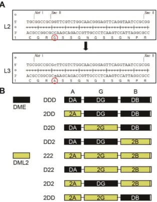
Cloning of domain swapping constructs between DME and DML2 The linker L2 of pEGFP-C1-D2D was replaced with a new synthetic linker L3 (CGRASSGNGSSGNPR) containing a single Sac II site for following cloning procedure (Figure 1-2A). Oligonucleotides DG276 and DG277 was annealed and digested with Not I and Sac II restriction enzymes. The digested linker fragment was inserted into the corresponding sites of pEGFP-C1-D2D to produce pEGFP-C1-D2D with L3. A partial fragment of D2D was inserted into the Pst I and Eco RI of pBS-DMECTDΔ1 (pBlueScript II KS) to produce pBS-D2D. Finally, the D2D fragment with L3 from pBS-D2D was cloned into the Bam HI and Xho I sites of the pLM302 vector to generate pLM302-D2D with L3. Note that all DME-DML2 swapping constructs contain L3 instead of L2 between the glycosylase domain and domain B.
Domain boundaries of DML2 were determined based on secondary structure prediction and amino acid sequence homology. The domain A and B of DML2 were amplified from Arabidopsis cDNA with primer pairs DG278-DG279, and DG280-DG281, respectively. Each fragment was
26
substituted into the domain A and B of pLM302-D2D to produce DME-DML2 swapping constructs, pLM302-22D and pLM302-D22, respectively. At the same time, the central glycosylase domain of DME was inserted into pLM302-D2D to generate pL302-DDD that harbors L3. In the same manner, the glycosylase domain of DME and domain B of DML2 were cloned into pLM302-22D, resulting pLM302-2DD and pLM302-222, respectively. Domain A of DML2 was cloned into the DDD resulting pLM302-2DD. The glycosylase domain of DME was cloned into the pLM302-222 producing pLM302-2D2. Finally, the glycosylase domain of D22 was replaced with that of DME to produce pLM302-DD2. Taken together, 8 possible combinations of constructs between DME and DML2 were obtained (Figure 1-2B).
Protein expression and purification
Constructs were transformed into E. coli Rosetta2 (DE3) strain (EMD Millipore) and cells were grown at 28°C in LB medium containing 50 μg/mL each of kanamycin and chloramphenicol antibiotics until the OD600 reached 0.4. Protein expression was induced with 0.1 mM IPTG at
18°C for 16 h. Cells were harvested by centrifugation at 7,000 rpm for 20 min at 4°C and resuspended in 30 mL of lysis buffer (50 mM Tris-HCl, pH
27
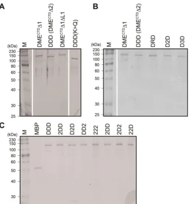
7.4, 100 mM NaCl, 10% glycerol, 0.1 mM dithiothreitol, 0.1 mM PMSF). Cells were sonicated (0.5x duty cycle) on ice for 5 min and cell extracts were clarified by centrifugation at 12,000 rpm for 25 min at 4°C. The supernatant was filtered through nylon membrane with 0.45 μm pore (Advantec) and the lysate was sequentially purified by three different types of columns: affinity column (His trap FF 5 mL, GE Healthcare), ion exchange column (Heparin HP 5 mL, GE Healthcare) and size exclusion (Superdex 200, GE Healthcare). The final eluted fractions were concentrated and aliquoted with 50% glycerol and stored in a storage buffer (20 mM Tris-HCl, pH 7.4, 40 mM NaCl, 4% glycerol, 0.1 mM dithiothreitol) at -80°C until use. Note that all the proteins used in this study are tagged with an MBP (Figure 1-3).
DNA substrate for radiolabeling
The 30-mer oligonucleotide containing 5mC in the middle of the sequence (5′- CTGTGTGATACTAT[5mC]GAATTCAGTATGATC -3′) and its complementary strand were synthesized (Integrated DNA Technologies). Forty pmol of oligonucleotide was radiolabeled by T4 polynucleotide kinase (Takara) with 30 μCi of [γ-32P]ATP (Perkin Elmer). The labeled oligonucleotide was purified by Qiaquick Nucleotide Removal
28
Kit (Qiagen) and annealed with complementary oligonucleotides to produce double-stranded DNA substrate. The mixture was boiled in water for 10 min and slowly cooled down for annealing to room temperature for at least 3 h.
In vitro DNA glycosylase assay
Radiolabeled oligonucleotide substrate (25 nM) was incubated with purified enzymes (100 nM) in the glycosylase reaction buffer (10 mM Tris-HCl, pH 7.4, 50 mM NaCl, 0.5 mM dithiothreitol, 0.2 mg/mL Bovine serum albumine) at 37°C for 1 h. Reaction was terminated by adding an equal
volume of stop solution (98% formamide, 10 mM
ethylenediaminetetraacetic acid (EDTA), 0.2% Xylen cyanol FF, 0.2% of bromophenol blue) and boiled at 95°C for 10 min. Reaction products were separated on a 15% denaturing polyacrylamide gel containing 7.5 M urea and 1x TBE (Tris/ Borate/ EDTA) at 1,200 V for 4 h. The gel was exposed to X-ray film (Fujifilm) at -80°C.
Single turnover kinetics of DME
The same reaction condition described in the In vitro DNA glycosylase assay was used for kinetics study but reactions were terminated at a various time point by adding 100 mM NaOH and boiling for 10 min.
29
And then one volume of stop solution (98% formamide, 10 mM EDTA, 0.2% Xylen cyanol FF, 0.2% of bromophenol blue) was added and reactions were boiled at 95°C for additional 10 min. Reaction products were loaded on a 15% polyacrylamide gel containing 7.5 M Urea and 1x TBE. After electrophoresis at 1,200 V for 4 h, the gel was exposed to a phosphorimager screen (Fujifilm) and the radioactivity was measured using the Fujifilm BAS-5000 (Fujifilm). The intensity of the amount of products relative to that of substrate was measured using the Multi Gauge V2.2 (Fujifilm). Estimation of single turnover kinetics (kcat-st) was accorded to Begley et al.
(2003). Briefly, the catalytic rate constant was calculated using the following equation: [product] = A[1 – exp[(-kcat-st) x min]], where A
represents the amplitude of the exponential phase.
Electrophoretic mobility shift assay
Radiolabeled oligonucleotide (100 nM) containing 5mC was incubated with increased amount of purified enzymes (0, 7, 35, 175 nM) in the DNA binding buffer (10 mM Tris-HCl, pH 8.0, 150 mM NaCl, 0.05% Triton X-100, 0.1 mg/mL BSA, 10% glycerol, 10 mM dithiothreitol) at 25°C for 15 min. Reactions were separated on a native polyacrylamide gel
30
(4% acrylamide, 2.5% glycerol and 0.5x TBE) at 50 V for 2 h. The gel was exposed to X-ray film at -80°C.
Sequence alignment and phylogenetic analysis
The glycosylase domain sequences of DME family proteins were obtained from TAIR (www.arabidopsis.org) and amino acid alignment was performed using ClustalX v2.1 (Larkin et al., 2007). The sequences were visualized by GeneDoc v2.7.00 (Nicholas and Nicholas, 1997), and the phylogenetic tree was constructed by the neighbor-joining method using MEGA v6 (Tamura et al., 2013) with 2,000 bootstrap replications.
31
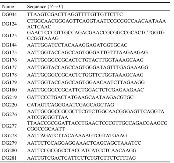
Table 1-1. Oligonucleotides used in this study.
Name Sequence (5′→3′) DG044 TTAAGTCGACTTAGGTTTTGTTGTTCTTC DG124 CTGGCAACGGGAGTTCAGGTAATCCGCGGCCAACAATAAA ACTCAAC DG125 GAACTCCCGTTGCCAGACGAACCGCGGCCGCACTCTGGTG CCGGTAAAG DG144 AATTGGATCCTACAAAGGAGATGGTGCAC DG175 AATTGGTACCAGCCAGTGGGATTGTTTAAGAAGAG DG176 AATTGCGGCCGCACTCTGTACTTGGTAAAGCAAG DG177 AATTGGTACCAGCCAGTGGGATAGTTTGAGAAAGG DG178 AATTGCGGCCGCACTCTGGTTCTGGTAAAGCAAG DG179 AATTGGTACCAGCCAGTGGAACAATCTTAGAAGG DG180 AATTGCGGCCGCATTCTGGACTCTCGAGAAGAAC DG219 GATTCCCTGACTATGAAGCAATAAGACGTGC DG220 CATAGTCAGGGAATCGAGCAGCTAG DG276 AATTGCGGCCGCGCTTCGTCTGGCAACGGGAGTTCAGGTA ATCCGCGGTTAA DG277 TTAACCGCGGATTACCTGAACTCCCGTTGCCAGACGAAGCG CGGCCGCAATT DG278 AATTAGATCTTACAAAAAGTCGTATGAAG DG279 AATTCTGCAGGAGGAAACTCAGCAGCTAAATCC DG280 AATTCCGCGGCCTACCATCATCCTCAACAAGG DG281 AATTGTCGACTCATTCCTCTGTCTTCTCTTTAG
32
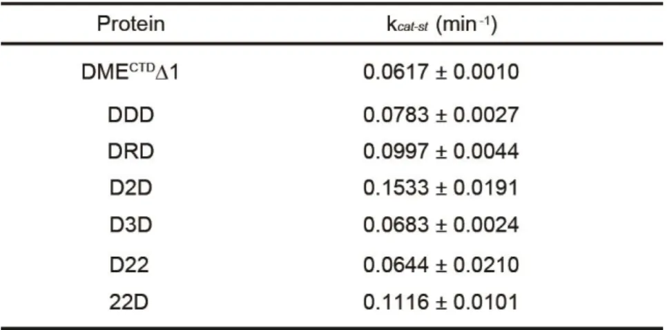
Figure 1-1. Structures of manipulated DME fragments and chimeric proteins between DME and other family members.
(A) Structures of full-length and manipulated DME fragments. DMECTD has C-terminal domains without N-terminal 935 amino acids. DMECTDΔ1 has deletions of N-terminal 935 amino acids and interdomain region IDR1, with the latter being replaced by a synthetic linker L1. DMECTDΔ2 consists of three core domains tethered with linkers L1 and L2, instead of IDR1 and IDR2. NVR, N-terminal variable region. (B) The DDD (DMECTDΔ2) cassette of the minimal catalytic core and its chimeric constructs, DRD, D2D and D3D.
33
Figure 1-2. Sequences of the synthetic linkers and structures of chimeric fragments generated by domain swapping between DME and DML2.
(A) Sequences of the synthetic linkers L2 and L3. A single amino acid substitution of L2 generated L3, used for following construction of DME-DML2 chimeric fragments. (B) Structures of possible combinations of chimeric fragments generated by domain swapping between DME and DML2. Domains of DME and DML2 were represented with black and yellow boxes, respectively.
34
Figure 1-3. SDS-PAGE analysis of purified proteins used for in vitro 5mC excision assay.
N-terminal MBP tagged purified proteins (200 ng) for experiment in (A) Figure 1-4, (B) Figure 1-6, (C) Figure 1-7 were electrophoresed on a 10% SDS-PAGE gel. The sizes of the protein marker (kDa) were indicated to the left. M, size marker.
35
RESULTS
Three conserved domains of DME comprise the minimal entity for 5mC excision in vitro.
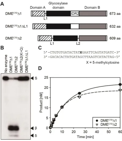
It was previously shown that three conserved domains of DME and ROS1 are essential, but most of the other variable regions are dispensable for 5mC excision activity in vitro (Mok et al., 2010; Hong et al., 2014). Deletion studies of DME showed that both DME without the entire N-terminal region (designated as DMECTD in Figure 1-1A) and the manipulated DME fragment mostly comprising the three conserved C-terminal domains but still harboring interdomain region IDR2 (designated as DMECTDΔ1 in Figure 1-1A) were able to remove 5mC in vitro (Mok et al., 2010). These results indicate that the core structure of DME mainly comprises the three domains as discrete modules. Homology modeling analysis supports this idea by predicting a model, in which the 5mC binding pocket of DME is composed of amino acid residues derived from domain A (AD) and the glycosylase domain (GD) (Brooks et al., 2014). To verify this interdomain 5mC binding pocket model, I directly tethered AD and GD without a synthetic linker L1 to produce DMECTDΔ1ΔL1 (Figure 1-4A). The in vitro 5mC excision analysis showed that DMECTDΔ1ΔL1 completely lost
36
5mC excision activity (Figure 1-4B), whereas DMECTDΔ1 with a synthetic dodecapeptide linker sequence between AD and GD excised 5mC from hemimethylated oligonucleotide substrates in vitro (Figure 1-4C). This indicates that the flexibility between AD and GD is required to configure a catalytic core structure suitable for 5mC excision, thus the variable region between AD and GD is required for function in vitro, and its sequence must at least confer flexibility.
To further test the modular structure of DME with interdomain flexibility, all variable regions including both interdomain regions IDR1 and IDR2 were removed and connected with flexible synthetic linkers L1 and L2 to produce a DMECTDΔ2 fragment consisting only of AD, GD, and domain B (BD) of DME (Figure 1-4A). In vitro analysis revealed that DMECTDΔ2 excised 5mC with the single turnover rate constant (kcat-st =
0.0783 ± 0.0027) comparable to that of DMECTDΔ1 (kcat-st = 0.0617 ± 0.0010)
(Figures 1-4D and Table 1-2), suggesting that the three conserved domains comprise the minimal entity required for 5mC excision in vitro, but that a flexible sequence between AD and GD, and likely also BD and GD, are required for function.
37
Figure 1-4. The in vitro 5mC excision activity of minimal DME catalytic core with three conserved domains.
(A) Diagrams of compact DME fragments consisting of three conserved domains. DMECTDΔ1 has a synthetic linker L1 replacing IDR1, whereas DMECTDΔ1ΔL1 has the first two domains directly tethered together. DMECTDΔ2 has synthetic linkers L1 and L2 replacing IDR1 and IDR2, respectively. (B) In vitro 5mC glycosylase assay with DME catalytic core fragments. Positions of substrate (S) and β- and δ-elimination products (β, δ) are indicated to the right of the panel. K>Q, a catalytic mutant with a K1544Q substitution. (C) Oligonucleotide substrate used in (B). (D)
38
Kinetics analysis with DMECTDΔ1 and DMECTDΔ2 under single turnover conditions. Standard deviations were calculated from three independent experiments and plotted with error bars.
39
Table 1-2. Catalytic rate constants (kcat-st) of purified proteins
40
Conserved domains of DME family proteins are catalytically compatible in vitro.
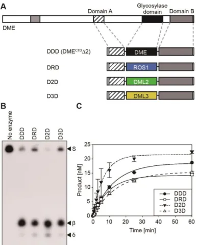
The DME family members in Arabidopsis share highly conserved domain structures, but each possesses distinct 5mC excision activity in vitro (Figure 1-5). In particular, DML2 displayed no discernable 5mC excision product unlike the other DME family members (Penterman et al., 2007), which raised the possibility that the activity of GD of DML2 might be restricted by its flanking domains AD and BD. In order to investigate whether these domains have functional compatibility between family members, I performed domain swapping experiments among the family members in a cassette configuration, in which the GD of the DMECTDΔ2 platform (referred to as DDD hereafter) was replaced with that of ROS1, DML2 and DML3, and the resulting chimeric fragments were designated as DRD, D2D and D3D, respectively (Figure 1-6A). All chimeric recombinant proteins were found to efficiently excise 5mC in vitro with a substantial amount of reaction products (Figure 1-6B), albeit they were reported to have different catalytic efficiencies in the native configuration (Penterman et al., 2007). Notably, the catalytic rate constant of D2D (kcat-st = 0.1533 ± 0.0191)
was about 2-fold higher than that of DDD (kcat-st = 0.0783 ± 0.0027), which
41
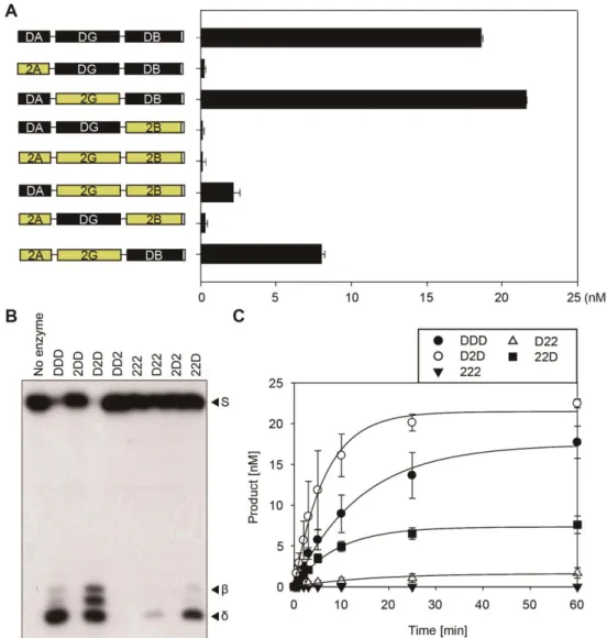
5mC excision activity of GD of DML2, supporting the hypothesis that intrinsic glycosylase activity of DML2 could be restricted by flaking domains AD and BD (Figure 1-6C and Table 1-2). These results not only indicate that the central GDs of DME family proteins retain equivalent structures and activities, but also suggest that DNA glycosylase domains of DME family proteins are catalytically compatible in vitro.
Besides the well-known central GD, the function of AD and BD is poorly understood, even though the previous random mutagenesis study revealed that a number of single amino acid substitutions abolishing the catalytic activity of DME in vitro are largely confined to the conserved domains AD and BD (Mok et al., 2010). To further investigate the function of flanking domains AD and BD, I performed domain swapping experiments between functionally distinguishable DME and DML2. Using the previously produced minimal DDD cassette configuration, 8 possible constructs of DME-DML2 swapping combination were created, and all chimeric proteins were purified for biochemical assay. Consistent with the previous report (Penterman et al., 2007), the 222 protein which consists of three domains of DML2 with flexible linkers, displayed no 5mC excision activity as expected (Figures 1-7A and 1-7B). Replacement of AD or BD of DDD cassette with that of DML2 produced 2DD or DD2 chimeric proteins,
42
both of which showed extremely reduced DME activity (Figures 1-7A and 1-7B). This result suggests that DML2 has impaired AD and BD, which inhibit intrinsic DNA glycosylase activity of central GD of DML2. Conversely, introduction of DME domains into the 222 cassette generated D22, 2D2 and 22D chimeric proteins. Except for 2D2, both D22 and 22D proteins restored the 5mC excision activity of DML2. Notably, 22D more efficiently catalyzed 5mC excision compared to D22, which is supported by the kinetics study that the catalytic rate constant of 22D (kcat-st = 0.1116 ±
0.0101) was 1.7-fold higher than that of D22 (kcat-st = 0.0644 ± 0.0210)
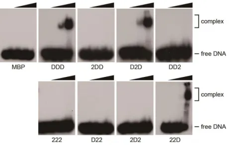
(Figure 1-7C and Table 1-2). Furthermore, the electrophoretic mobility shift assay revealed that different biochemical efficiencies of these chimeric proteins are correlated with DNA binding activity (Figure 1-8). These results demonstrate that the BD appears to be more critical to DME function than AD, which probably binds to DNA directly or serves as a structural role to facilitate DNA binding activity of DME.
43
Figure 1-5. The conserved motifs of the DME family proteins in Arabidopsis.
Schematic diagrams of four DME family members in Arabidopsis. Conserved motifs are denoted in colored boxes. In vitro 5mC excision activity of DME family proteins estimated from the previous study (Penterman et al., 2007) is indicated to the right.
44
Figure 1-6. The in vitro 5mC excision activity of chimeric proteins generated by swapping glycosylase domains between DME family members.
(A) Structures of chimeric proteins between DME and other family members in a minimal cassette configuration. The central glycosylase domain of DMECTDΔ2 (shown as DDD) was replaced with that of ROS1, DML2 or DML3 to produce chimeric proteins DRD, D2D and D3D, respectively. (B) In vitro 5mC glycosylase assay with the chimeric proteins. The radiolabeled DNA substrate (25 nM) containing 5mC was incubated with 100 nM each of MBP-DDD, DRD, D2D and D3D at 37°C for 1 h.
45
Positions of the oligonucleotide substrate (S), and the reaction products (β, δ) were indicated to the right of the panel. (C) Kinetics analysis with the chimeric proteins under single turnover conditions. Reactions were terminated at various time points (0.5, 1, 2, 3, 5, 10, 25 and 60 min), and the amounts of reaction products were plotted over time. Standard deviations were calculated from three independent experiments and plotted with error bars.
46
Figure 1-7. The in vitro 5mC excision activity of DME-DML2 chimeric proteins.
(A) In vitro 5mC glycosylase assay with the DME-DML2 chimeric proteins. Structures of chimeric proteins are represented to the left of the panel. The amount of the reaction products from experiment (B) was estimated and plotted as a bar graph. Standard deviations were calculated from three independent experiments and plotted with error bars. (B) In vitro 5mC
47
excision activity of the chimeric proteins. The radiolabeled DNA substrate (25 nM) containing 5mC was incubated with 100 nM each of chimeric proteins at 37°C for 1 h. Reactions were terminated by adding 100 mM NaOH with heat denaturing. Positions of the oligonucleotide substrate (S), and the reaction products (β, δ) were indicated to the right of the panel. (C) Kinetics analysis with the chimeric proteins under single turnover conditions. Reactions were terminated at various time points (0.5, 1, 2, 3, 5, 10, 25 and 60 min), and the amounts of reaction products were plotted over time. Standard deviations were calculated from three independent experiments and plotted with error bars.
48
Figure 1-8. Electrophoretic mobility shift assay of DME-DML2 chimeric proteins.
The radiolabeled oligonucleotide substrate containing 5mC (100 nM) was incubated with increased amount (0, 7, 35, 175 nM) of MBP or MBP-tagged DME-DML2 chimeric proteins at 25°C for 15 min. Free DNA substrate and protein-DNA complex are indicated to the right.
49
Functional motifs in the conserved domains of DME
The DME family proteins are variable in size but share similar domain structures, notably for the conserved three domains AD, GD and BD tethered by highly variable unstructured regions (Figures 1-5 and 1-9). Especially, the central GD is known to be catalytically essential for 5mC excision, which is supported by the observation that GD showed high sequence similarities among the DME family members (Figure 1-9A). Although phylogenetic analysis on GD implies that DME and ROS1 are the closest to each other, while DML3 is less related to the other members (Figure 1-9C), DME family members are predicted to have diverged from a common ancestor, and they all share several motifs in the conserved domains (Figure 1-5). The core of DME family proteins comprises the helix-hairpin-helix (HhH) motif and a glycine/proline-rich loop with a conserved aspartic acid (GPD) followed by the 4Fe-4S cluster loop (FCL) motif, a permutated CXXC motif, and a divergent version of an RNA Recognition Motif (RRM) fold (Figures 1-9A and 1-9B). The catalytic pocket containing HhH and FCL motifs is predicted to span AD and GD (Brooks et al., 2014), whereas the CXXC motif and RRM fold are present in the C-terminal half of BD (Figure 1-9B). In particular, the CXXC motif between the FCL motif and RRM fold is found in many chromatin
50
modifiers in mammals such as DNMT1, Methyl-CpG-binding domain protein 1, and notably, mammalian DNA demethylase, ten-eleven translocation 1 (TET1) (Rhee et al., 2002; Tahiliani et al., 2009; Long et al., 2013). Recent study also reported that the CXXC motif is required for DNA binding and target selection (Xu et al., 2018), which allows us to presume a biological role of the CXXC motif in plants.
51
Figure 1-9. Sequence alignment of the conserved domains of DME family proteins.
(A) Amino acid sequence alignment of the glycosylase domain of the DME family proteins. Catalytic residues (K1544 and D1562 for DME) in the HhH-GPD motif and four cysteine residues of the 4Fe-4S cluster are denoted as red, blue and green, respectively. (B) Amino acid sequence alignment of the domain B of DME family proteins. The CXXC motif and the RNA recognition motif are denoted above the sequence. Four cysteine residues comprising the CXXC motif are colored in yellow. (C) The phylogenetic tree was constructed with the Neighbor-Joining method using the sequences of the glycosylase domains of the DME family proteins.