저작자표시-비영리-변경금지 2.0 대한민국 이용자는 아래의 조건을 따르는 경우에 한하여 자유롭게 l 이 저작물을 복제, 배포, 전송, 전시, 공연 및 방송할 수 있습니다. 다음과 같은 조건을 따라야 합니다: l 귀하는, 이 저작물의 재이용이나 배포의 경우, 이 저작물에 적용된 이용허락조건 을 명확하게 나타내어야 합니다. l 저작권자로부터 별도의 허가를 받으면 이러한 조건들은 적용되지 않습니다. 저작권법에 따른 이용자의 권리는 위의 내용에 의하여 영향을 받지 않습니다. 이것은 이용허락규약(Legal Code)을 이해하기 쉽게 요약한 것입니다. Disclaimer 저작자표시. 귀하는 원저작자를 표시하여야 합니다. 비영리. 귀하는 이 저작물을 영리 목적으로 이용할 수 없습니다. 변경금지. 귀하는 이 저작물을 개작, 변형 또는 가공할 수 없습니다.
A Thesis
for the Degree of Doctor of Philosophy
Regulation of CD4
+
CD8
–
CD25
+
and
CD4
+
CD8
+
CD25
+
T cells by gut
microbiota in chicken
닭 장내 미생물에 의한 CD4
+CD8
–CD25
+및
CD4
+CD8
+CD25
+T 세포 조절 기전 연구
February 2017
By
In Kyu Lee
School of Agricultural Biotechnology
Graduate School
농 학 박 사 학 위 논 문
Regulation of CD4
+
CD8
–
CD25
+
and
CD4
+
CD8
+
CD25
+
T cells by gut
microbiota in chicken
닭 장내 미생물에 의한 CD4
+CD8
–CD25
+및
CD4
+CD8
+CD25
+T 세포 조절 기전 연구
지도교수 윤 철 희
이 논문을 농학 박사학위논문으로 제출함
2017 년 2 월
서울대학교 대학원
농생명공학부 동물생명공학 전공
이 인 규
이인규의 박사학위논문을 인준함
2017 년 2 월
위 원 장 한 승 현 (인)
부위원장 윤 철 희 (인)
위 원 박 병 철 (인)
위 원 강 석 성 (인)
위 원 박 태 섭 (인)
I
Summary
Gut microbiota in chicken has long been studied and considered for mostly growth performance point of view. And therefore, immunological studies regarding gut homeostasis in chicken have been insufficiently achieved. Regulatory T cells (Tregs) are a notable subtype of CD4+ T cells playing an
important role to maintain gut homeostasis in humans and animals. Intestinal Tregs are induced by gut microbiota, such as, Clostridium spp. cluster IV and XIVa strains, altered Schaedler flora (ASF), or Bacteroides fragilis in mice. Although it has been suggested that CD4+CD25+ T cells act as Tregs, there are
no such studies showing the relationship between gut microbiota and Tregs in chickens.
The first, I established the model for ABX-treated chickens by the administration of various concentrations of antibiotic cocktail consisting of ampicillin, gentamycin, neomycin, metronidazole, and vancomycin in water. Cecal contents from chickens treated with antibiotic cocktail consisting of 100 g/ml of ampicillin, gentamycin, neomycin and metronidazole, and 50 g/ml of vancomycin for 7 days eliminated colony forming unit (CFU) over 99%. These chickens treated by certain concentration of antibiotics cocktail (ABX) were referred as ‘ABX-treated chickens’. There were no changes on physiological traits, for example, weight of body and immune organs (spleen, bursa and liver), length of intestine (duodenum, jejunum, ileum and large intestine) and the concentration of glucocorticoid in the serum. Furthermore,
II
the population and MHC class II expression on B cells and macrophages in the cecal tonsils and spleen were not changed. I concluded that physiological traits, B cells and macrophages were not changed in ABX-treated chickens.
The second, I examined whether subtype of CD4+ T cells was changed in
ABX-treated chickens. In cecal tonsil, CD4+CD8–CD25+ and
CD4+CD8+CD25+ T cells were significantly decreased in ABX-treated
chickens, however these cells in the spleen were not changed. The expression of IL-10 and IFN- was significantly decreased in CD4+CD8–CD25+ T cells
from cecal tonsils of ABX-treated chickens. It was noting that CD4+CD8–
CD25+ and CD4+CD8+CD25+ T cells from ABX-treated chickens did not
suppress the proliferation of CD4+CD25– T cells. The reduction of CD4+CD8–
CD25+ and CD4+CD8+CD25+ T cells in cecal tonsils from ABX-treated
chickens expressed high level of CD5hi. Interestingly, the percentage of thymic
CD4+CD8+CD25+ T cells was not changed in ABX-treated chickens.
Conclusively, the population and suppressive function of peripheral CD4+CD8–
CD25+ and CD4+CD8+CD25+ T cells decreased in ABX-treated chickens.
The third, I examined what factors affected the population of CD4+CD8–
CD25+ and CD4+CD8+CD25+ T cells. ABX-treated chickens co-housed with
wild type chickens recovered the number of gut microbiota, and the proportion of CD4+CD8–CD25+ or CD4+CD8+CD25+ T cells in cecal tonsils to similar
levels as those of wild type chickens. The results further showed that Gram-positive bacteria appeared to be responsible for the changes of CD4+CD8–
III
short chain fatty acids, in ABX-treated chickens recovered CD4+CD8–CD25+ T
cells and CD4+CD8+CD25+ T cells in cecal tonsils. Both butyrate and
propionate did not show the effect to recover these cells. Interestingly, GPR43 mRNA level was highly expressed in CD4+CD8–CD25+ T cells.
Conclusively, my study demonstrated that gut microbiota can regulate the population and suppressive function of CD4+CD8–CD25+ or CD4+CD8+CD25+
T cells, and acetate can induce CD4+CD8–CD25+ T cells in cecal tonsils via
IV
Contents
Summary... I Contents... IV List of Figures... VII List of Table... VIII List of Abbreviations... IX I. Review of Literature... 1 1. Gut homeostasis... 1 1.1. Regulatory T cells... 1 1.2. T helper 17 cells... 3 1.3. Immunoglobulin A... 4
1.4. Innate lymphoid cells... 6
2. Gut microbiota in chicken... 7
2.1. Intestine... 7
2.2. Establishment of gut microbiota... 8
2.3. Gut microbiota on growth performance... 9
2.4. Effects of gut microbiota on immunological aspect... 10
II. Introduction... 13
III. Materials and Methods... 16
1) Experimental animal and ABX treatment... 16
2) Measurement of colony forming unit... 17
V
4) Flow cytometric analysis for immune cells... 18
5) Measurement of mRNA level using RT-qPCR... 19
6) Changes on the subtype of CD4+ T cells treated with antibiotics in vitro... 21
7) T cell suppression assay... 21
8) Co-housing experiment... 22
9) The elimination of Gram positive and negative bacteria... 23
10) Administration of short chain fatty acids... 23
11) Statistical Analysis... 23
IV. Results... 25
1) Elimination of gut microbiota in chicken... 25
2) Verification of physiological alteration in ABX-treated chickens... 28
3) Change of B cells and macrophages in ABX-treated chickens... 30
4) Change of CD4+ T cells in ABX-treated chickens... 32
5) Change of IL-10 and IFN- from subtype of CD4+ T cells in ABX-treated chickens... 36
6) Direct effect of antibiotics on the change of CD4+CD8–CD25+ and CD4+CD8+CD25+ T cells... 38
7) Changes of CD4+CD8–CD25+ and CD4+CD8+CD25+ T cells in periphery of ABX-treated chickens... 40
8) Suppressive function of CD4+CD8–CD25+ and CD4+CD8+CD25+ T cells in ABX-treated chickens... 43
9) Changes of CD4+CD8–CD25+ and CD4+CD8+CD25+ T cells in ABX-treated chickens after co-housing with control chickens... 45
10) Effect of Gram-positive or negative bacteria on the population changes of CD4+CD8–CD25+ and CD4+CD8+CD25+ T cells... 47
VI
11) Effect of SCFAs on CD4+CD8–CD25+ and CD4+CD8+CD25+ T
cells... 49
V. Discussion... 51
VI. Literature Cited... 60
VII
List of Figures
Figure 1. Induction of Tregs by host cells, gut microbiota and their
products... 2 Figure 2. Elimination of gut microbiota in chickens treated with
antibiotics... 26 Figure 3. No physiologic changes in ABX-treated chickens... 29 Figure 4. No changes of B cells and macrophages in cecal tonsils and
spleen in ABX-treated chickens... 31 Figure 5. Gating strategy to analyze subtype of CD4+ T cells... 33
Figure 6. CD4+CD8–CD25+ and CD4+CD8+CD25+ T cells were reduced in
cecal tonsils from ABX-treated chickens... 34 Figure 7. CD4+CD8–CD25+ and CD4+CD8+CD25+ T cells were not
changed in spleen from ABX-treated chickens... 35 Figure 8. Expression of IL-10 and IFN-γ mRNA among CD4+ T cell
subsets in cecal tonsils from ABX-treated chickens... 37 Figure 9. No changes of CD4+CD8–CD25+ and CD4+CD8+CD25+ T cells in
chicken splenocytes treated with antibiotics... 39 Figure 10. Reduction of CD5hi cells in CD4+CD8–CD25+ and
CD4+CD8+CD25+ T cells in ABX-treated chickens... 41
Figure 11. CD5hi cells of CD4+CD8+CD25+ T cells were not changed in
thymus of ABX-treated chickens... 42 Figure 12. Elimination of gut microbiota caused reduction of suppressive
ability of CD4+CD8–CD25+ and CD4+CD8+CD25+ T cells... 44
Figure 13. Changes of CFU, and CD4+CD8–CD25+ and CD4+CD8+CD25+
T cells in cecal tonsils from ABX-treated chickens after co-housing with control chickens... 46 Figure 14. Elimination of Gram positive bacteria is responsible for the
change of CD4+CD8–CD25+ and CD4+CD8+CD25+ T cells in
VIII
List of Table
Table 1. Elimination of gut microbiota in chickens administered with various concentration of antibiotics in drinking water for 7 days... 27 Figure 15. Changes of CD4+CD8–CD25+ and CD4+CD8+CD25+ T cells in
chickens administered with acetate... 50 Supplementary Figure 1. No differences in Maf and Ahr gene expression
IX
List of Abbreviations
ABX Antibiotics cocktailAhr Aryl hydrocarbon receptor APCs Antigen presenting cells APRIL A proliferation-inducing ligand ASF Altered Schaedler flora
BAFF B cell activating factor CFU Colony forming unit
c-Maf Cellular homolog of the avian virus oncogene musculoaponeurotic fibrosarcoma
CTs Cecal tonsils CTV CellTrace™ Violet DCs Dendritic cells
EHEC Enterohemorrhagic Escherichia coli EPEC Enteropathogenic Escherichia coli Foxp3 Forkhead box P3
GALT Gut-associated lymphoid tissue GC Germinal center
GPR G protein coupled receptor H3K27 H3 lysine 27
HDAC Histone deacetylase
IBD Inflammatory bowel disease IELs Intraepithelial lymphocytes IgA Immunoglobulin A
IL Interleukin
X iNOS Inducible nitric oxide synthase LP Lamina propria
Maf v-maf avian musculoaponeurotic fibrosarcoma oncogene homolog
PPs Peyer’s patches pTregs Peripheral Tregs RA Retinoic acid
RegIII Regenerating islet-derived protein 3 SAA Serum amyloid A protein
SCFAs Short chain fatty acids
SFB Segmented filamentous bacteria SIgA Secreted IgA
TCRs T cell receptors
TGF Transforming growth factor Th17 T helper 17
TLR Toll-like receptor TNF Tumor necrosis factor Tr1 Type 1 regulatory T Tregs Regulatory T cells
1
I. Review of Literature
1. Gut homeostasis
Mouse and human studies suggested that the microbiota continuously interact with the intestinal immune system for the balance between pro-inflammatory and tolerogenic immune responses, called gut homeostasis [1-3]. Various immune cells and their products are associated with gut homeostasis including forkhead box P3 (Foxp3)+ regulatory T cells (Tregs), T helper 17
(Th17) cells, IgA+ B cells, innate lymphoid cells (ILCs), transforming growth
factor (TGF)- and interleukin (IL)-10. Gut homeostasis of chicken has not been fully understood and it is assumed to be similar to that of mammals [4].
1.1. Regulatory T cells
Tregs are a subset of CD4+ T cells that exist in peripheral organs and intestine,
where they help to maintain gut homeostasis. The absence of Tregs results in the abnormal expansion of CD4+ T cells expressing commensal
bacteria-specific T cell receptors (TCRs) resulting in intestinal inflammation [5]. The development of peripheral Tregs is known to partly depend on the gut
2
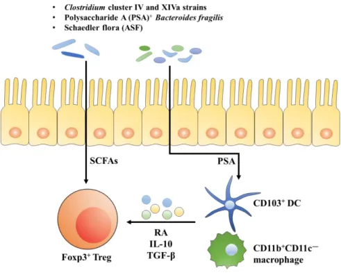
microbiota [2, 6]. As shown in Fig. 1, Tregs are induced by specific populations of commensal bacteria which comprise Clostridium spp. cluster IV and XIVa strains [2], altered Schaedler flora (ASF) [6], or Bacteroides fragilis [3] and/or short chain fatty acids (SCFAs) [7] produced as gut microbial product through IL-10, TGF- or retinoic acid (RA) expressing antigen presenting cells (APCs), such as, CD103+ dendritic cells (DCs) and CD11b+CD11c– macrophages.
Figure 1. Induction of Tregs by host cells, gut microbiota and their products. Tregs are known to be induced by specific populations of commensal bacteria together with their product, SCFAs and PSA, through IL-10, TGF- or RA produced by CD103+ DC and CD11b+CD11c– macrophages.
3
Besides Foxp3+ Tregs, CD4+Foxp3– type 1 regulatory T (Tr1) cells, one of
Treg subsets, could contribute to gut homeostasis by suppressing inflammatory condition. In SCID mice with inflammatory bowel disease (IBD), antigen specific Tr1 cells (pre-induced in vitro with IL-10) prevented to the progression of colitis [8]. It has been suggested that Bifidobacterium breve and B. longum, as probiotics, induced Tr1 cells in mouse and alleviated the development of intestinal inflammation [9]. CD4+CD25+ T cells of chicken are known as Tregs
[10], which are absent of Foxp3 gene [11, 12], unlike their mammalian counterpart. Furthermore, chicken CD4+CD25+ T cells migrated preferentially
cecal tonsils rather than spleen and lung [13]. Chicken Tregs are not fully investigated, for example, intrinsic and extrinsic factors conditions to induce Tregs.
1.2. T helper 17 cells
Th17 cells are one of CD4+ T cell lineages, producing IL-17A, IL-17F and
IL-22, which have a role for host defense and development of autoimmune disease in human and mouse [14]. Intestinal Th17 cells are significantly reduced in antibiotic-treated or germ-free mice [1, 15-18], suggesting that the microbiota play a crucial role to develop Th17 cells in gut. Segmented filamentous bacteria (SFB), one of Clostridia-related bacteria, induces the
4
generation of Th17 cells [1, 16, 17]. SFB stimulates the host epithelium to upregulate serum amyloid A protein (SAA) production, which is known to promote IL-6 and IL-23 from CD11c+ lamina propria (LP) DCs [1]. ATP
produced by gut microbiota, but not much of pathogens, for instance,
Salmonella typhimurium which secreted ATP lesser than gut microbiota
resulting in induction of Th1 cells, promotes LP CD70hiCD11clow cells to
develop Th17 cells [15]. Regenerating islet-derived protein 3 g (RegIIIg), as a C‑type lectin antimicrobial peptide, from Th17 cells prevents the intestinal infection by pathogens including Citrobacter rodentium and Listeria
monocytogenes [19-21]. In chicken studies, IL-17 is assumed as Th17 response,
simply because anti-chicken 17 antibody to measure the cells secreting IL-17 directly is not available. Salmonella enterica serovar Enteritidis infection in chicken induced IL-17 in ceca [22]. It has been suggested that IL-17 was increased in chicken infected with Eimeria tenella, a protozoan parasite [23, 24]. The role of Th17 cells in the gut of chicken has yet to be fully understood.
1.3. Immunoglobulin A
The relationship between gut microbiota and gut-specific B cell responses, for instance, immunoglobulin A (IgA) secretion, is closely associated. IgA is an active component involving host protection and a major class of
5
immunoglobulin in the intestine. IgA exists as a polymeric IgA in the intestinal lumen [25]. Secreted IgA (SIgA) can recognize commensal bacteria and soluble antigens to inhibit penetration into the lamina propria [25]. IgA regulates the composition of the gut microbiota [26, 27]. Activation-induced cytidine deaminase, which is known to be essential enzyme for class switching, deficiency mice showed increase anaerobic bacteria, including
Peptostreptococcus, Bacteroidacease, Eubacterium and Bifidobacterium in
small intestine, whereas cecum microbiota was not changed [28]. Furthermore, gut microbiota regulates IgA production, as the number of IgA-producing cells in the intestine, for example, is decreased significantly in germ-free mouse [25]. Commensal bacteria induces various effector molecules, such as tumor necrosis factor (TNF), inducible nitric oxide synthase (iNOS), B cell activating factor (BAFF) and a proliferation-inducing ligand (APRIL) that are involved in the induction of IgA+ B cells in lamina propria [29, 30]. It is probable that gut
microbiota stimulates DCs in lamina propria to induce IgA+ B cells, and in
return, SIgA regulates the function and composition of the gut microbiota to maintain gut homeostasis. In chicken, IgA expression of ileum, ceca and cecal tonsils was burst at 7 days post hatching [31]. Probiotics mixture, consisting of
Lactobacillus acidophilus, Bifidobacterium bifidum, and Streptococcus faecalis,
induced natural IgA from intestinal contents reacting tetanus toxoid and
Clostridium perfringens alpha-toxin [32]. However, there is no molecular
6
1.4. Innate lymphoid cells
ILCs are known as immune cells involved in innate immune responses [33, 34] in human and mouse studies. It has been suggested that gut microbiota is required for the differentiation of ILCs and the production of IL-22 [35, 36]. In other study, gut microbiota suppressed IL-22 production by RORγt+ ILCs [37].
A role of IL-22 in the gut is to promote antimicrobial peptide production by intestinal epithelial cells. IL-22 induces the expression of the C-type lectin antimicrobial peptides, for example, RegIII, which protect the host from the infection of pathogens, for example, enterohemorrhagic Escherichia coli (EHEC) and enteropathogenic E. coli (EPEC) and C. rodentium [19]. RegIII limited the number of surface-associated Gram-positive bacteria, Firmicutes phylum (Eubacterium rectale, and SFB), and activation of adaptive immunity, for instance, IgA and IFN-+ cells [21]. Conclusively, ILCs regulate not only
both commensal and harmful bacteria but also host immunity in the gut. There is no report about chicken ILCs.
7
2. Gut microbiota in chicken
2.1. Intestine
The intestine is important in converting the feed into the nutrients for animals’ maintenance and growth. Digestive tract of chicken is composed of beak/mouth, esophagus, crop, proventriculus, gizzard, small intestine, ceca, large intestine and cloaca. During the digestion, morphology and chemical composition of feed change as they passing through several organs. Since chicken does not have teeth, they pick up feed with beak and it enters the mouth without chewing. Crop, an out-pocketing of the esophagus, is located in the neck region and stores feed and water [38]. When crop is empty, or near empty, hunger signal transmit to the brain [39]. Very little digestion occurs in crop by amylase secreted in mouth [40].
Proventriculus plays as the true stomach and begins to digest feed with hydrochloric acid and pepsin [41]. However, feed is not yet ground at this point. Gizzard is a unique digestive organ in chicken. It is referred to as ‘mechanical stomach’ since strong muscles of gizzard as acts like the bird’s teeth [42]. Furthermore, feed is grinding, mixing, and mashing with digestive enzymes in gizzard [41].
8
Small intestine in chicken is consisted of the duodenum, jejunum and ileum similar to that of a mammal. Duodenum secrets digestive enzymes and bicarbonate to counter the hydrochloric acid [43]. Digestive enzymes produced by the pancreas are primarily involved in protein digestion. Digestion of lipids and absorption of fat-soluble vitamins, such as, vitamins A, D, E and K in here occurs with bile [44]. Nutrients are absorbed mainly in the jejunum and ileum.
Ceca are two blind pouches located and a joint point of the small and large intestine. Water in fecal material is reabsorbed and fermentation of indigestible materials at here. It is known that the fermentation produces short fatty acids and vitamin B [45, 46]. Large intestine in chicken is much shorter than the small intestine in chicken. The last of water re-absorption occurs in here. In cloaca, feces are mixed with urine from urates.
2.2. Establishment of gut microbiota
Microorganisms in animal gut has evolved with host [47]. Microorganisms are abundant in the colon and ceca of chicken [48]. Domestic birds, including chicken, duck, and turkey, have about 1 × 10¹¹ cells/g in ceca [49, 50].
The chicks are initially exposed to microbes from the surrounding environment. Therefore, the early stage of the post hatching period would be critical for the formation of gut microbial community. The density of gut
9
microbiota in chicken increased rapidly within 24 h post hatching [51, 52]. Aerobes such as Enterobacteriaceae, Lactobacillus, and Streptococcus colonized initially in small intestine show a positive oxidation at hatching [53, 54]. Then, oxygen consumption by aerobes causes more anaerobic conditions in lower gut environment, which facilitates growth and colonization of the obligate anaerobes [49, 55, 56].
Ceca contain a more diverse community of gut microbiota, including
Bacteroides, Bifidobacterium, Clostridium, Enterococcus, Escherichia, Fusobacterium, Lactobacillus, Streptococcus and Campylobacter in chicken
[56-58]. Density of gut microbiota increases throughout the digestive tract, for example, duodenum, ileum and ceca contain 103–105, 108–109, <1012 colony
forming unit (CFU) gram-1 of digesta by microscope-counts, respectively [56,
58].
2.3. Gut microbiota on growth performance
The role of gut microbiota in chicken has long been interested for research scientists, industry and the field, because of its impact on growth performance. Probiotics have several positive effects in chicken, (1) improvement of weight gain and feed utilization, (2) decrease mortality through preventing enteric pathogens (3) to attach and colonize in intestine [59, 60]. Non-degradable
10
complex carbohydrates from diet in the small intestine, such as non-starch polysaccharides and resistant starch, are the main sources of carbon and energy for the commensal bacteria [61]. The metabolites are derived from fermentation by intestinal bacteria, which are consumed as the energy source for host [62]. For example, SCFAs, which are the metabolites by anaerobic microbes utilizing carbohydrates, are considerable energy source in animal [63]. Several pathways associated with production of SCFAs were detected in a meta-genomic analysis of cecal microbiota in chicken [64]. SCFAs from fermentation of non-hydrolysable oligo- and polysaccharides feeding may provide extra energy and a better feed conversion ratio in chicken.
2.4. Effects of gut microbiota on immunological aspect
Gut immune homeostasis in chicken, although seemingly similar to that of mammals, has not yet been fully understood. However, there are some studies about the relationship between gut microbiota and immune system in chicken. The complexity of gut microbiota impacts the repertoire of TCR in the gut [65] and the kinetics on the expression of immune-associated genes during the maturation of gut immune system [66]. It has been suggested that gut microbiota also indirectly affects the development of B cells in the bursa. When the bursal duct is ligated during embryonic development to preclude the normal
11
traffic of gut-derived molecules into the bursa, cortico-medullary structure in bursal follicle fails to develop normally after hatching [67, 68]. The mechanism of the phenomenon is explained that these gut-derived molecules, probably and mostly bacterial mitogens, could directly induce maturation and proliferation of bursal B cells [69, 70], or indirectly stromal cells to produce cytokines, perhaps via Toll-like receptor (TLR) signaling, for B cell development [71].
2.5. Cecal tonsils
The gut-associated lymphoid tissue (GALT) consists of multiple lymphoid follicles and these are made up of cecal tonsils (CTs), Peyer’s patches (PPs), the bursa of Fabricius, Meckel’s diverticulum located along the digestive tract in chicken [72]. Especially, CTs are located on the entrance of each cecum, which consist of a pair of fingerlike pockets located in end of small intestine. CTs are histologically [73, 74] and immunologically [75] as secondary lymphoid tissue, similar to the spleen. CTs consist of a cryptosporidians, diffuse lymphoid follicles and germinal centers [76]. Considering cellular and morphological features, a role in antigen sampling of CTs could be similar to mammalian PPs [72, 77]. Within organized lymphoid structures, such as CTs, CD4+ T cells and B cells exist [78, 79], whereas T cells predominate in
12
The development of CTs during embryogenesis has not been described in detail. CTs are appeared at near hatching [76, 81], unlike lymphoid cells infiltrate at presumptive sites of PPs [72]. During embryogenesis, clusters of MHC class II+ cells, a few scattered Bu-1+ cells and IgM+ cells were observed
at E13. At E17, MHC class II+ cells were widely and densely expended, and
Bu-1+ cells and IgM+ cells are increased more than those at E13 [82]. It suggests
that MHC class II+ cells, presumably antigen presenting cells including
13
II. Introduction
Tregs are a subtype of CD4+ T cells, known to play an important role in
maintaining gut immune homeostasis since the gastrointestinal tract is constantly exposed to inflammatory condition by a huge microbial components [84]. In mouse and human, Foxp3 is the master transcription factor for Tregs [8, 85]. Common surface molecule and cytokines as makers for Tregs are high expression of CD25 (IL-2 receptor ), and IL-10 and TGF-, respectively [86]. Non-Foxp3 Tregs, also called Tr1 cells [87], induced by chronic activation of CD4+ T cells with antigen and IL-10 [8] are also reported. Although the master
transcription factor is unknown for Tr1 cells yet, unique features of cytokines are suggested as IL-10++, TGF-+, interferon (IFN)-+, IL-5+, IL-4– and IL-2low/–
[8, 88]. CD4+CD25+ T cells in chicken has been reported as Tregs [10],
although no Foxp3 orthologue gene exists [11].
Certain gut microbiota, including Clostridium spp. cluster IV and XIVa strains, ASF, or Bacteroides fragilis, are known to induce Foxp3+ Tregs in mice
and human [2, 3]. These bacteria alleviate the symptom of IBD by inducing Tregs [2, 3, 6]. However, no such studies on the relationship between gut microbiota and Tregs are reported in chicken.
Gut immune homeostasis is largely regulated by microbiota in not only a direct [3] but also indirect manner. Induction [2] and function [3] of Tregs are
14
affected by gut microbiota related factors, such as SCFAs [89-91] including acetate (C2), propionate (C3), and butyrate (C4), which are generated especially by Firmicutes and Bacteroidetes, after fermenting undigested carbohydrates [84]. It has been shown that acetate, propionate and butyrate exist in 3:1:1 ratio, respectively at 150 mM in colon of mouse [92], whereas 50-70 mM of acetate, 5-30 mM of butyrate and 5-10 mM of propionate are contained in chicken ceca [93-95].
It has been shown in mouse experiments that several G protein coupled receptors (GPRs) on immune and non-immune cells recognize SCFAs [96, 97]. Activation of GPR43 using SCFAs promotes the number and function of IL-10+Foxp3+ Tregs, and propionate directly increases Foxp3 expression and
IL-10 production [91]. GPRIL-109a, expressed on colonic epithelial cells, DCs and macrophages [98], is known to be activated by butyrate.
IL-10 and RA produced by mostly antigen presenting cells treated with SCFAs [99] could induce the differentiation of naïve T cells into Foxp3+ Tregs
and Tr1 cells [99]. SCFAs are also known to act as a histone deacetylase (HDAC) inhibitor. For instance, butyrate enhances acetylation at histone H3 lysine 27 (H3K27) of the Foxp3 promoter causing the differentiation of naïve T cells into Tregs [89]. Acetate, on the other hand, induces the acetylation of p70 S6 kinase and phosphorylation rS6, resulted in Tr1 cell induction [100]. There are not only a very few studies on immunological effects of SCFAs in chicken but also no reports about the factors regulating gut homeostasis.
15
Germ-free mouse model has been a critical tool to carry out the research on immune homeostasis in the mucosal tissues as well as peripheral organs for decades [101-103]. Gut immune balance is the result of interaction among various immune cells including Tregs, Th17 cells, IgA secreting B cells, and innate immune cells [103]. In indigenous germ-free mice, peripheral Tregs (pTregs) are scarce in the lamina propria of the intestine [2, 104]. Antibiotics cocktail (ABX) treatment is an alternative way to make an intestinal germ-free animal. ABX-treated mice showed closely resembling indigenous germ-free mice in terms of immunological changes in not only the gut but also peripheral organs [105-107]. The presence of intestinal Th17 cells is dramatically reduced in ABX-treated mice [16]. Although Foxp3+ Tregs are still detectable, they are
significantly decreased in colonic lamina propria [2]. Unfortunately, there is no report, at the best of my knowledge, on immunological researches in germ-free or gut microbiota-free chicken model.
In the present study, the model for studying gut immune homeostasis in chicken treated with ABX was established. The main goal of the study was to (1) examine the changes in population and function of immune cells in ABX-treated chickens and (2) find the factors regulate gut homeostasis.
16
III. Materials and Methods
1) Experimental animal and ABX treatment
Fertile eggs of White Leghorn were provided by Animal Farm, Seoul National University, Pyeong-Chang, Korea. Fertile eggs were incubated at 37.5-38°C incubator (Rcom, Korea) for 21d. The condition of cage sustained 28-30°C and filte
red air. Care room maintained 23-25°C, 20-40% of humidity and positive pressure. The experiment was approved by Institutional Animal Care and Use Committee of Seoul National University (IACUC No., SNU-150327-2). Crumble feed was supported by SeoulFeed company and sterilized by -radiation by GREENPIA TECHNOLOGY company. For antibiotics treated group, chickens at hatching were treated with various concentrations of antibiotics in drinking water ad libitum for 7 days. I defined dilution factor (DF) 1 as an antibiotics containing ampicillin, gentamycin, neomycin (all from Sigma-Aldrich, St. Louis, MO) and metronidazole (Abcam, Cambridge, MA) for 1 mg/ml each, and vancomycin (Sigma-Aldrich) for 0.5 mg/ml. DFs were tested 1:1, 1:2, 1:10, and 1:20. As control (Con) group. ABX-treated chickens
17
were referred by treatment of 1:10 diluted antibiotics for 1-3 weeks.
2) Measurement of colony forming unit
Cecal contents from chickens treated with ABX were dissolved in PBS to adjust at 1 mg/ml. Dissolved cecal contents from Con group were diluted in 100-1000 times with PBS while those from ABX-treated group were used without dilution. All dissolved cecal contents were spread on Brain Heart Infusion (BHI) agar media (BD Biosciences, San Jose, CA) and then incubated at 37°C for 12 hr. CFU was determined by counting the number of colony.
3) Examination of physiological changes in ABX-treated chickens
Body weight changes were monitored in chickens every day for 7 days. At the end of the experiment, major immune organs (liver, spleen and bursa) were taken and briefly semi-dried by tapping on paper towel, and the weight was examined. Length of intestine was segmented to jejunum (J), duodenum and ileum (D+I), Ceca (C) and large intestine (L), and measured with millimeter scale. Blood samples from a wing vein were taken at 7 days after the ABX treatment. Amount of glucocorticoid in serum, which was obtained by
18
centrifugation at 1,000 × g at 4°C for 20 min, was measured by chicken glucocorticoid ELISA kit (MyBioSource, San Diego, CA). In brief, 50 l of serum per well, diluted to 1:50 (pre-determined, data not shown), along with standard samples, was added to a 96-well microplate pre-coated with glucocorticoid specific antibodies. After the wash with PBS, 50 μl of secondary antibody, conjugated with HRP, was added into each well, and color was developed by the addition of 90 μl of tetramethylbenzidine. The reaction was stopped by the addition of 50 μl of stop solution. Absorbance was measured at 450 nm using an ELISA microplate reader (Molecular Device, Sunnyvale, CA) and the amount of glucocorticoid was calculated from the standard curve.
4) Flow cytometric analysis for immune cells
Chunked spleen or longitudinally cut cecal tonsils after wash were minced with the flat end of a 3 ml syringe plunger through a 40 m cell strainer (BD Biosciences, San Jose, CA) into a 50 ml conical tube (SPL, Pocheon, Korea). In order to purify immune cells, red blood cells were lysed using ACK buffer (BD Biosciences) for 3 min at room temperature, and then washed.
For examination of B cells and macrophages, anti-chicken MHC class II-FITC (clone 2G11), Monocyte/Macrophage-PE (clone KUL01), and Bu-1-Alexa Flour 647 (AV20) (all from Southern Biotec, Birmingham, AL) were
19
used. In order to examine CD4+ subtype T cells, anti-chicken CD4-FITC (clone
CT-4), CD8 -PE (clone CT-8), CD5-biotin (clone 2-191) (all from Southern Biotec) and CD25-Alexa Fluor 647 (clone 13504; AbD Serotec, Puchheim, Germany), and Brilliant Violet 605 streptavidin (BioLegend, San Diego, CA) were used.
Flow cytometric data, acquired by flow cytometry (FACS Canto II, BD Biosciences), were analyzed with FlowJo software (Tree Star, San Carlos, CA). Total cell number was determined by automatic cell counter TC10 (Bio-Rad, Hercules, CA). Each number of immune cells was calculated from total cell number and the proportion of immune cells.
5) Measurement of mRNA level using RT-qPCR
CD4+ subtype T cells (CD4+CD8–CD25–, CD4+CD8–CD25+,
CD4+CD8+CD25– and CD4+CD8+CD25+) B cells (Bu-1+) and APCs (KUL01+,
MHC class II+Bu-1–KUL01–) were sorted by using ARIAII FACS sorter (BD
Biosciences). Total RNA of each CD4+ subtype T cells was extracted by
miRNeasy Micro Kit (QIAGEN, Hilden, Germany). The concentration of total RNA was quantified with NanoDrop (Amersham Biosciences, Piscataway, NJ) at A260. Subsequently, 100 ng of purified RNA was reverse transcribed to cDNA using M-MLV Reverse Transcriptase (Invitrogen, Carlsbad, CA)
20 according to the manufacturer’s instruction.
The real-time quantitative PCR was performed on cDNA using a StepOne Plus real-time PCR system (Applied Biosystems, Foster City, CA). SYBR green PCR master Mix was used according to manufacturer’s specification (Applied Biosystems). The PCR reaction was carried out in a 96-well reaction plate with 10 l SYBR PCR master mix, 0.5 l per primer (2 pM), 1-2 l cDNA template and 7-8 l nuclease-free water. Each reaction involved a pre-incubation at 95 °C for 10 min, followed by 45 thermal cycles at 95 °C for 15 s, 55 °C for 30 s, and elongation at 72 °C for 30 s. Relative quantification of target genes was calculated using the 2-Δ ΔCt method.
Target gene expression was normalized to -actin mRNA level. Primers for IL-10 (forward: AGCTGACGGTGGACCTATTATT-5’, reverse: 3’-GGCTTTGCGCTGGATTC-5’), IFN- (F: 3’-CGGGAGCTGAGGGTGAA-5’,
R: 3’-GTGAAGAAGCGGTGACAGC-5’), Ahr (F: 3’-
CAGGTCCCTGAAAACCTTGACT-5’, R: 3’-
ACGGCACCTGCATAACATGTT-5’), Maf (F: 3’-
CCCCGTTACCTGAGGTCAGA-5’, R: 3’-
GTCTTCGTGCCAGAACGTTGT-5’), G-coupled protein receptor 43 (F: 3’-CTCTTTATGGCTGCCCTCAG-5’, R: 3’-GTAGCCCAGGCTTGGTTGG-5’) and -actin (F: CAACACAGTGCTGTCTGGTGGTA-5’, R: 3’-ATCGTACTCCTGCTTGCTGATCC-5’) were synthesized from Bioneer Inc. (Daejeon, Korea).
21
6) Changes on the subtype of CD4
+T cells treated with antibiotics
in vitro
Spleens from 2-3 weeks old-chickens were taken and single cells were produced. Splenocytes (1 × 10⁵ cells/well) in a 96-well culture plate (Nunc,
Roskilde, Denmark) were treated with 100 g/ml of ampicillin (A), gentamycin (G), metronidazole (M), neomycin (N) and 50 g/ml of vancomycin (V) for 24 h. Change of CD4+ subtype T cells was analyzed by flow cytometry with
anti-chicken CD4-FITC, CD8-PE and CD25-Alexa Fluor 647. Total cell numbers were determined by automatic cell counter TC10. Cell number of each CD4+
subtype T cells was calculated from total cell number and the proportion of CD4+ subtype T cells was analyzed by using FlowJo software.
7) T cell suppression assay
Splenocytes from Con and ABX group were stained with anti-chicken CD4 antibody followed by the incubation with anti-mouse IgG bead (Miltenyi Biotec, Auburn, CA) for 30 min. CD4+ T cells were sorted by MACS magnetic
22
CellTrace™ Violet (CTV) dye (Invitrogen, Carlsbad, CA) for 20 min at 37°C, and then washed 3 times with pre-warm complete RPMI. CD4+ T cells stained
with CTV were cultured with anti-chicken CD3 and CD28 antibodies for 3 d. The cells were stained with anti-chicken CD4-FITC, CD8-PE and CD25-Alexa Fluor 647 and the proliferation of CD4+CD25– T cells was determined
by flow cytometry and FlowJo software.
8) Co-housing experiment
Co-housing experiment was performed for 7 days at the end of ABX treatment. Cecal contents and cecal tonsils were taken at 6 h, 1, 3, 5 and 7 days after co-housing. Cecal contents were dissolved at 1 mg/ml. Dissolved cecal contents from Con and ABX group were diluted by 10-1000 times to adjust into proper range of colony numbers (data not shown). Then, the contents were spread on BHI agar media and incubated at 37°C for 12 hr. CFU was determined by counting the number of colony. For analysis for CD4+ subtype T cells,
longitudinally cut cecal tonsils were processed into single cells. Anti-chicken CD4-FITC, CD8-PE and CD25-Alexa Flour 647 were used for examination of CD4+ subtype T cells. All flow cytometric data, acquired by flow cytometry,
were analyzed with FlowJo software. A total cell number was determined by automatic cell counter TC10. Each cell number of CD4+ subtype T cells was
23
calculated with total cell number and the percentage of CD4+ subtype T cells.
9) The elimination of Gram positive and negative bacteria
Chickens at hatching were treated with vancomycin (100 g/ml; Van) for the removal of Gram positive bacteria or polymyxin B (10 g/ml; PolyB) for Gram negative bacteria for 7 days. CFU of cecal contents (1 mg/ml) was measured. Gram staining was performed by using the kit (BD Biosciences).
10) Administration of short chain fatty acids
Chickens at hatching were fed with a diet containing SCFAs, acetate (50 mM), butyrate (30 mM), and propionate (10 mM) (concentration pre-determined, data not shown), and ABX as a positive control for 7 days. Cecal tonsils were taken and subtype of CD4+ T cells was analyzed with anti-chicken
CD4-FITC, CD8a-PE and CD25-Alexa 647. All flow cytometric data were analyzed with FlowJo software.
24
Using SAS 9.3, statistical differences were determined using T-test and one-way ANOVA with Turkey’s test. Differences were considered significant at P ≤ 0.05.
25
IV. Results
1) Elimination of gut microbiota in chicken
Elimination of gut microbiota in chickens administered with a various concentration of ABX in drinking water [108] containing ampicillin, gentamycin, metronidazole, neomycin, and vancomycin (Table 1) ad libitum for 7 days was examined. Colony from cecal tonsils of chicken treated with ABX (1:10) was not observed (Fig. 2). The result demonstrated that gut microbiota of chickens treated with ABX at 1:10 were eliminated. Therefore, ABX-treated chickens, hereafter, will be referred as ABX (1:10) treatment.
26
Figure 2. Elimination of gut microbiota in chickens treated with antibiotics. Chickens were treated with distilled water (control, Con) or cocktail of antibiotics (ABX, 1:10) for 7 days. Nine ceca were taken and cecal contents diluted with autoclaved distilled water were plated on BHI agar plate for 12 hr at 37C°. (A) CFU was determined by counting the number of colonies on the plate. (B) One representative picture from ten similar results is shown.
27
Table 1. Elimination of gut microbiota in chickens administered with various concentration of antibiotics in drinking water for 7 days.
DF A G M N V Unit Elimination of microbes (%) 1:1 1 1 1 1 0.5 g/L (mg/ml) 99 > 1:2 500 500 500 500 250 mg/L (μg/ml) 99 > 1:10 100 100 100 100 50 99 > 1:20 50 50 50 50 25 97 >
* DF: Dilution factor, A: Ampicillin, G: Gentamycin, M: Metronidazole, N: Neomycin, V: Vancomycin
28
2) Verification of physiological alteration in ABX-treated chickens
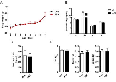
It is critical that no side effects or physiologic changes are observed after the elimination of gut microbiota in chickens. No significant differences on body weight, and the length of distinct regions of small intestine (duodenum, jejunum and ileum) and large intestine (Fig. 3A-B) were observed. Amount of glucocorticoid in serum, as a stress marker, was not changed (Fig. 3C). Furthermore, the weight of major organs including spleen, bursa and liver was not altered (Fig. 3D). Taken together, ABX treatment in chicken model in the present study did not alter physiological traits.
29
Figure 3. No physiologic changes in ABX-treated chickens. ABX in drinking water was treated to chickens at hatching for 7 days. (A) Body weight was measured daily. (B) The length of intestine (D: duodenum, J: jejunum, I: ileum), (C) amount of glucocorticoid by ELISA and (D) the weight of major immune organs were measured.
30
3) Change of B cells and macrophages in ABX-treated chickens
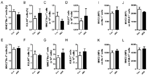
I examined whether the elimination of gut microbiota affects the population of B cells (MHC2+Bu-1+ cells) and macrophages (KUL01+ cells) in cecal
tonsils and spleen in chickens. No significant changes on the percentage and absolute number of B cells and macrophages in cecal tonsils (Fig. 4A-D) and spleen (Fig. 4E-H) were observed. Furthermore, MHC class II (MHC2) expression on B cells and macrophages in cecal tonsils (Fig. 4I and J) and spleen (Fig. 4K and L) was not significantly changed. Taken together, population and expression of MHC2 of B cells and macrophages did not alter by the elimination of gut microbiota.
31
Figure 4. No changes of B cells and macrophages in cecal tonsils and spleen in ABX-treated chickens. Chickens, treated with antibiotics for 7 days, were sacrificed, and then (A-D and I, J) cecal tonsils and (E-H and K, L) spleen were taken. Single cells produced from each organ were stained with anti-chicken MHC class II (MHC2), KUL01 (for macrophages), and Bu-1 (for B cells) antibodies. The percentage of (A and E) B cells and (B and F) macrophages, and absolute number of (C and G) B cells and (D and H) macrophages was calculated from their percentages. MFI of MHC2 on (I and K) B cells and (J and L) macrophages were examined using flow cytometry.
32
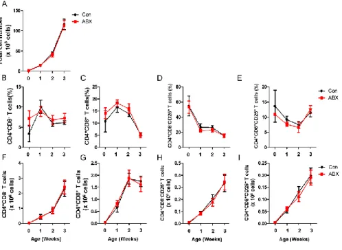
4) Change of CD4
+T cells in ABX-treated chickens
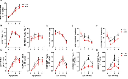
To examine the percentage and absolute number of CD4+ subtype T cells in
cecal tonsils, flow cytometry analysis after the staining of the cells with anti-chicken TCR, CD3, CD4, CD8, and CD25 antibodies was performed. CD3+TCR– cells were pre-gated, and then, CD4+ T cells were divided into
CD4+CD8– and CD4+CD8+ T cells. Finally, CD25+ cells were analyzed (Fig. 5).
Total cell number of cecal tonsils showed no significant changes in ABX-treated chickens (ABX) when compared to control (Fig. 6A). Furthermore, there were no changes on T cells (Fig. 6B and G), and CD4+CD8– (Fig. 6C
and H) and CD4+CD8+ (Fig. 6D and I) T cells. Interestingly, in CD4+CD8–
CD25+ (Fig. 6E and J) and CD4+CD8+CD25+ T cells (Fig. 6F and K) T cells
from cecal tonsils were significantly reduced in ABX compared with those of control (Con). Interestingly, however, no significant changes on CD4+CD8–
33
Figure 5. Gating strategy to analyze subtype of CD4+ T cells. Chickens at
hatching were given water containing antibiotics for 7 days and cecal tonsils were taken. Single cells produced from cecal tonsils were, then, stained with anti-chicken TCR, CD3, CD4, CD8, and CD25 antibodies. CD3+TCR–
cells gated were regarded as T cells, and then, CD4+CD8–CD25+ and
34
Figure 6. CD4+CD8–CD25+ and CD4+CD8+CD25+ T cells were reduced in
cecal tonsils from ABX-treated chickens. Chickens at hatching were given water containing antibiotics for 7 days and cecal tonsils were taken. Single cells from cecal tonsils were, then, stained with anti-chicken TCR, CD3, CD4, CD8, and CD25 antibodies. (A) Total number of cells in cecal tonsils is shown. The percentage of (B) CD3+TCR– cells, (C) CD4+CD8– T cells, (D)
CD4+CD8+ T cells, (E) CD4+CD8–CD25+ and (F) CD4+CD8+CD25+ T cells,
and absolute number of (G) CD3+TCR– T cells, (H) CD4+CD8– T cells, (I)
CD4+CD8+ T cells, (J) CD4+CD8–CD25+ and (K) CD4+CD8+CD25+ T cells was
calculated with the percentage of these cells. Significant differences were shown as asterisks between Con and ABX at P ≤ 0.05.
35
Figure 7. CD4+CD8–CD25+ and CD4+CD8+CD25+ T cells were not changed
in spleen from ABX-treated chickens. Chickens at hatching were given water containing antibiotics for 7 days and spleens were taken. Single cells from spleens were, then, stained with anti-chicken CD4, CD8, and CD25 antibodies. (A) Total number of cells in spleen is shown. The percentage of (B) CD4+CD8–
T cells, (C) CD4+CD8+ T cells, (D) CD4+CD8–CD25+ and (E)
CD4+CD8+CD25+ T cells, and absolute number of (F) CD4+CD8– T cells, (G)
CD4+CD8+ T cells, (H) CD4+CD8–CD25+ and (I) CD4+CD8+CD25+ T cells was
36
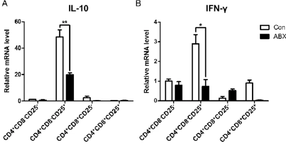
5) Change of IL-10 and IFN- from subtype of CD4
+T cells in
ABX-treated chickens
Chicken CD4+CD25+ T cells expressed high IL-10 and played a role as Tregs
[10]. I examined whether the elimination of gut microbiota affects mRNA expression of cytokines in subset of CD4+ T cells. Interestingly, both IL-10 (Fig.
8A) and IFN- (Fig. 8B) mRNA in CD4+CD8–CD25+ T cells were significantly
37
Figure 8. Expression of IL-10 and IFN-γ mRNA among CD4+ T cell subsets
in cecal tonsils from ABX-treated chickens. Chickens at hatching were given water containing antibiotics for 7 days and cecal tonsils were taken. Single cells from cecal tonsils were, then, stained with anti-chicken CD4, CD8 and CD25 antibodies. Each subset of CD4+ T cells was sorted by using ARIA II FACS
sorter. The mRNA was extracted from each subset and the level of (A) IL-10 and (B) IFN- was determined by RT-qPCR. Relative quantification of target genes was calculated using the 2–ΔΔCt method and normalized to actin mRNA level. Significant differences were shown as asterisks between Con and ABX at P ≤ 0.05.
38
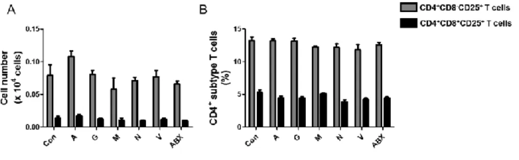
6) Direct effect of antibiotics on the change of CD4
+CD8
–CD25
+and CD4
+CD8
+CD25
+T cells
In order to examine the possibility for the change of these T cells by direct effect of antibiotics, I performed in vitro experiment where pre-determined (data not shown) each antibiotic or combination was treated to splenocytes for 24 h. There were no significant differences on the cell number (Fig. 9A) and the proportion of these cells (Fig. 9B) when compared with control. These results suggested that the reduction of CD4+CD8–CD25+ T cells in ABX-treated
39
Figure 9. No changes of CD4+CD8–CD25+ and CD4+CD8+CD25+ T cells in
chicken splenocytes treated with antibiotics. Spleens were taken from two week-old chickens and splenocytes were treated with pre-determined concentration of each antibiotic or mixed antibiotics (ABX). (A) Cell number and (B) proportion of CD4+CD8–CD25+ and CD4+CD8+CD25+ T cells were
examined using anti-chicken CD4-FITC, CD8a-PE and CD25-Alexa647 antibodies by flow cytometry. Con, non-treatment; A, ampicillin (100 g/ml); G, gentamycin (100 g/ml); M, metronidazole (100 g/ml); N, neomycin (100 g/ml); V; vancomycin (50 g/ml); and ABX, antibiotics cocktail as mentioned in the Materials and Methods.
40
7) Changes of CD4
+CD8
–CD25
+and CD4
+CD8
+CD25
+T cells in
periphery of ABX-treated chickens
I further examined the reduction of CD4+CD8–CD25+ and CD4+CD8+CD25+
T cells in periphery organs in ABX-treated chickens. It has been suggested that CD5hiCD4+CD25–Foxp3– T cells preferentially develop into peripheral Foxp3+
Tregs in mice [109]. The present results showed that CD5hi cells were deceased
in both CD4+CD8–CD25+ and CD4+CD8+CD25+ T cells in cecal tonsils of
ABX-treated chickens (Fig. 10).
CD4+CD25+ T cells migrate from thymus to cecal tonsils preferentially [110].
The reduction of CD4+CD8–CD25+ and CD4+CD8+CD25+ T cells in cecal
tonsils could be resulted by lesser migration from thymus. The results showed that CD4+CD8+ T cells are the major population of CD4+ T cells in chicken
thymus (Fig. 11A). Furthermore, there was no change on CD5 expression onCD4+CD8+CD25+ T cells in the thymus from ABX-treated chickens (Fig.
11B). Taken together, the reduction of CD4+CD8–CD25+ and CD4+CD8+CD25+
41
Figure 10. Reduction of CD5hi cells in CD4+CD8–CD25+ and
CD4+CD8+CD25+ T cells in ABX-treated chickens. Chickens, at hatching,
were given water containing antibiotics for 7 days and cecal tonsils were taken. Single cells from cecal tonsils were, then, stained with anti-chicken CD4, CD5, CD8, and CD25 antibodies. (A) The percentage of CD5hi and CD5low/– cells
was analyzed in CD4+CD8–CD25+ and CD4+CD8+CD25+ T cells in cecal
tonsils by using flow cytometry. (B) CD5hi and CD5low/- cells in CD4+CD8– and
CD4+CD8+CD25+ T cells were evaluated by Grandparents analysis by using
FlowJo. Significant differences were shown as asterisks between Con and ABX at P ≤ 0.05.
42
Figure 11. CD5hi cells of CD4+CD8+CD25+ T cells were not changed in
thymus of ABX-treated chickens. Chickens at hatching were given water containing antibiotics for 7 days and thymus was taken. Single cells from thymus were, then, stained with anti-chicken CD4, CD8, and CD25 antibodies. (A) Thymocytes were analyzed by dot plot based on CD4 and CD8 expression. (B) Total cell number was obtained from a thymic lobe. Cell number of (C) CD4+CD8+CD25+ T cells and (D) CD5hi of CD4+CD8+CD25+ T cells, and the
percentage of (E) CD4+CD8+CD25+ T cells and (F) CD5hi of CD4+CD8+CD25+
43
8)
Suppressive
function
of
CD4
+CD8
–CD25
+and
CD4
+CD8
+CD25
+T cells in ABX-treated chickens
The elimination of gut microbiota caused reduction of IL-10 mRNA in CD4+CD8–CD25+ T cells (Fig. 8A). It could be postulated that the reduction of
IL-10 expression caused to change the function of CD4+CD8–CD25+ T cells
since it is known as an immune suppressive cytokine [111]. I examined whether the elimination of gut microbiota affected the suppressive function of CD4+CD8–CD25+ and CD4+CD8+CD25+ T cells. CD4+CD8–CD25– and
CD4+CD8+CD25– T cells from ABX-treated chickens were proliferated more
than those of Con (Fig. 12) suggesting that the elimination of gut microbiota caused a significant reduction of CD4+CD8–CD25– and CD4+CD8+CD25– T
44
Figure 12. Elimination of gut microbiota caused reduction of suppressive ability of CD4+CD8–CD25+ and CD4+CD8+CD25+ T cells. Spleens were
taken from two week-old chickens administered with water (Con) or water containing antibiotics (ABX) for two weeks. Splenic CD4+ T cells were sorted
by magnetic bead sorting. CD4+ T cells, stained with CellTrace™ Violet (CTV)
dye, were stimulated with anti-chicken CD3 and CD28 antibodies for 3 d. Proliferation of CD4+CD8–CD25– and CD4+CD8+CD25– T cells were
determined by flow cytometry. Significant differences were shown as an asterisk between Con and ABX at P ≤ 0.05.
45
9) Changes of CD4
+CD8
–CD25
+and CD4
+CD8
+CD25
+T cells in
ABX-treated chickens after co-housing with control chickens
CD4+CD8–CD25+ and CD4+CD8+CD25+ T cells were significantly reduced
in ABX-treated chickens (Fig. 6). Therefore, the reconstitution of gut microbiota may concordant with recovery of CD4+CD8–CD25+ and
CD4+CD8+CD25+ T cells in ABX-treated chickens was examined after
co-housing with wild type chickens. The CFU was observed as early as 6 h post co-housing and reached at the similar level as ABX-untreated control at 1 d post co-housing (Fig. 13A). Interestingly, the number of CD4+CD8–CD25+ and
CD4+CD8+CD25+ T cells was gradually increased to the similar level as control
at 7 days post co-housing (Fig. 13B) suggesting that gut microbiota influence the number and function of CD4+CD8–CD25+ and CD4+CD8+CD25+ T cells.
46
Figure 13. Changes of CFU, and CD4+CD8–CD25+ and CD4+CD8+CD25+
T cells in cecal tonsils from ABX-treated chickens after co-housing with control chickens. Chickens at hatching were treated with ABX for 7 days and then co-housed with ABX-untreated control (Con) chickens for 7 days at the normal condition. (A) CFU was measured from cecal contents (1 mg/ml) at 6 h, 1 d, 3 d and 5 d after co-housing. (B) Proportion and cell number of CD4+CD8–CD25+ and CD4+CD8+CD25+ T cells in cecal tonsils were analyzed
by flow cytometry after co-housing for 1 d, 3 d and 7 days. Significant differences were shown as asterisks between Con and ABX at P ≤ 0.05.
47
10) Effect of Gram-positive or negative bacteria on the population
changes of CD4
+CD8
–CD25
+and CD4
+CD8
+CD25
+T cells
Next, I examined whether Gram-positive or Gram-negative bacteria influenced the change of CD4+CD8–CD25+ and CD4+CD8+CD25+ T cells.
Selective deletion of bacteria by using vancomycin (Van) for eliminating Gram-positive bacteria and polymyxin B (PolyB) for Gram-negative bacteria [2], was performed. The total CFU of Van and PolyB was slightly higher than that of Con (Fig. 14A). PolyB eliminated Gram-negative bacteria completely. Van eliminated Gram-positive bacteria from 33% to 7% (Fig. 14B). Surprisingly, CD4+CD8–CD25+ and CD4+CD8+CD25+ T cells were significantly decreased
by Van, but not PolyB treatment (Fig. 14C). In order to make sure the effect of Van, I have examined another group, ABX without vancomycin, Without Van, and the result showed no significant differences (Fig. 14D) indicating the change was caused by Gram-positive bacteria. Taken together, Gram-positive bacteria have a critical role to induce CD4+CD8–CD25+ and CD4+CD8+CD25+
48
Figure 14. Elimination of Gram positive bacteria is responsible for the change of CD4+CD8–CD25+ and CD4+CD8+CD25+ T cells in ABX-treated
chickens. Chickens, at hatching, were treated with vancomycin (Van; 50 mg/ml), antibiotics without vancomycin (Without van; ampicillin 100 mg/ml, gentamycin 100 mg/ml, metronidazole 100 mg/ml, neomycin 100 mg/ml), or polymyxin B (PolyB; 10 mg/ml) for 7 days and co-housed with ABX-untreated control (Con) chickens for 7 days. (A) CFU of cecal contents was measured from Van and PolyB groups and, (B) The composition of colonies was averaged with Gram positive or negative colonies pre-determined by Gram staining. (C and D) Proportion of CD4+CD8–CD25+ and CD4+CD8+CD25+ T cells in cecal
tonsils were analyzed in chickens treated with vancomycin (Van), polymyxin B (PolyB), or antibiotics without vancomycin (Without Van) using flow cytometry and FlowJo. Significant differences were shown as a different alphabet at P ≤ 0.05.
49
11) Effect of SCFAs on CD4
+CD8
–CD25
+and CD4
+CD8
+CD25
+T
cells
It has been suggested that short SCFAs are one of the factors to induce Tregs or Tr1 in mice [91]. We, therefore, examined whether SCFAs affect the population of CD4+CD8–CD25+ and CD4+CD8+CD25+ T cells in chickens. It
was intriguing that ABX-treated chickens administered with acetate recovered CD4+CD8–CD25+ T cells in cecal tonsils (Fig. 15A). CD4+CD8+CD25+ T cells
showed a tendency of recovery without significant (Fig. 15B). Other SCFAs, butyrate and propionate, did not show such effect (Fig. 15C-F). GPR43 is known as a receptor for acetate [112]. GPR43 mRNA expression on CD4+CD8–
CD25+ T cells was significantly higher than other immune cells (Fig. 15G)
strongly suggest that the recovery of CD4+CD8–CD25+ T cells by acetate
administration in ABX-treated chickens could be associated with high GPR43 expression on CD4+CD8–CD25+ T cells.
50
Figure 15. Changes of CD4+CD8–CD25+ and CD4+CD8+CD25+ T cells in
chickens administered with acetate. SCFAs (acetate 50 mM, butyrate 30 mM, propionate 10 mM) or ABX was treated to chickens at hatching with drinking water for 7 days. Cell number of (A, C and E) CD4+CD8–CD25+ and (B, D and
F) CD4+CD8+CD25+ T cells of cecal tonsils was calculated with total cell
number and proportion of CD4+ subtype T cells. (G) Each subset of CD4+ T
cells, B cells (Bu-1+) and APCs (KUL01+, MHC class II (MHC2)+KUL01–
Bu-1–) were sorted by using ARIA II FACS sorter. The mRNA was extracted from each subset and the level of GPR43 was determined by RT-qPCR. Significant differences were shown as a different alphabet at P ≤ 0.05.