INTRODUCTION
In vitro production (IVP) system of porcine embryos is important for production of transgenic pigs that are used
for human disease and xenograft models (Jeon et al., 2014). Especially, oocytes acquire intrinsic capabilities during oocyte maturation, including nuclear and cyto-plasmic maturation mechanisms involved in subsequent
Alpha-linolenic acid enhances maturation and
developmental competence via regulation of
glutathione, cAMP and fatty acid accumulation
during
in vitro maturation of porcine oocytes
Ye-Eun Jeon, Yong Hwangbo, Sun-Young Kim and Choon-Keun Park*
Department of Animal Life Sciences, Kangwon National University, Chuncheon 24341, KoreaOriginal Article
pISSN: 2671-4639 • eISSN: 2671-4663
https://doi.org/10.12750/JARB.35.4.357
JARB
Journal of Animal Reproduction and Biotechnology
Received November 5, 2020 Revised December 16, 2020 Accepted December 21, 2020 *Correspondence Choon-Keun Park E-mail: [email protected] ORCID https://orcid.org/0000-0003-2786-8814
ABSTRACT The aim of present study was to investigate regulatory mechanism of alpha-linolenic acid (ALA) during in vitro maturation (IVM) on nuclear and cytoplasmic maturation of porcine oocytes. Basically, immature cumulus-oocyte complexes (COCs) were incubated for 22 h in IVM-I to which hormone was added, and then further incubated for 22 h in IVM-II without hormone. As a result, relative cumulus expansion was increased at 22 h after IVM and it was enhanced by treatment of ALA compared with control group (p < 0.05). During IVM process within 22 h, cAMP level in oocytes was decreased at 6 h (p < 0.05) and it was recovered at 12 h in ALA-treated group, while oocytes in control group recovered cAMP level at 22 h. In cumulus cells, it was reduced in all time point (p < 0.05) and ALA did not affect. Treatment of ALA enhanced metaphase-I (MI) and MII population of oocytes compared with oocytes in control group at 22 and 44 h, respectively (p < 0.05). Intracellular GSH levels in ALA group was increased at 22 and 44 h after IVM (p < 0.05), whereas it was increased in control group at 44 h after IVM (p < 0.05). In particular, the GSH in ALA-treated oocytes during 22 h of IVM was higher than control group at 22 h (p < 0.05). Lipid amount in oocytes from ALA group was higher than control group (p < 0.05). Treatment of ALA did not influence to absorption of glucose from medium. Cleavage and blastocyst formation of ALA-treated oocytes were enhanced compared with control group (p < 0.05). These findings suggest that supplementation of ALA could improve oocyte maturation and development competence through increasing GSH synthesis, lipid storage, and regulation of cAMP accumulation during early 22 h of IVM, and these might be mediated by cumulus expansion.
Keywords: alpha-linolenic acid, cyclic adenosine monophosphate, glutathione, In vitro maturation, porcine oocytes
developmental processes (Ferreira et al., 2009). Nuclear and cytoplasmic maturation and developmental compe-tence of oocytes can be improved by regulating cyclic adenosine monophosphate (cAMP) levels during IVM. Intercellular communication within the ovarian follicles are regulated by arrest and resumption of meiosis in vivo. High cAMP levels were maintained continuously through gap junctions of cumulus and granule cells, maintain mei-otic arrest in oocytes within the follicles (Bornslaeger et al., 1986). However, during needle aspiration for IVM, the cumulus-oocyte complexs (COCs) are separated from the follicle, contact between oocyte and follicle cell are halted and induces spontaneous resumption of the COCs itself, producing oocytes with low developmental capacity.
In pigs, the ability of oocytes to develop in vitro is still low compared to in vivo maturation. Therefore, oocytes matured by in vitro are less likely to develop into the blastocyst stage (Gilchrist and Thompson, 2007) and in-duces higher rates of miscarriage compare with oocytes matured in vivo (Buckett et al., 2008). Accordingly, many studies have investigated the effect of supplements during in vitro oocyte maturation to improve embryonic devel-opment competence (Kwak et al., 2012; Lee et al., 2018).
Among them, alpha-linolenic acid (ALA) is one of polyunsaturated fatty acids (PUFAs), present in sper-matozoa, oocytes and follicles in mammals (Homa and Brown, 1992). The ALA improved embryonic develop-ment through the expansion of cumulus cells essential for maturation in pigs (Lee et al., 2018). In addition, ALA-treated COCs significantly increased the PGE2 concentra-tion in the medium and regulated meiotic arrest at the germinal vesicle (GV) (Lee et al., 2018). There have been many studies related to the maturation and development of oocytes by ALA addition during IVM, but studies have not evaluated to explain the regulatory effects of ALA on development of COCs (Lee et al., 2018). This study was hypothesized that ALA will only regulate nuclear and cy-toplasmic maturation, but also affect embryonic develop-ment by regulating cAMP levels through the action of oo-cytes. Therefore, the aim of this study was to investigate the regulation of ALA mechanisms during IVM of porcine oocytes. In this study was also evaluated the effects of ALA on cAMP levels, nuclear maturation, GSH, fatty acid content, glucose amount, and subsequent embryonic de-velopment.
MATERIALS AND METHODS
Chemicals
Medium-199 and CellTrackerTMRed were purchased from
Invitrogen (Invitrogen, Carlsbad, CA, USA). cAMP com-plete ELISA kit (ADI-900-163) and Glucose Colorimetric/ Fluorometric Assay Kit (K606) were purchased from Enzo Life Sciences and BioVision, respectively. Other materials including ALA, luteinizing hormone (LH), follicle-stimu-lating hormone (FSH), epidermal growth factor (EGF) were obtained from Sigma-Aldrich (Sigma-Aldrich, St. Louis, MO, USA).
In vitro maturation
All procedures that involved the use of animals were approved by the Kangwon National University Institu-tional Animal Care and Use Committee (KIACUC-19-047). Ovaries were collected from pre-pubertal gilts in a local slaughterhouse, placed in a thermos containing 0.9% (w/v) saline, and transferred to the laboratory within 2 h. COCs were aspirated from antral follicles and only COCs with up to three layers of cumulus cells were collected under stereo-microscope. Collected immature COCs were in-cubated in 4-well plate with 650 μL medium-199 supple-mented with 10 ng/mL EGF, 10 μg/mL LH, 0.5 μg/mL FSH, 10 IU/mL human chorionic gonadotropin (hCG), and 10% (v/v) porcine follicular fluid (pFF) with or without 50 μM ALA for 22 h at 38.5℃ and 5% CO2 air condition, then,
they were subsequently matured in hormone-free IVM medium with or without 50 μM ALA for 22 h.
Assessment of cumulus expansion
To evaluate effect of ALA on cumulus expansion, twenty to thirty COCs in each group were used to assess cumulus expansion at 22 h after IVM. Image J software (version 1.46; National Institutes of Health, Bethesda, MD, USA) was used to measure the area of each COCs, and all of cumulus expansion data were normalized to non-ALA group at 0 h (Park et al., 2020).
Measurement of cAMP concentration
The cAMP complete ELISA kit was used to measure cAMP level in oocytes and cumulus cells according to the manufacturer’s instructions. Cumulus cells from fifty COCs at 0, 6, 12, and 22 h after IVM were removed from oocytes and collected. Both of denuded oocytes and
cu-mulus cells were washed using PBS-PVA and stored in 100 μL of 0.1 M HCl at -80℃ until analysis. Oocytes and cumulus cell lysates were acetylated and added to 96-well plate. Absorbance of each samples was read at 405 nm using microplate reader.
Evaluation of nuclear maturation
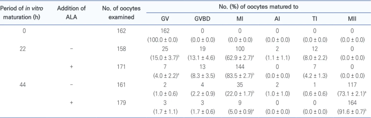
The effect of ALA on nuclear maturation was evaluated at 0, 22, and 44 h after IVM. Cumulus cells around mature COCs were removed by gently pipetting in IVM medium with 0.1% hyaluronidase. The denuded oocytes were mounted on slides and fixed in acetic alcohol solution (acetic acid:ethanol = 1:3; v/v) for 48 h at room tempera-ture. Then, they were stained with 1% (w/v) aceto-orcein solution at room temperature for 7 min and morphology of nuclear was observed under a light microscope. Oo-cytes were classified into germinal vesicle (GV), germinal vesicle breakdown (GVBD), metaphase-I (MI), anaphase-I (Aanaphase-I), telophase-anaphase-I (Tanaphase-I), and Manaphase-Ianaphase-I stages according to pre-viously described morphological criteria of chromatin staining (Motlík and Fulka, 1976; Park et al., 2020).
Measurement of intracellular GSH level
After 0, 22 and 44 h after IVM, twenty mature COCs from each group were randomly selected for measurement of intracellular GSH levels, respectively. The cumulus cells were denuded by gently pipetting in maturation medium containing 0.1% (w/v) hyaluronidase and denuded oocytes were fixed in 4% (v/v) paraformaldehyde solution at room temperature for 7 min. Then, they were stained with 5 μM CellTrackerTM for 30 min in a dark room (Park et al.,
2020). Stained oocytes were washed three times with PBS-PVA and were observed under an epifluorescence micro-scope with optical filter (ex: 510-560 nm, em: 590 nm). Fluorescent intensity was measured using Image J soft-ware.
Staining of lipid droplet
Nile red was used to observe the morphology of lipid droplet and to measure amount of lipid in oocytes. De-nuded oocytes at 0, 22, and 44 h after IVM were fixed 4% (v/v) paraformaldehyde solution at room temperature for 30 min. Twenty oocytes were stained using 10 μg/mL Nile red at room temperature for 30 min, and were washed three times using PBS-PVA. Stained oocytes with Nile red were observed under an epifluorescence microscope with
optical filter (ex: 510-560 nm, em: 590 nm) and fluores-cent intensity was measured using Image J software.
Glucose absorption
Concentration of glucose in IVM medium was mea-sured using Glucose Colorimetric/Fluorometric Assay Kit according to the manufacturer’s instructions. Medium, which was used to mature COCs, was collected at 22 and 44 h after IVM, and fresh medium was used as positive control. Samples were reacted with glucose reaction mix at 37℃ for 30 min and absorbance was read at 570 nm using microplate reader.
In vitro fertilization and culture
Mature COCs were placed in IVM medium containing 0.1% (w/v) hyaluronidase to weaken adhesion between cumulus cells for 5 min and fifty COCs were transferred to 4-well culture plate with 250 μL of modified Tris-buffered medium (mTBM) supplemented with 0.4% (w/v) bovine serum albumin (BSA) without caffeine. Fresh boar semen was washed twice using Modena B and resuspended with mTBM containing 0.2% (w/v) BSA and caffeine to a final concentration of 6 × 10⁵ spermatozoa/mL. Then, 250
μL aliquot of sperm was inseminated into COCs and co-incubated at 38.5℃ in 5% CO2 for 6 h.
After 6 h of fertilization, spermatozoa and cumulus cells binding to the zona pellucida were removed by gentle pipetting. Fifty putative zygotes were placed in 4-well plates with 500 μL of porcine zygote medium-3 (PZM-3) supplemented with 0.3% (w/v) BSA and cultured for 48 h at 38.5℃ in 5% CO2. Then, the culture medium was changed
for fresh medium and the putative zygotes were incubated for 5 days (Park et al., 2020). Cleavage rate and blastocyst formation were evaluated at 48 and 168 h after insemina-tion.
Statistical analysis
The data were presented as the mean ± standard error of the mean and Statistical Analysis System Software (SAS, version 9.3) was used to analyze all numerical data rep-resenting each parameter. All of percentage values were transformed into arcsine to obtain a normal distribution. Duncan’s multiple range test was used assess relative cumulus expansion, GSH and ROS levels, nuclear matu-ration, and embryonic development and a generalized linear model (GLM) was used to compare parameters. A
value of p < 0.05 was considered to indicate a statistically significant difference.
RESULTS
Changes of cumulus expansion and cAMP level during
first 22 h of IVM by ALA treatment
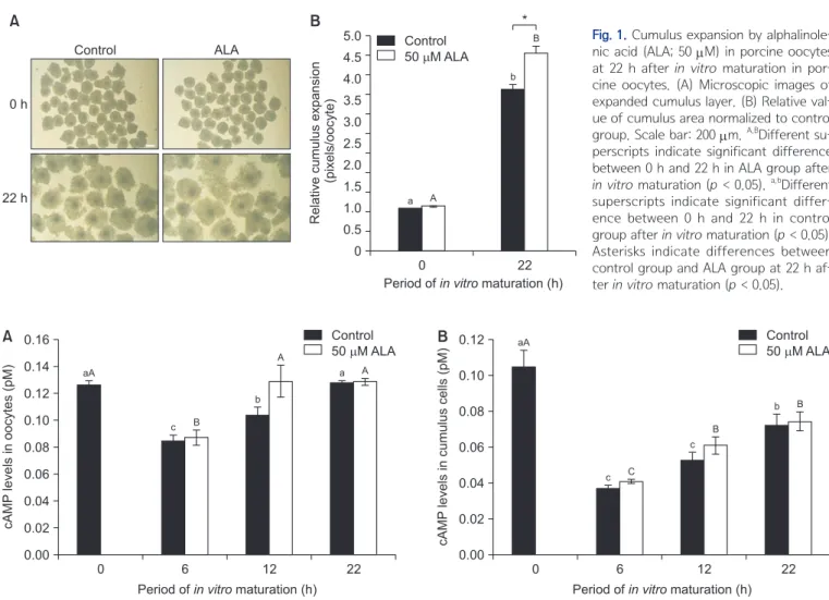
In both oocyte populations, the area of cumulus oopho-rus was increased after 22 h of IVM (p < 0.05; Fig. 1). In particular, the treatment of ALA enhanced cumulus ex-pansion compared with control group at 22 h after IVM (p < 0.05; Fig. 1). The cAMP levels in control and ALA-treated group was reduced at 6 h after IVM (p < 0.05; Fig. 2A). The cAMP levels in ALA-treated oocytes was completely recovered after 12 h of IVM, whereas cAMP in control group was still lower than in the treatment group (p
< 0.05; Fig. 2A). Similar with cAMP in oocyte, cAMP levels were reduced in cumulus cells at 6 h after IVM and was recovered after 6 h of IVM (p < 0.05; Fig. 2B). However, ALA treatment did not influence to cAMP levels in cumu-lus cells at all of time points (Fig. 2B).
Effect of ALA on nuclear maturation at 22 and 44 h
after IVM
Treatment of ALA during IVM affected to nuclear status of porcine oocytes (Table 1). All oocytes collected at 0 h were arrested at germinal vesicle (GV) stage. After 22 h of IVM, population of oocytes at GV stage was lower in ALA treatment group than non-ALA group, while ALA enhanced population of MⅠ reached oocytes (p < 0.05). The MⅡ reached oocytes were higher and MⅠ stage arrested oocytes was lower in ALA group than ALA absent groups (p < 0.05).
Fig. 1. Cumulus expansion by
alphalinole-nic acid (ALA; 50 μM) in porcine oocytes
at 22 h after in vitro maturation in por-cine oocytes. (A) Microscopic images of expanded cumulus layer. (B) Relative val-ue of cumulus area normalized to control
group. Scale bar: 200 μm. A,BDifferent
su-perscripts indicate significant difference between 0 h and 22 h in ALA group after
in vitro maturation (p < 0.05). a,bDifferent
superscripts indicate significant differ-ence between 0 h and 22 h in control group after in vitro maturation (p < 0.05). Asterisks indicate differences between control group and ALA group at 22 h af-ter in vitro maturation (p < 0.05).
B * A 0 h 22 h Control ALA 0 5.0 4.5 4.0 3.5 3.0 2.5 2.0 1.5 1.0 0.5 22 Relative cumulus expansion (pixels/oocyte)
Period ofin vitromaturation (h) 0 A a B b Control 50 M ALA
Fig. 2. Effect of alpha-linolenic acid (ALA; 50 μM) on cyclic AMP content in oocytes (A) and cumulus cells (B) on in vitro maturation of
por-cine oocytes. A-CDifferent superscripts indicate significant difference between 0 h, 22 h and 44 h in ALA group after in vitro maturation (p <
0.05). a-cDifferent superscripts indicate significant difference between 0 h, 22 h and 44 h in control group after in vitro maturation (p < 0.05).
0 6 12 22 0.16 0.14 0.12 0.10 0.08 0.06 0.04 0.02 0.00 cAMP levels in oocytes (pM)
Period ofin vitromaturation (h)
A Control B 50 M ALA aA c B b A a A 0 6 12 22 0.12 0.10 0.08 0.06 0.04 0.02 0.00 cAMP levels in cumulus cells (pM)
Period ofin vitromaturation (h)
Control 50 M ALA aA c C c B b B
Intracellular GSH level and fatty acid content
Intracellular GSH level in oocytes during IVM was in-creased (Fig. 3). In oocytes from non-ALA groups, GSH was increased after 44 h of IVM, whereas ALA enhanced GSH level at 22 and 44 h after IVM (p < 0.05; Fig. 3). Es-pecially, cAMP levels were higher in ALA-treated oocytes than control groups at 22 h of IVM (p < 0.05; Fig. 3). This study observed data showed that lipid content in oocytes was higher in ALA present groups at 22 h than control groups at 0 and 22 h of IVM (p < 0.05; Fig. 4).
Change of glucose level in maturation medium and
embryonic development by ALA
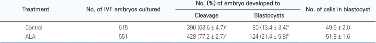
Glucose concentration in maturation medium, which
were used to each IVM stages, was not differed between ALA treated and non-treated groups (Fig. 5). Both of cleavage and blastocyst formation were increased by ALA treatment during IVM (p < 0.05; Table 2).
DISCUSSION
Numerous studies have attempted to improve the quality of IVM oocytes through substance supplementation, and the IVP system of porcine embryos has been improved by many researchers. However, due to stressful environment during in vitro maturation, the developmental ability of mature oocytes in vitro is still lower than that of mature oocytes in vivo (Guérin et al., 2001). Fatty acid (FA) is
Table 1. Effects of alpha-linolenic acid (ALA; 50 μM) during in vitro maturation on nuclear maturation in porcine oocytes
Period of in vitro maturation (h) Addition of ALA No. of oocytes examined
No. (%) of oocytes matured to
GV GVBD MI AI TI MII 0 162 162 0 0 0 0 0 (100.0 ± 0.0) (0.0 ± 0.0) (0.0 ± 0.0) (0.0 ± 0.0) (0.0 ± 0.0) (0.0 ± 0.0) 22 − 158 25 19 100 2 12 0 (15.0 ± 3.7)b (13.1 ± 4.6) (62.9 ± 2.7)a (1.1 ± 1.1) (8.0 ± 2.2) (0.0 ± 0.0) + 171 7 13 144 0 7 0 (4.0 ± 2.2)a (8.3 ± 3.5) (83.5 ± 2.7)b (0.0 ± 0.0) (4.2 ± 1.3) (0.0 ± 0.0) 44 − 161 2 4 35 2 1 117 (1.0 ± 0.6) (2.2 ± 0.9) (22.0 ± 1.7)b (1.0 ± 1.0) (0.6 ± 0.6) (73.1 ± 2.1)a + 179 3 3 9 0 0 164 (1.7 ± 1.1) (1.7 ± 0.6) (5.0 ± 0.9)a (0.0 ± 0.0) (0.0 ± 0.0) (91.6 ± 0.7)b
Data were presented as number (means ± SEM) from four replications.
a,bDifferent superscripted letters indicate a significant difference in the same time (p < 0.05).
GV : germinal vesicle; GVBD : germinal vesicle break down; MI : metaphase I; AI : anaphase I; TI : telophase I; MII : metaphase II.
Fig. 3. Effect of alpha-linolenic acid
(ALA; 50 μM) treatment on intra-oocyte
GSH content during in vitro matura-tion of porcine oocytes. (A) Fluorescent microscopic images of porcine oocytes. (B) Fluorescence intensity of GSH levels.
Scale bar: 200 μm. a-cDifferent
super-scripts indicate significant difference between 0 h, 22 h and 44 h after in vitro maturation (p < 0.05). Asterisks indicate differences between control group and ALA group at 22 h after in vitro maturation (p < 0.05). B A - + * 0 1.6 1.4 1.2 1.0 0.8 0.6 0.4 0.2 22 Intracellular GSH level (pixels/oocyte)
Period ofin vitromaturation (h) 0 a b a Control 50 M ALA ALA 0 h 22 h 44 h c c 44
contained in the cytoplasm of pig and bovine oocytes and is one of the energy sources (McKeegan & Sturmey, 2011). Many studies have reported that dietary supplementation of PUFA could improve fertility (Wakefield et al., 2008; Hoyos-Marulanda et al., 2019), and that adding oleic acid to the maturation medium could improve oocyte develop-ment by suppressing the adverse effects of saturated FAs (Yenuganti et al., 2016). Additionally, previous studies have shown that ALA supplementation increased intracel-lular GSH levels (Lee et al., 2016), cumulus cell expan-sion (Lee et al., 2018), and oocyte maturation (Lee et al., 2017) in porcine oocytes after IVM. The effect of ALA in mammalian oocytes has been suggested by studies (Lee et al., 2016; Lee et al., 2018), but the ALA action mecha-nism during the IVM stages on porcine oocytes has not been investigated. Therefore, we evaluated the regulatory mechanism of oocyte maturation by ALA treatment.
In our results, at the initial 22 h of IVM, cumulus cell expansion, nuclear maturation rate, GSH and fatty acid
contents were significantly increased in the ALA-treated group compared to the control group. Generally, nuclear maturation and cytoplasmic maturation are important for successful embryo development (Kishida et al., 2004). Even if nuclear maturation is complete, incomplete cy-toplasmic maturation reduces embryonic development. Lee et al. (2018) reported that treatment of 50 μM ALA increased cumulus expansion in porcine oocytes dur-ing initial 22 h IVM. Durdur-ing IVM, cumulus cells play an important role in the utilization of energy substrates (Heikinheimo et al., 1998) and the acquisition of the op-timum GSH levels via gap junction communication (Zhang et al., 1995). In addition, cumulus cells provide metabo-lites through gap junction to maintain average ATP level in mammalian oocytes (Webb et al., 2002).
Intracellular GSH is molecular marker of cytoplasmic maturation in pigs (Liu et al., 2002). In particular, the cysteine and glutamine transported from cumulus cells to oocytes by gap junctions are synthesized from oocytes to
Fig. 4. Effect of alpha-linolenic acid (ALA; 50 μM) on fatty acid
content during in vitro maturation of porcine oocytes. a,bDifferent
superscripts indicate significant difference between 0 h, 22 h and 44 h in ALA group after in vitro maturation (p < 0.05). Asterisk in-dicates differences between control group and ALA group at 22 h
after in vitro maturation (p < 0.05).
0 22 44 3.0 2.5 2.0 1.5 1.0 0.5 0 Relative fluorescent intensity
Period ofin vitromaturation (h) Control 50 M ALA b a * ab
Fig. 5. Effects of alpha-linolenic acid (ALA; 50 μM) on amount of glucose during in vitro maturation of porcine oocytes. Data show glucose consumption per cumulus–oocyte complex (COC) in each treatment group. Data are the mean ± SEM. Asterisks indicate differences between fresh medium and treatment groups at 22 h and 44 h after in vitro maturation (p < 0.05).
Fresh - + 5.0 4.5 4.0 3.5 3.0 2.5 2.0 1.5 1.0 0.5 0 Glucose concentration (ng/mL) 22 h 44 h Fresh -+ * * * *
Table 2. Effects of alpha-linolenic acid (ALA; 50 μM) during in vitro maturation (IVM) on developmental competence of porcine oocytes
Treatment No. of IVF embryos cultured No. (%) of embryo developed to No. of cells in blastocyst Cleavage Blastocysts
Control 615 390 (63.6 ± 4.7)a 80 (13.4 ± 3.4)a 49.6 ± 2.0
ALA 551 428 (77.2 ± 2.7)b 124 (21.4 ± 5.8)b 51.8 ± 1.6
Data from 4 replication are presented as mean ± SEM.
a,bDifferent superscripted indicate a significant difference within a same column (p < 0.05).
GSH, and the synthesis of GSH is an important indicator of cytoplasmic maturation (Mori et al., 2000). GSH par-ticipates directly or indirectly in cellular processes such as DNA, protein synthesis, metabolism and transport (Meister et al., 1983). Therefore, high GSH levels in oocytes follow-ing the ALA treatment improved subsequent embryonic and viability, and GSH was used as an important predic-tor of cellular maturation (Wang et al., 1997).
Lipid content is significant indicator of cytoplasmic maturation (Prates et al., 2013). Lipids are important sig-naling molecules that control the mechanisms regulating maturity and stimulate the acquisition of oocyte abilities (Holm, 2003). Therefore, lipid metabolism is an essential source of energy for oocytes and early embryo develop-ment (Niemann and Rath, 2001). In immature oocytes, triglycerides represent a major component of intracellular lipids and can be metabolized during oocyte maturation, fertilization and bovine embryonic division (Kim et al., 2001). In particular, there was a significant difference in the lipid content of the oocyte at 22 h, indicating that the oocyte maturation was improved during the initial 22 h. One report has demonstrated fatty acid accumulation in oocytes, especially neutral lipids in culture medium (Genicot et al., 2005). Therefore, it was presumed that the ALA treatment group had a positive effect on the oocytes maturation by rapidly recovering the cAMP levels and in-creasing the GSH and lipid accumulation.
In mammalian cells, cAMP is the second messenger of the intercellular signaling process and a wide range of cellular processes are regulated by the cAMP response. It has been well shown that oocyte meiotic resumption is associated with decreased concentration of intracellular cAMP and resulted inactivation of cAMP-dependent pro-tein kinase A (PKA) in various vertebrate species (Webb et al., 2002). When COC is aspirated from the follicle, control of the follicular environment is lost, resulting in a premature decrease in cAMP levels, which spontaneously resumes meiosis, accelerating and progressing to MII. Hu et al. (2019) study showed that ALA in human oocytes had a more cleared effect on the GV stage than on the M Ⅰ stage. Therefore, high cAMP levels at 0 h maintained meiosis arrest in oocytes. After that, the decrease in cAMP concentration allowed the oocyte to resume meiosis.
Unexpected, there was no significant difference in glucose in the culture medium compared to the control group. This is because there is a limit to the total amount
of glucose that oocytes can absorb from the culture solu-tion. Therefore, it is judged that there is no significant difference between the control group and the ALA-treated group in the amount of glucose remaining in the culture medium.
In conclusion, treatment with ALA during IVM quickly restores cAMP levels with lipid storage before the initial 22 h and is effective in cumulus expansion and nuclear and cytoplasm maturation, which are measures of oo-cytes maturation, and high maturation rate by ALA treat-ment later. It was also confirmed that the cleavage rate and blastocyst formation were positively affected. The results obtained from this study suggest the mechanism of maturation is regulated through cumulus expansion, GSH levels and lipid metabolism in porcine oocytes by ALA. It is thought that this can contribute to the development and improvement of an IVC system for successful IVM of mammalian oocytes. It is also expected to contribute to the advancement of basic knowledge about mammalian reproductive physiology.
CONFLICTS OF INTEREST
No potential conflict of interest relevant to this article was reported.
ACKNOWLEDGEMENTS
This work was supported by the National Research Foun-dation of Korea (NRF) grant funded by the Korea govern-ment (Ministry of Education) (2019R1A2C1004307).
AUTHOR CONTRIBUTIONS
Conceptualization: Yong HwangboData curation: Ye-Eun Jeon, Sun-Young Kim Formal analysis: Ye-Eun Jeon, Sun-Young Kim Investigation: Ye-Eun Jeon, Sun-Young Kim Methodology: Ye-Eun Jeon, Sun-Young Kim
Project administration: Yong Hwangbo, Choon-Keun Park Resources: Choon-Keun Park
Supervision: Choon-Keun Park Validation: Choon-Keun Park Visualization: Ye-Eun Jeon
Writing - original draft: Ye-Eun Jeon
AUTHOR’S POSITION AND ORCID NO.
YE Jeon, Grad Student,https://orcid.org/0000-0002-8979-9546 Y Hwangbo, Research Studeunt,
https://orcid.org/0000-0003-3636-3551 SY Kim, Grad Student,
https://orcid.org/0000-0003-2771-5981 CK Park, Professor,
https://orcid.org/0000-0003-2786-8814
REFERENCES
Bornslaeger EA, Mattei P, Schultz RM. 1986. Involvement of cAMP-dependent protein kinase and protein phosphoryla-tion in regulaphosphoryla-tion of mouse oocyte maturaphosphoryla-tion. Dev. Biol. 114:453-462.
Buckett WM, Chian RC, Dean NL, Sylvestre C, Holzer HE, Tan SL. 2008. Pregnancy loss in pregnancies conceived after in vitro oocyte maturation, conventional in vitro fertilization, and intracytoplasmic sperm injection. Fertil. Steril. 90:546-550.
Ferreira EM, Vireque AA, Adona PR, Meirelles FV, Ferriani RA, Navarro PA. 2009. Cytoplasmic maturation of bovine oocytes: structural and biochemical modifications and ac-quisition of developmental competence. Theriogenology 71:836-848.
Genicot G, Leroy JL, Soom AV, Donnay I. 2005. The use of a flu-orescent dye, Nile red, to evaluate the lipid content of single mammalian oocytes. Theriogenology. 63:1181-1194. Gilchrist RB and Thompson JG. 2007. Oocyte maturation:
emerging concepts and technologies to improve develop-mental potential in vitro. Theriogenology 67:6-15.
Guérin P, El Mouatassim S, Ménézo Y. 2001. Oxidative stress and protection against reactive oxygen species in the pre-implantation embryo and its surroundings. Hum. Reprod. Update 7:175-189.
Heikinheimo O and Gibbons WE. 1998. The molecular mecha-nisms of oocyte maturation and early embryonic develop-ment are unveiling new insights into reproductive medicine. Mol. Hum. Reprod. 4:745-756.
Holm C. 2003. Molecular mechanisms regulating hormone-sensitive lipase and lipolysis. Biochem. Soc. Trans. 31(Pt 6):1120-1124.
Homa ST and Brown CA. 1992. Changes in linoleic acid dur-ing follicular development and inhibition of spontaneous breakdown of germinal vesicles in cumulus-free bovine oo-cytes. J. Reprod. Fertil. 94:153-160.
Hoyos-Marulanda V, Alves BS, Rosa PRA, Vieira AD, Gasperin BG, Mondadori RG, Lucia Jr. T. 2019. Effects of polyunsatu-rated fatty acids on the development of pig oocytes in vitro following parthenogenetic activation and on the lipid
con-tent of oocytes and embryos. Anim. Reprod. Sci. 205:150-155.
Hu JJ, Li JX, Wang XL, Guan YC, Sun LJ, 2019. Preliminary research on the effect of linolenic acid on human oocyte maturation. Reprod. Dev. Med. 3:42-48.
Jeon Y, Yoon JD, Cai L, Hwang SU, Kim E, Zheng Z, Lee E, Kim DY, Hyun SH. 2014. Supplementation of zinc on oocyte in vitro maturation improves preimplatation embryonic devel-opment in pigs. Theriogenology 82:866-874.
Kim JY, Kinoshita M, Ohnishi M, Fukui Y. 2001. Lipid and fatty acid analysis of fresh and frozen-thawed immature and in vitro matured bovine oocytes. Reproduction 122:131-138. Kishida R, Lee ES, Fukui Y. 2004. In vitro maturation of porcine
oocytes using a defined medium and developmental capac-ity after intracytoplasmic sperm injection. Theriogenology 62:1663-1676.
Kwak SS, Cheong SA, Jeon Y, Lee E, Choi KC, Jeung EB, Hyun SH. 2012. The effects of resveratrol on porcine oocyte in vitro maturation and subsequent embryonic development after parthenogenetic activation and in vitro fertilization. Therio-genology 78:86-101.
Lee JE, Hwangbo Y, Cheong HT, Yang BK, Park CK. 2018. Alpha-linolenic acid: it contribute regulation of fertilization capac-ity and subsequent development by promoting of cumulus expansion during maturation. Dev. Reprod. 22:297-307. Lee JE, Yong H, Kim HY, Lee WH, Cheong HT, Yang BK, Park
CK. 2017. Effect of alpha-linolenic acid on oocyte matura-tion and embryo development in pigs. Dev. Reprod. 21:205-213.
Lee Y, Lee H, Park B, Elahi F, Lee J, Lee ST, Park CK, Hyun SH, Lee E. 2016. Alpha-linolenic acid treatment during oocyte maturation enhances embryonic development by influenc-ing mitogen-activated protein kinase activity and intraoo-cyte glutathione content in pigs. J. Anim. Sci. 94:3255-3263. Liu RH, Li YH, Jiao LH, Wang XN, Wang H, Wang WH. 2002.
Extracellular and intracellular factors affecting nuclear and cytoplasmic maturation of porcine oocytes collected from different sizes of follicles. Zygote 10:253-260.
McKeegan PJ and Sturmey RG. 2011. The role of fatty acids in oocyte and early embryo development. Reprod. Fertil. Dev. 24:59-67.
Meister A and Anderson ME. 1983. Glutathione. Annu. Rev. Biochem. 52:711-760.
Mori T, Amano T, Shimizu H. 2000. Roles of gap junctional communication of cumulus cells in cytoplasmic maturation of porcine oocytes cultured in vitro. Biol. Reprod. 62:913-919.
Motlík J and Fulka J. 1976. Breakdown of the germinal vesicle in pig oocytes in vivo and in vitro. J. Exp. Zool. 198:155-162. Niemann H and Rath D. 2001. Progress in reproductive
bio-technology in swine. Theriogenology 56:1291-1304.
Park JE, Lee SH, Hwangbo Y, Park CK. 2020. Porcine follicular fluid derived from > 8 mm sized follicles improves oocyte maturation and embryo development during in vitro matu-ration of pigs. Preprints. doi: 10.1017/S0967199420000398.
Prates EG, Nunes JT, Pereira RM. 2014. A role of lipid metabo-lism during cumulus-oocyte complex maturation: impact of lipid modulators to improve embryo production. Mediators Inflamm. 2014:692067.
Wakefield SL, Lane M, Schulz SJ, Hebart ML, Thompson JG, Mitchell M. 2008. Maternal supply of omega-3 polyunsatu-rated fatty acids alter mechanisms involved in oocyte and early embryo development in the mouse. Am. J. Physiol. En-docrinol. Metab. 294:E425-E434.
Wang WH, Abeydeera LR, Cantley TC, Day BN. 1997. Effects of oocyte maturation media on development of pig embryos produced by in vitro fertilization. J. Reprod. Fertil. 111:101-108.
Webb RJ, Marshall F, Swann K, Carroll J. 2002. Follicle-stimu-lating hormone induces a gap junction-dependent dynamic change in [cAMP] and protein kinase a in mammalian oo-cytes. Dev. Biol. 246:441-454.
Yenuganti VR, Viergutz T, Vanselow J. 2016. Oleic acid induces specific alterations in the morphology, gene expression and steroid hormone production of cultured bovine granulosa cells. Gen. Comp. Endocrinol. 232:134-144.
Zhang L, Jiang S, Wozniak PJ, Yang X, Godke RA. 1995. Cumulus cell function during bovine oocyte maturation, fertilization, and embryo development in vitro. Mol. Reprod. Dev. 40:338-344.