저작자표시-비영리-변경금지 2.0 대한민국 이용자는 아래의 조건을 따르는 경우에 한하여 자유롭게 l 이 저작물을 복제, 배포, 전송, 전시, 공연 및 방송할 수 있습니다. 다음과 같은 조건을 따라야 합니다: l 귀하는, 이 저작물의 재이용이나 배포의 경우, 이 저작물에 적용된 이용허락조건 을 명확하게 나타내어야 합니다. l 저작권자로부터 별도의 허가를 받으면 이러한 조건들은 적용되지 않습니다. 저작권법에 따른 이용자의 권리는 위의 내용에 의하여 영향을 받지 않습니다. 이것은 이용허락규약(Legal Code)을 이해하기 쉽게 요약한 것입니다. Disclaimer 저작자표시. 귀하는 원저작자를 표시하여야 합니다. 비영리. 귀하는 이 저작물을 영리 목적으로 이용할 수 없습니다. 변경금지. 귀하는 이 저작물을 개작, 변형 또는 가공할 수 없습니다.
- 1 -
A Doctoral Dissertation
Protective effect of 7, 8-dihydroxyflavone against
oxidative stress in high glucose-damaged neuronal cells
Suk Ju Cho, M.D.
Department of Medicine
GRADUATE SCHOOL
JEJU NATIONAL UNIVERSITY
- 2 -
고포도당으로 손상된 신경세포의
산화적 스트레스에 대한
7, 8-dihydroxyflavone의 보호 효과
지도교수 현 진 원
조 석 주
이 논문을 의학 박사학위 논문으로 제출함
2015년 2월
조석주의 의학 박사학위 논문을 인준함
심사위원장
印
위 원
印
위 원
印
위 원
印
위 원
印
제주대학교 대학원
2015년 2월
- 3 -
Protective effect of 7, 8-dihydroxyflavone against oxidative
stress in high glucose-damaged neuronal cells
Suk-Ju Cho
(Supervised by professor Jin-Won Hyun)
A thesis submitted in partial fulfillment of the requirement for the
degree of Doctor of Philosophy in Medicine
2015 . 2.
This thesis has been examined and approved.
Date
Department of Medicine
GRADUATE SCHOOL
JEJU NATIONAL UNIVERSITY
- 4 -
CONTENTS
ABSTRACT... 5 INTRODUCTION... 6 MATERIALS AND METHODS... 9 1. Materials
2. Cell culture and drug treatment 3. Determination of cell viability 4. Intracellular ROS measurement
5. Measurement of lipid peroxidation assay 6. Mitochondrial membrane potential analysis 7. Western blot analysis
8. Hoechst 33342 staining 9. Statistical analysis
RESULTS... 14 1. Determination of 8-isoprostane
2. High glucose cytotoxicity dose response
3. Effect of 7, 8-DHF against high glucose-induced cytotoxicity 4. 7,8-DHF effect on high glucose-induced ROS production 5. 7,8-DHF effect on apoptosis-related proteins
6. 7,8-DHF effect on glucose-induced mitochondrial membrane potential depolarization 7. Glucose induces apoptosis via the mitochondria-dependent pathway
DISCUSSION... 30
- 5 -
LIST OF FIGURES
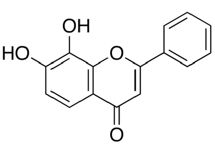
Figure 1. Chemical structure of 7, 8-dihydroxyflavone ... 10
Figure 2. Serum 8-isoprostane levels in patients with type 2 diabetes and healthy controls….. 16
Figure 3. Glucose cytotoxicity in SH-SY5Y cells………...………... 18
Figure 4. Protective effect of 7,8-DHF against high glucose-induced cell damage…………...21
Figure 5. Effect of 7,8-DHF on high glucose-induced intracellular ROS generation... 23
Figure 6. Effects of 7,8-DHF on apoptosis-related proteins…….…………...………... 25
Figure 7. Effects of 7, 8-DHF on glucose-induced mitochondrial membrane potential
depolarization……….………..27
- 6 -
ABSTRACT
Oxidative stress is considered to be a major contributor in the pathogenesis of diabetic neuropathy and in diabetes complications such as nephropathy and cardiovascular diseases. Diabetic neuropathies, which are the most frequent complications of diabetes, affect sensory, motor, and autonomic nerves. Recently, 7,8-dihydroxyflavone (7,8-DHF) has been reported to act as a neuroprotective agent against glutamate- or 6-hydroxydopamin-induced toxicity via its antioxidant effects. However, the effects of 7,8-DHF against high glucose-induced toxicity in neuronal cells have not been reported. This study was designed to investigate whether 7,8-DHF may protect SH-SY5Y neuronal cells against high glucose-induced toxicity. 7,8-DHF restored cell viability, which was decreased by high glucose treatment, and scavenged reactive oxygen species (ROS) generated by the high glucose treatment. High glucose-induced apoptosis via ROS generation was assessed in terms of apoptotic body formation and mitochondrial membrane depolarization, and 7,8-DHF attenuated both the processes. Moreover, high glucose significantly modulated the expression of apoptosis-associated proteins. It reduced the expression levels of B cell lymphoma-2 (Bcl-2), increased the expression levels of Bcl-2-associated X protein, and induced the activation of caspase-9 and caspase-3. However, these effects were reversed by 7,8-DHF, which protected neuronal cells against high glucose-induced apoptosis. These results suggest that 7,8-DHF protects SH-SY5Y cells against high glucose-induced cytotoxicity through its antioxidant effects. Thus, 7,8-DHF may be developed into a promising candidate for the treatment of diabetic neuropathy.
- 7 -
INTRODUCTION
Diabetes mellitus, also known as diabetes, is a group of metabolic diseases in which blood sugar levels remain high over a prolonged period of time, due to defects of insulin action, insulin secretion, or both [1]. The morbidity and mortality of diabetes is due to the development of both macrovascular and microvascular complications [2]. Macrovascular complications, including myocardial infarction, stroke, and large vessel peripheral vascular disease are 2–4 times more prevalent in individuals with diabetes than in those without diabetes. The underlying common factor in macrovascular complications is the acceleration of atherogenesis due to the diabetic condition. Atherogenesis is a multifactorial response of vessels to injury. Primary triggers of atherogenic injury are insulin resistance and elevated lipid levels [3]. While macrovascular complications are common in patients with diabetes, diabetes-specific microvascular complications will eventually affect nearly all individuals with diabetes [2]. Hyperglycemia induces serious diabetic microvascular complications such as neuropathy, nephropathy, and retinopathy. Diabetic neuropathies, affecting the autonomic, sensory, and motor peripheral nervous system are among the most frequent diabetes complications. Greater than half of all patients with diabetes develop neuropathy, resulting from progressive deterioration of peripheral and autonomic nerves. Diabetic neuropathy is a serious consequence of long-term intracellular glucose metabolism that leads to neuronal damage, resulting in neuronal and neural diabetic complications [4].
Glucose neurotoxicity in patients with diabetes may be due to oxidative stress, which is promoted by the free radical generation and impaired free radical scavenging. Hydrogen peroxide is produced by the action of superoxide dismutase on superoxide and is generated by increased oxidative metabolism of glucose in mitochondria [2].
Both in vivo and in vitro models of diabetes show that hyperglycemia activates a number of glucose metabolism pathways, which are implicated in the development of neuropathy. These pathways
- 8 -
involve sorbitol and fructose accumulation, NAD(P)-redox imbalance, protein kinase C activation, hexosamine upregulation, superoxide overproduction, and reduced levels of key antioxidative enzymes, all of which result in elevated cellular oxidative stress [5]. Clinical evidence shows that hyperglycemia-induced oxidative stress predisposes to complications in patients with diabetes and its inhibition may block the initiation and progression of neuropathy [6, 7]. Little is known about the direct toxic effect of high glucose concentrations on neuronal cells. Apoptosis could be proposed as a possible mechanism for high glucose-induced neuronal cell death. It has been established that apoptosis contributes to neuronal loss in most neurodegenerative diseases [8]. Apoptosis is a gene-regulated phenomenon that is important in both physiological and pathological conditions, and it is characterized by distinct morphological features, including chromatin condensation, cell and nuclear shrinkage, membrane blebbing, and oligonucleosomal DNA fragmentation [9]. Two major apoptotic pathways have been identified, 1) the death receptor-mediated pathway and 2) the mitochondrial apoptotic pathway. The mitochondrial pathway involves release of mitochondrial apoptotic proteins such as cytochrome c [10]. Although the mechanism that underlies the release of mitochondrial apoptotic proteins remains uncertain, the B cell lymphoma-2 (Bcl-2) family members play a central role in regulating changes in the mitochondrial outer membrane permeability. Studies have shown that the anti-apoptotic Bcl-2 family members such as Bcl-2, Bcl-XL, and Mcl-1 appear to preserve the integrity of the outer mitochondrial membrane by binding to mitochondrial channels. Apoptosis proceeds when the proapoptotic proteins such as Bcl-2-associated X protein (Bax) and Bcl-2 homologous antagonist killer bind to the mitochondrial outer membrane, where they initiate changes in the mitochondrial outer membrane permeability [11].
Flavonoids are natural polyphenolic compounds found in fruits, vegetables, and tea. Growing evidence shows that some flavonoids are neuroprotective, although the underlying mechanisms have not been fully understood. Recently, 7,8-dihydroxyflavone (7,8-DHF), a member of the flavonoid family, has been identified as a selective TrkB agonist and provides protection against neuronal injury involved in Parkinson’s disease, Alzheimer’s disease, and stroke [12]. However, mechanisms, other
- 9 -
than TrkB activation, may also be involved in the neuroprotective effect of 7,8-DHF. Several studies have demonstrated that 7,8-DHF may act as a potent antioxidant and protect various cells, including retinal ganglions, RGC-5 cells, hippocampal HT-22 cells, and Chinese hamster lung fibroblast cells against glutamate or H2O2-induced oxidative injury [13]. However, very few reports have addressed
the mechanisms underlying the effect of 7,8-DHF on DNA repair of oxidative DNA lesions. Our study provides new evidence and indicates that 7,8-DHF protects SH-SY5Y neuroblastoma cells against high glucose-induced cytotoxicity through its antioxidant activity.
- 10 -
MATERIALS AND METHODS
1. Materials
7,8-DHF was purchased from Tokyo Chemical Industry Co. (Chuo-ku, Tokyo, Japan; Figure 1) and dissolved in dimethylsulfoxide (DMSO). The final concentration of DMSO did not exceed 0.02%. The 5,5-dimethyl-1-pyrroline-N-oxide was purchased from Sigma-Aldrich (St. Louis, MO, USA). The 5,5′,6,6′-tetrachloro-1,1′,3,3′-tetraethyl-benzimidazolylcarbocyanine iodide (JC-1) was purchased from Invitrogen (Carlsbad, CA, USA). The primary antibodies against Bcl-2, Bax, phospho Bcl-2, and cytochrome c were purchased from Santa Cruz Biotechnology (Santa Cruz, CA, USA). Primary antibodies against caspase 9 and caspase 3 antibodies were purchased from Cell Signaling Technology (Beverly, MA, USA). 2′,7′-Dichlorofluorescin diacetate (DCF-DA) and Hoechst 33342 were purchased from Sigma-Aldrich.
- 11 -
2. Cell culture and drug treatment
Catecholaminergic neuroblastoma SH-SYSY cells were grown in Dulbecco's modified Eagle medium supplemented with 10% fetal bovine serum, 100 U/mL penicillin, and 100 L/ml streptomycin. Cells were maintained in a humidified atmosphere of 5% CO2, at 37ºC and cultured in
polystyrene tissue culture dishes. The medium was changed every other day and cells were plated at an appropriate density for each experiment.
3. Determination of cell viability
7,8-DHF effect on the viability of SY-SH5Y cells was determined by using the MTT assay, which is based on the cleavage of a tetrazolium salt by mitochondrial dehydrogenase in viable cells [14]. SY-SH5Y cells were seeded in a 96-well plate at a concentration of 1 105 cells/mL and, 16 h after plating, cells were treated with 7,8-DHF at 5 μg/mL and N-acetylcysteine, a ROS scavenger, at 2 mM. One hour later, glucose was added to the plate and cells were incubated for an additional 24 h at 37ºC. Fifty microliter of MTT stock solution (2 mg/mL) was then added to each well (total reaction volume: 200 μL). After 4 h of incubation, the plate was centrifuged at 800 g for 5 min and the supernatants aspirated. The formazan crystals in each well were dissolved in 150 μL DMSO and the absorbance at 540 nm was measured by using a scanning multi-well spectrophotometer.
4. Intracellular ROS measurement
To detect intracellular ROS, the DCF-DA method was used. DCF-DA diffuses into cells where it is hydrolyzed by intracellular esterase to polar 2',7'-dichlorodihydrofluorescein. This non-fluorescent fluorescein analog is trapped in cells and can be oxidized to the highly fluorescent 2',7'-dichlorofluorescein by intracellular oxidants [15]. The SY-SH5Y cells were seeded in a 96-well plate at a concentration of 1 105 cells/mL and treated with 5 μg/mL of 7,8-DHF 16 h after plating. Thirty minutes later, glucose was added to the plate. Cells were incubated for an additional 30 min at 37ºC.
- 12 -
The fluorescence of 2',7'-dichlorofluorescein was detected at 485 nm excitation and at 535 nm emission using a PerkinElmer LS-5B spectrofluorometer. The intracellular ROS scavenging activity (%) was calculated as [(optical density of glucose)-(optical density of glucose with 7,8-DHF treatment)]/(optical density of glucose) 100.
5. Measurement of lipid peroxidation assay
The human biospecimens used in this study were provided by the Jeju National University Hospital Biobank, a member of the National Biobank of South Korea supported by the Ministry of Health and Welfare. All samples were obtained with written informed consent, according to the Institutional Review Board (IRB) protocol (No. 2014-10-004), which was approved by the Institutional Review Board of the Jeju National University. Blood samples were obtained from 10 patients with diabetes and healthy volunteers. Blood was allowed to clot for 30 min before centrifugation at approximately 1000 g. Separated serum were stored at 70C until assayed for 8-isoprostane by enzyme linked immunosorbent assay (ELISA) (Cayman chemical, Ann Arbor, MI, USA). Briefly, 100 μL of standards and serum samples were placed in a 96-well plate that was precoated with a polyclonal antibody against 8-isoprostane. The 8-isoprostane in the sample or an alkaline phosphatase molecule covalently bound to 8-isoprostane were allowed to bind in a competitive manner. After a simultaneous incubation at room temperature, the excess reagents were washed away and substrate was added. After a short incubation time, the enzyme reaction was stopped and the generated yellow color was measured on a microplate reader at 405 nm.
6. Mitochondrial membrane potential analysis
Δψm analysis was determined by confocal image analysis and flow cytometry. Cells were treated
with 7,8-DHF at 5 μg/mL. After 1 h, glucose was added to the plate and the mixture was incubated for 24 h. Cells were then harvested and, after changing the media, JC-1 was added to each well and
- 13 -
incubated for an additional 30 min at 37ºC. After washing with phosphate buffered saline (PBS), the stained cells were mounted onto microscope slides in mounting medium (DAKO, Carpinteria, CA, USA). Microscopic images were collected using the Laser Scanning Microscope 5 PASCAL program (Carl Zeiss, Jena, Germany) on a confocal microscope [16]. In addition, Δψm was also determined
by flow cytometry. The cells were harvested, washed, and suspended in PBS containing JC-1 (10 μg/mL). After incubation for 15 min at 37ºC, the cells were washed, suspended in PBS, and analyzed by flow cytometry [17].
7. Western blot analysis
The SY-SH5Y cells were placed in a plate at 1 105 cells/mL. Sixteen hours after plating, the cells were treated with 5 μg/mL of 7,8-DHF. The cells were then harvested at the indicated times and washed twice with PBS. The harvested cells were then lysed on ice for 30 min in 100 μL of lysis buffer [120 mM NaCl, 40 mM Tris (pH 8), 0.1% NP 40] and centrifuged at 13,000 g for 15 min. Supernatants were collected from the lysates and protein concentrations were determined. Aliquots of the lysates (40 μg of protein) were boiled for 5 min and electrophoresed in 10% SDS-polyacrylamide gel. Proteins were transferred onto nitrocellulose membranes (Bio-Rad, Hercules, CA, USA), which were then incubated with the primary antibodies. The membranes were further incubated with secondary immunoglobulin G horseradish peroxidase conjugates (Pierce, Rockford, IL, USA) and then exposed to X-ray film. Protein bands were detected using an enhanced chemiluminescence Western blotting detection kit (Biosciences, Amersham, Buckinghamshire, UK).
8. Hoechst 33342 staining
Chromosome staining of SH-SY5Y cells was performed using Hoechst 33342. Briefly, cells were washed with PBS and then fixed with methanol at 37ºC for 15 min. After incubation, Hoechst 33342 was added to each well and further incubated at 37ºC for 30 min in the dark. Control and apoptotic
- 14 -
SH-SY5Y cells were visualized under a fluorescent microscope equipped with a CoolSNAP-Pro color digital camera to examine the degree of nuclear condensation.
9. Statistical analysis
Results are represented as the mean ± standard error (SE). The results were subjected to an analysis of the variance (ANOVA) using the Tukey post hoc test to analyze the difference. P < 0.05 was considered significant.
- 15 -
RESULTS
1. Determination of 8-isoprostane
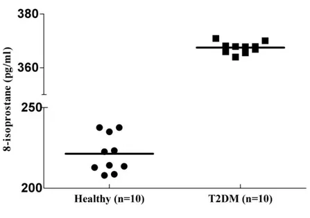
Serum 8-isoprostane levels in patients with type 2 diabetes mellitus (T2DM) (median 366 pg/mL; range 364–368 pg/mL) were significantly higher than those of healthy controls (median 221 pg/mL; range 207–237 pg/mL; P < 0.05; Fig. 2). Remarkably, 100% (10/10) of the patients with T2DM exhibited elevated 8-isoprostane levels, while none of the healthy individuals showed increased levels. Thus, all patients with diabetes exhibited elevated 8-isoprostane levels that could discriminate patients with T2DM from healthy controls. The finding that there was a significant association between patients with T2DM and 8-isoprostane levels suggests that oxidative stress is related to diabetes.
- 16 -
Figure 2: Serum 8-isoprostane levels in patients with type 2 diabetes and healthy controls
- 17 -
2. High glucose cytotoxicity dose response
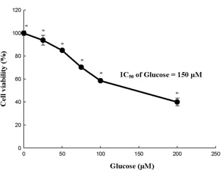
The effect of different concentrations of glucose was assessed on 1 104 cells/well to determine the concentration of glucose, which resulted in 50% of cell viability inhibition (IC50 value). The results
showed a significant decline of cell viability following 24 h of incubation of SH-SY5Y cells with an increasing concentration of glucose (0–200 μM). The glucose concentration, which resulted in 50% SH-SY5Y cell viability inhibition was 150 μM (150 2) (Fig. 3A and B). After 24 h of incubation, 10 μg/mL of 7,8-DHF was cytotoxic. Thus, a concentration of 5 μg/mL of 7,8-DHF was used for pretreatment (Fig. 3C).
- 18 -
A
Figure 3: Glucose cytotoxicity in SH-SY5Y cells
(A) Dose–response curve. SH-SY5Y cells (1 105 cells/well) were incubated with different concentrations of glucose (0–200 μM) for 24 h and cell viability was evaluated. The viability of SH-SY5Y cells was determined by using the MTT reduction assay. Values are the percentages of viable cells, with the viability of untreated control cells taken as 100%. * P < 0.05, relative to control cells.
- 19 -
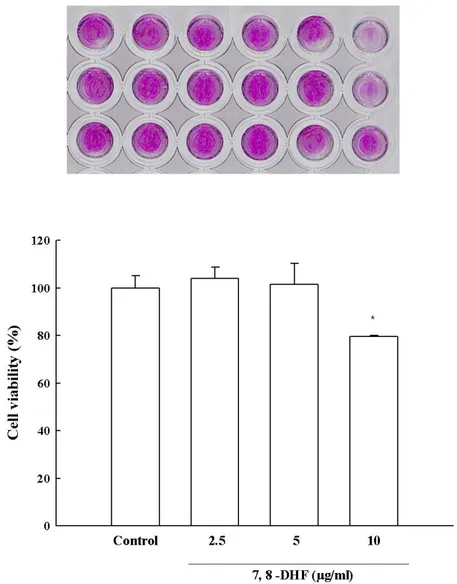
B
C
Figure 3 (continued): (B) The effect of glucose treatment was visualized by MTT assay. (C) Effect of 7,8-DHF on glucose-induced cytotoxicity in SH-SY5Y cells. Cells were pretreated with different concentrations of 7,8-DHF (2.5, 5, and 10 μg/mL). 7,8-DHF was cytotoxic at 10 μg/mL.
* Significantly different from the control group (P < 0.05).
- 20 -
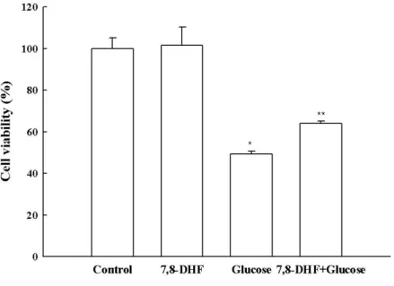
3. Effect of 7,8-DHF against high glucose-induced cytotoxicity
7,8-DHF protective effect on cell survival was assessed using glucose (150 μM)-treated SH-SY5Y cells. Cells were treated with 7,8-DHF at 5 μg/mL for 24 h, prior to the addition of glucose. Cell viability was determined 24 h later by MTT assay. As shown in Figure 3, 7,8-DHF treatment enhanced cell survival rate to 102% compared to 100% for the control, indicating that 7,8-DHF at 5 μg/mL was not cytotoxic for SH-SY5Y cells. Combination of 7,8-DHF at 5 μg/mL and glucose (150 μM) increased the cell survival rate to 64% compared to 49% for glucose (150 μM)-treated cells.
- 21 -
Figure 4:Protective effect of 7,8-DHF against high glucose-induced cell damage
The viability of SH-SY5Y cells treated with glucose, 7,8DHF, or both was determined by MTT assay.
* Significantly different from the control group (P < 0.05). ** Significantly different from the glucose
- 22 -
4. 7,8-DHF effect on high glucose-induced ROS production
To determine the role of ROS in mediating glucose toxicity, levels of intracellular ROS were measured using DCF-DA assay in SH-SY5Y cells. The level of intracellular ROS detected by flow cytometry revealed a fluorescence intensity of 184 in glucose-treated cells in presence of 7,8-DHF at 5 μg/mL compared to a fluorescence intensity of 347 in glucose-treated cells (Fig.5.), reflecting a reduction in ROS generation. The flow cytometry data revealed that glucose treatment increased the level of mitochondrial ROS compared to control. However, treatment with 7,8-DHF at 5 μg/mL attenuated the glucose-induced ROS increase. These data suggest that 7,8-DHF is a scavenger of intracellular ROS.
- 23 -
A
B
Figure 5: Effect of 7,8-DHF on high glucose-induced intracellular ROS generation
The intracellular ROS generated was detected by (A) flow cytometry and (B) confocal microscopy after DCF-DA treatment. The representative confocal images illustrate the increase in red fluorescence intensity of DCF produced by ROS (original magnification 400). The measurements were made in duplicate and the values are expressed as means ± SE. * Significantly different from
- 24 -
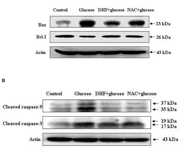
5. 7,8-DHF effect on apoptosis-related proteins
To further understand the protection mechanism of 7,8-DHF on the glucose-induced apoptotic process, the expression of proteins involved in mitochondria-related apoptosis was investigated. Beforehand, changes in Bcl-2 expression, an anti-apoptotic protein, and Bax expression, a pro-apoptotic protein, were examined. As shown in Figure 6, 7,8-DHF treatment induced an increase in Bcl-2 expression and a decrease in Bax expression in glucose-treated cells. Next, caspase-9 and caspase-3 expression was examined by western blot since these enzymes are activated due to mitochondrial membrane disruption [18]. As shown in Figure 6, 7,8-DHF treatment inhibited glucose-induced active form of 9 (39 and 37 kDa) and 3 (19 and 17 kDa), a target of caspase-9. These results suggest that 7,8-DHF protects cells from apoptosis by inhibiting the mitochondrial caspase-dependent pathway.
- 25 -
A
B
Figure 6: Effects of 7,8-DHF on apoptosis-related proteins
Cell lysates were electrophoresed and (A) Bax, Bcl-2, (B) Cleaved (active) caspase-9, caspase-3 as well as actin were detected by using specific antibodies.
- 26 -
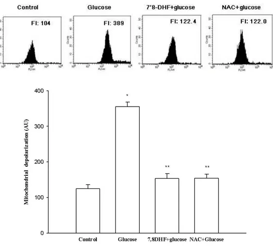
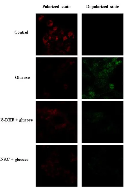
6. 7,8-DHF effect on glucose-induced Mitochondrial membrane potential analysis depolarization
Change in Δψm was examined to improve our understanding of 7,8-DHF protection mechanism
against the glucose-induced apoptotic process in terms of mitochondrial involvement. Mitochondria are instrumental in oxidative phosphorylation, cell death regulation, and ROS production [19]. JC-1 is a cationic dye that indicates mitochondrial polarization by shifting its fluorescence emission from green (approximately 525 nm) to red (approximately 590 nm). As shown in Figure 7, control cells and 7,8-DHF-treated cells exhibited strong red fluorescence (JC-1 aggregated form, indicative of mitochondrial polarization) in the mitochondria. However, glucose treatment resulted in reducing the red fluorescence and increasing the green fluorescence (JC-1 monomer form, indicative of mitochondrial depolarization) in mitochondria. 7,8-DHF treatment blocked the glucose effect. Image analysis data were consistent with flow cytometry data. The level of Δψm loss increased in
glucose-treated cells, as substantiated by an increase in fluorescence with the JC-1 dye. However, 7,8-DHF recovered the level of Δψm loss (Fig. 7A and B), suggesting that 7,8-DHF inhibited the
- 27 -
A
Figure 7: Effects of 7, 8-DHF on glucose-induced mitochondrial membrane potential depolarization Δψm depolarization was analyzed by (A) flow cytometry. * Significantly different from control cells
- 28 -
B
- 29 -
7. Glucose induces apoptosis via the mitochondria-dependent pathway
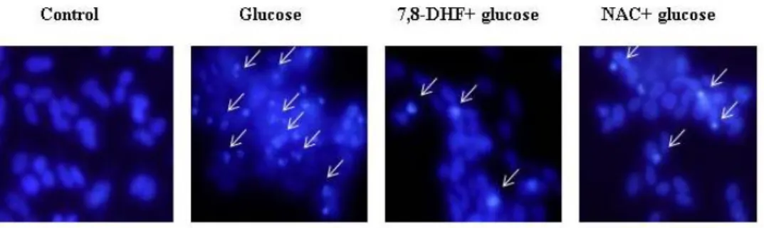
To evaluate the cytoprotective effects of 7,8-DHF with respect to apoptosis induced by glucose, the nuclei of SH-SY5Y cells were stained with Hoechst 33342 and analyzed by microscopy. The microscopic images in Figure 8A revealed that the control cells had intact nuclei, while the glucose-treated cells showed significant nuclear fragmentation, which is indicative of apoptosis. However, pretreatment of glucose-treated cells with 7,8-DHF induced a decrease in nuclear fragmentation. These results suggest that 7,8-DHF protects cells by inhibiting glucose-induced apoptosis.
- 30 -
Figure 8: Effect of 7,8-DHF against glucose-induced apoptosis
Apoptotic body formation was observed under a fluorescent microscope after Hoechst 33342 staining. The apoptotic bodies are indicated with white arrows. Apoptotic body formation was quantified using a microscope. * Significantly different from control cells (P < 0.05), ** Significantly different from
- 31 -
DISCUSSION
Oxidative stress plays a pivotal role in cellular injury induced by hyperglycemia. High glucose level can stimulate free radical production. Thus, a weak defense system becomes unable to counteract the enhanced ROS generation and, as a result, an imbalance occurs which leads to oxidative stress [20, 21]. In the present study, we demonstrated that 7,8-DHF dramatically inhibited high glucose-induced cell death, apoptosis, and mitochondrial dysfunction. 7,8-DHF protective effect might be attributed to its powerful antioxidant action, as evidenced by the markedly reduced ROS.
SH-SY5Y, a neuroblastoma cell line, is commonly used to study diabetic neuropathy [22, 23]. It is a thrice cloned sub-line of the neuroblastoma cell line SK-N-SH established in 1970 from a metastatic bone tumor [24]. These cells, when treated with high glucose, show results similar to those observed by using dorsal root ganglia neurons and Schwann cells [25]. In addition, SH-SY5Y cells have also been effectively used to demonstrate changes in signal transduction [23]. Pathways that lead to abnormal nitric oxide production and changes in the activity of the Na+/K+ pump have also been studied [23]. Neuroblastoma cells have also been extensively used to screen the efficacy of uncouplers [25] and insulin-like growth factor-1 [26] as neuroprotectants and to investigate their mechanism of action. Therefore, high glucose treated SH-SY5Y cells provide a suitable cell model to investigate 7,8-DHF antioxidant activity.
Hyperglycemia causes tissue damage through multiple mechanisms, including increased flux of glucose and other sugars through the polyol pathway, increased intracellular formation of advanced glycation end products (AGEs), increased the expression levels of the receptor for AGEs and its activating ligands, activation of protein kinase C isoforms, and upregulation of the hexosamine pathway [27].Excess glucose can be converted to sorbitol by aldose reductase [28]. This first step in the polyol pathway is linked to the oxidation of NADPH to NADP+. This leads to NADPH depletion, needed for regenerating glutathione [28]. Thus, early after the induction of diabetes, metabolic defects
- 32 -
lead to loss of NADPH, which limits the nerve ability to scavenge ROS, promoting a vicious cycle of oxidative stress. Sorbitol accumulation can also result in cellular osmotic stress that may alter the anti-oxidant potential of the cell and increase ROS [29]. Aldose reductase inhibitors that penetrate the nerve, decrease nerve sorbitol levels. In one clinical trial, there is evidence of enhanced axonal regeneration [30]. In this study, high glucose treatment increased the level of mitochondrial ROS in SH-SY5Y cells compared to the control cells. However, treatment with 7,8-DHF at 5 μg/mL attenuated the glucose-induced ROS increase.
Recent evidence suggests that oxidative stress is responsible for the development and progression of neuropathy [31]. Blocking oxidative stress in diabetic animals prevents the development of neuropathy and restores sciatic and saphenous nerve conduction velocities in streptozotocin diabetic rats [31-33].
Our data indicate that increased concentrations of glucose rapidly induced ROS production. This can be modulated by reducing electron flux along the electron transfer chain and generation of superoxide anions using 7,8-DHF. In turn, 7,8-DHF is able to reduce the mitochondrial membrane depolarization observed with high glucose and block the induction of caspase cleavage. This most likely results from the ability of 7,8-DHF to reduce ROS generation and to stabilize the Δψm. Both in vivo and in vitro,
mitochondrial swelling is observed in dorsal root ganglion neurons and Schwann cells exposed to high glucose and, ultimately, the Δψm, is decreased, an event that, in other paradigms, initiates
programmed cell death [34]. It has been previously shown that release of cytochrome c into the cytosol is associated with formation of a cytochrome c/caspase-9/Apaf-1 complex (the apoptosome) and cleavage of downstream effector caspases such as caspase-3 and caspase-7 [34]. In human and rat neurons, hyperglycemia induces mitochondrial dysfunction and apoptosis, and similar changes are observed in another important cell type in the peripheral nervous system, namely, the Schwann cells [22, 35]. In nerves from 12-month diabetic rats, an increase in caspase-9 cleavage and caspase-3 cleavage is observed in cultured Schwann cells exposed to high glucose. Similar to the changes observed in rat neurons, human Schwann cells show evidence of both mitochondrial swelling and
- 33 -
apoptosis when cultured in presence of high glucose.
In summary, 7,8-DHF could effectively protect SH-SY5Y cells against high glucose-induced apoptosis through its antioxidant action. By free radical scavenging, 7,8-DHF markedly inhibited high glucose-induced oxidative injury, preventing apoptosis and mitochondrial dysfunction. Our study suggests that 7,8-DHF is a promising agent in the treatment of diabetic neuropathy.
- 34 -
REFERENCE
1. American Diabetes A: Diagnosis and classification of diabetes mellitus. Diabetes Care 2014, 37 Suppl 1:S81-90.
2. Brownlee M: Biochemistry and molecular cell biology of diabetic complications. Nature 2001, 414:813-820.
3. Assmann G, Carmena R, Cullen P, Fruchart JC, Jossa F, Lewis B, Mancini M, Paoletti R: Coronary heart disease: reducing the risk: a worldwide view. International Task Force for the Prevention of Coronary Heart Disease. Circulation 1999, 100:1930-1938.
4. Tomlinson DR, Gardiner NJ: Glucose neurotoxicity. Nature Reviews Neuroscience 2008, 9:36-45. 5. Zaltzberg H, Kanter Y, Aviram M, Levy Y: Increased plasma oxidizability and decreased erythrocyte and plasma antioxidative capacity in patients with NIDDM. The Israel Medical Association journal: IMAJ 1999, 1:228-231.
6. Greene DA, Sima AA, Stevens MJ, Feldman EL, Lattimer SA: Complications: neuropathy, pathogenetic considerations. Diabetes care 1992, 15:1902-1925.
7. Mousavi S, Tayarani N, Parsaee H: Protective effect of saffron extract and crocin on reactive oxygen species-mediated high glucose-induced toxicity in PC12 cells. Cellular and molecular neurobiology 2010, 30:185-191.
8. Waldmeier PC, Tatton WG: Interrupting apoptosis in neurodegenerative disease: potential for effective therapy? Drug discovery today 2004, 9:210-218.
9. Wyllie AH: Apoptosis: an overview. British Medical Bulletin 1997, 53:451-465.
10. Ashkenazi A, Dixit VM: Death receptors: signaling and modulation. Science 1998, 281:1305-1308. 11. Shimizu S, Narita M, Tsujimoto Y, Tsujimoto Y: Bcl-2 family proteins regulate the release of apoptogenic cytochrome c by the mitochondrial channel VDAC. Nature 1999, 399:483-487.
- 35 -
and BACE1 elevation in a mouse model of Alzheimer's disease. Neuropsychopharmacology 2011, 37:434-444.
13. Gupta VK, You Y, Li JC, Klistorner A, Graham SL: Protective effects of 7, 8-dihydroxyflavone on retinal ganglion and RGC-5 cells against excitotoxic and oxidative stress. Journal of Molecular Neuroscience 2013, 49:96-104.
14. Carmichael J, DeGraff WG, Gazdar AF, Minna JD, Mitchell JB: Evaluation of a tetrazolium-based semiautomated colorimetric assay: assessment of chemosensitivity testing. Cancer research 1987, 47:936-942.
15. Rosenkranz AR, Schmaldienst S, Stuhlmeier KM, Chen W, Knapp W, Zlabinger GJ: A microplate assay for the detection of oxidative products using 2′, 7′-dichlorofluorescin-diacetate. Journal of immunological methods 1992, 156:39-45.
16. Cossarizza A, Baccaranicontri M, Kalashnikova G, Franceschi C: A new method for the cytofluorometric analysis of mitochondrial membrane potential using the J-aggregate forming lipophilic cation 5, 5′, 6, 6′-tetrachloro-1, 1′, 3, 3′-tetraethylbenzimidazolcarbocyanine iodide (JC-1). Biochemical and biophysical research communications 1993, 197:40-45.
17. Troiano L, Ferraresi R, Lugli E, Nemes E, Roat E, Nasi M, Pinti M, Cossarizza A: Multiparametric analysis of cells with different mitochondrial membrane potential during apoptosis by polychromatic flow cytometry. Nature protocols 2007, 2:2719-2727.
18. Perkins CL, Fang G, Kim CN, Bhalla KN: The role of Apaf-1, caspase-9, and bid proteins in etoposide-or paclitaxel-induced mitochondrial events during apoptosis. Cancer research 2000, 60:1645-1653.
19. Orrenius S, Gogvadze V, Zhivotovsky B: Mitochondrial oxidative stress: implications for cell death. Annu Rev Pharmacol Toxicol 2007, 47:143-183.
20. Halliwell B: Free radicals and other reactive species in disease. Wiley Online Library; 2005. 21. Pandey KB, Mishra N, Rizvi SI: Protein oxidation biomarkers in plasma of type 2 diabetic patients. Clinical biochemistry 2010, 43:508-511.
- 36 -
22. Russell J, Feldman E: Insulin-like growth factor-I prevents apoptosis in sympathetic neurons exposed to high glucose. Hormone and metabolic research 1999, 31:90-96.
23. Shindo H, Thomas TP, Larkin DD, Karihaloo AK, Inada H, Onaya T, Stevens MJ, Greene DA: Modulation of basal nitric oxide-dependent cyclic-GMP production by ambient glucose, myo-inositol, and protein kinase C in SH-SY5Y human neuroblastoma cells. Journal of Clinical Investigation 1996, 97:736.
24. Biedler JL, Roffler-Tarlov S, Schachner M, Freedman LS: Multiple neurotransmitter synthesis by human neuroblastoma cell lines and clones. Cancer research 1978, 38:3751-3757.
25. Vincent AM, Brownlee M, Russell JW: Oxidative stress and programmed cell death in diabetic neuropathy. Annals of the New York Academy of Sciences 2002, 959:368-383.
26. Leinninger G, Russell J, Van Golen C, Berent A, Feldman E: Insulin-like growth factor-I regulates glucose-induced mitochondrial depolarization and apoptosis in human neuroblastoma. Cell Death & Differentiation 2004, 11:885-896.
27. Brownlee M: The pathobiology of diabetic complications a unifying mechanism. Diabetes 2005, 54:1615-1625.
28. NEUROPATHY PD: Treatment of diabetic neuropathy. 1994.
29. Obrosova IG, Minchenko AG, Vasupuram R, White L, Abatan OI, Kumagai AK, Frank RN, Stevens MJ: Aldose reductase inhibitor fidarestat prevents retinal oxidative stress and vascular endothelial growth factor overexpression in streptozotocin-diabetic rats. Diabetes 2003, 52:864-871. 30. Greene D, Arezzo J, Brown M: Effect of aldose reductase inhibition on nerve conduction and morphometry in diabetic neuropathy. Neurology 1999, 53:580-580.
31. Stevens MJ, Obrosova I, Cao X, Van Huysen C, Greene DA: Effects of DL-alpha-lipoic acid on peripheral nerve conduction, blood flow, energy metabolism, and oxidative stress in experimental diabetic neuropathy. Diabetes 2000, 49:1006-1015.
32. Low PA, Nickander KK, Tritschler HJ: The roles of oxidative stress and antioxidant treatment in experimental diabetic neuropathy. Diabetes 1997, 46:S38-S42.
- 37 -
33. Cameron NE, Cotter MA: Metabolic and vascular factors in the pathogenesis of diabetic neuropathy. Diabetes 1997, 46:S31-S37.
34. Green DR, Reed JC: Mitochondria and apoptosis. Science-AAAS-Weekly Paper Edition 1998, 281:1309-1311.
35. Russell JW, Sullivan KA, Windebank AJ, Herrmann DN, Feldman EL: Neurons undergo apoptosis in animal and cell culture models of diabetes. Neurobiology of disease 1999, 6:347-363.
- 38 -
ABSTRACT IN KOREAN
당뇨병성 신경증은 당뇨병 환자에서 흔한 만성 합병증 중의 하나로 신경세포의 손상 및 신경세포에 혈액을 공급하는 미세혈관 변성에 의한 말초 순환장애로 인해 유발되는 것으 로 보고되고 있다. 이에 대한 원인으로는 고혈당증으로 알려져 있고 이러한 당뇨병성 신 경증은 다양한 신호전달계의 활성에 의해서 발병하는 것으로 알려져 있다. 그 중 산화성 스트레스의 증가는 당뇨병에서 신경증을 포함한 여러 미세 혈관계의 합병증을 유발하는 데 주요한 역할을 하는 것으로 알려져 있다. 이 연구에서는 당뇨병 환자의 혈청을 이용 하여 산화적 스트레스의 정도를 지질과산화의 직접적인 지표인 8-isoprostane을 측정하 여 유의하게 증가 되어있음을 확인하였다. 이러한 활성산소를 억제하기 위해 항산화제로 보고된 플라보노이드 계열인 7,8-dihydroxyflavone가 고농도의 포도당에 의해 유발된 SH-SY5Y 세포의 산화적 손상과 세포자연사에 대해 항산화 효과와 신경세포 보호 효과 를 확인 하였다. 먼저 SH-SY5Y neuroblastoma cells에 산화적 스트레스를 유도하기 위 해 고농도의 포도당을 처리하여 포도당이 증가할수록 세포의 생존율이 감소하였고 활성 산소가 증가하였으며, 7,8-DHF가 유의하게 차단 작용을 보여 주었다. 미토콘드리아에서 cytochrome c의 방출 및 미토콘드리아의 막 전위차를 조절한다고 알려져 있는 Bax, Bcl-2 단백질의 발현 양상은 고농도로 포도당을 처리한 군에서 pro-apoptotic protein 인 Bax가 증가하였고, anti-apoptotic protein인 Bcl-2가 감소하였으며, 7,8-DHF를 처 리한 군에서는 상반되는 반응을 보였다. 세포고사와 관련된 단백질의 발현을 알아보기 위해 고농도로 포도당을 처리한 군에서 caspase-9 protease와 caspase-3 protease활 성이 증가 하였고 7,8-DHF처리한 군에서는 감소되는 현상을 보였다. 또한 세포고사가 미토콘드리아의 기능 변화와 관련되었는지의 여부를 알아보고자 미토콘드리아의 막 전위- 39 -
차 변화(membrane potential transition;MPT)를 확인 하기 위하여 JC-1 염색후에 형광 현미경을 통해 관찰하여 고농도 포도당을 처리한 군에서 자연사가 일어난 세포질임을 보 여주었다. 세포고사 기전에 의해 매개되었는지 여부와 이러한 세포고사에 ROS가 관여 하였는지 여부를 알아보고자, 세포고사의 형태학적인 특징과 관련된 단백질의 발현을 확 인하기 위해 세포의 핵을 Hoechst 33342 염색시, 고농도의 포도당군은 세포수가 적어지 고 핵이 여러 조각으로 분절된 형광염색을 나타내었고, 7,8DHF 처리한 군은 세포핵의 모양이 타원형의 균일한 형광염색을 나타내었다. 7,8-DHF은 이러한 경로를 통하여 산화 적 손상으로 유도된 세포자연사를 억제하는 것으로 확인하였다. 본 연구는 고농도의 포도당으로 유도되는 SH-SY5Y 신경세포의 산화적 스트레스에 대한 7, 8-dihydroxyflavone의 보호 효과를 확인할 수 있었다.
- 40 -
ACKNOWLEDGEMENTS
First and foremost, I would like to express my sincere appreciation and gratitude to my advisor, Jin-Won Hyun, for her academic guidance and enthusiastic encouragement throughout the research. Besides completing a PhD dissertation, she stimulated and directed me in publishing conference papers, journal papers and a book. During these projects, she was always there to provide necessary assistance and academic resources. Without her support, I could not have done what I was able to do. She was very generous in sharing her experiences on academic life and beyond. These experiences will be my primary guide during my life and academic career.
I owe my sincere gratitude to Prof. Jong Cook Park, Prof. Ji Hoon Kang, Prof. Maeng Young Hee at the school of medicine in Jeju National University and Hee Sun Kim at Ewha Womans University who gave me the instruction to complete this thesis. In addition, I greatly appreciate all the professors who have instructed me in the school of medicine.
Last but not least, I would like to give my special thanks to my wife, Sang ah Lee and my daughter, Yuhong cho who are the origin of my power.