저작자표시-비영리-변경금지 2.0 대한민국 이용자는 아래의 조건을 따르는 경우에 한하여 자유롭게 l 이 저작물을 복제, 배포, 전송, 전시, 공연 및 방송할 수 있습니다. 다음과 같은 조건을 따라야 합니다: l 귀하는, 이 저작물의 재이용이나 배포의 경우, 이 저작물에 적용된 이용허락조건 을 명확하게 나타내어야 합니다. l 저작권자로부터 별도의 허가를 받으면 이러한 조건들은 적용되지 않습니다. 저작권법에 따른 이용자의 권리는 위의 내용에 의하여 영향을 받지 않습니다. 이것은 이용허락규약(Legal Code)을 이해하기 쉽게 요약한 것입니다. Disclaimer 저작자표시. 귀하는 원저작자를 표시하여야 합니다. 비영리. 귀하는 이 저작물을 영리 목적으로 이용할 수 없습니다. 변경금지. 귀하는 이 저작물을 개작, 변형 또는 가공할 수 없습니다.
Predictive biomarkers for inhibition of
B-cell receptor signaling pathway in
activated B-cell (ABC) diffuse large
B-cell lymphoma (DLBCL)
Hyewon Lee
Department of Medicine
The Graduate School, Yonsei University
[UCI]I804:11046-000000523511
[UCI]I804:11046-000000523511
Predictive biomarkers for inhibition of
B-cell receptor signaling pathway in
activated B-cell (ABC) diffuse large
B-cell lymphoma (DLBCL)
Hyewon Lee
Department of Medicine
Predictive biomarkers for inhibition of
B-cell receptor signaling pathway in
activated B-cell (ABC) diffuse large
B-cell lymphoma (DLBCL)
Directed by Professor Jin Seok Kim
Doctoral Dissertation
submitted to the Department of Medicine,
the Graduate School of Yonsei University
in partial fulfillment of the requirements for the degree of
Doctor of Philosophy
Hyewon Lee
This certifies that the Doctoral
Dissertation of Hyewon Lee is approved.
---
Thesis Supervisor : Jin Seok Kim
---
Thesis Committee Member#1 : Woo Ick Yang
---
Thesis Committee Member#2 : Jong Rak Choi
---
Thesis Committee Member#3: Hyoung-Pyo Kim
---
Thesis Committee Member#4: Hyeon-Seok Eom
The Graduate School
Yonsei University
ACKNOWLEDGEMENTS
I would like to express my special gratitude to Prof. Jin Seok
Kim, who consistently guided me in every step of the thesis with
a professional, warm and delicate attention. Also, great thanks to
Prof. Woo-Ick Yang, Prof. Jong Rak Choi, Prof Hyoung-Pyo
Kim, and Prof. Hyeon-Seok Eom who offered precious advises
and supports for me to finalize the thesis. I also appreciate of
education from Prof. Yoo Hong Min and Prof. June-Won
Cheong in my hematology training, generous help from the
staffs of Center for Hematologic Malignancies, National Cancer
Center, Korea, in my daily work, and special advises and
supports from Dr. Yuri Kim in the early step of this research.
Secondly I would like to give my thanks and love to my husband,
Dr. Namsu Ku, and lovely son, Yoon Seong Ku, who are my
pride and joy. I pray for my parents and parents-in-law, who
have always encouraged and supported me with love.
I want to dedicate this paper to all with all of my heart.
<TABLE OF CONTENTS>
ABSTRACT ··· 1
I. INTRODUCTION ··· 3
II. MATERIALS AND METHODS ··· 6
1. Patients ··· 6
2. Immunohistochemistry ··· 7
3. RNA extraction and NanoString assay ··· 9
4. Analysis of Lymph2Cx and additional ABC-related genes ··· 9
5. Statistical methods ··· 11
III. RESULTS ··· 11
1. Patient characteristics ··· 11
2. Cell of origin analyses ··· 12
3. DEL in high risk and ABC type DLBCL ··· 12
4. Clinical outcomes of all patients ··· 14
5. Role of upfront ASCT in subgroups ··· 16
6. Gene expression profiles and biomarkers associated with survival
outcomes ··· 17
IV. DISCUSSION ··· 23
V. CONCLUSION ··· 26
REFERENCES ··· 28
LIST OF FIGURES
Figure 1. Cell of origin by Hans criteria and Lymph2Cx algorithm ··· 13
Figure 2. Survival outcomes of all patients ··· 15
Figure 3. Survival outcomes according to upfront autologous stem cell
transplantation ··· 15
Figure 4. Clinical
o
utcomes according to upfront ASCT status and gender
··· 17
Figure 5. Clinical
o
utcomes according to upfront ASCT and response to
the first line chemotherapy ··· 17
Figure 6. RAB7L1 expression and survival
o
utcomes in non-GCB type
DLBCL ··· 21
Figure 7. S1PR2 gene expression and survival
o
utcomes in non-GCB
type DLBCL ··· 21
LIST OF TABLES
Table 1. Patient characteristics and the upfront ASCT ··· 8
Table 2. Genes included in the Lymph2Cx code sets ··· 10
Table 3. Frequency of DEL according to COO ··· 13
Table 4. Univariate analyses of factors affecting OS and PFS: all patient
cohort ··· 14
Table 5. Gene expression profiles in patients with non-GCB subtype
DLBCL ··· 18
Table 6. Univariate analyses of ABC subtype-specific genes associated with
survival ··· 19
Table 7. Multivariate analyses of factors affecting survival adjusted for
LDH, EOT response, and upfront ASCT: ABC subgroup ··· 20
Table 8. Multivariate analyses of factors affecting surviva
l:
all patient
cohort ··· 22
Table 9. Multivariate analyses of factors affecting survival: ABC
subgroup ··· 23
.
1
ABSTRACT
Predictive biomarkers for inhibition of B-cell receptor signaling pathway
in activated B-cell (ABC) diffuse large B-cell lymphoma (DLBCL)
Hyewon Lee
Department of Medicine
The Graduate School, Yonsei University
(Directed by Professor Jin Seok Kim)
Background: Gene expression profiling (GEP) has been used to distinguish
molecular subtypes of diffuse large B-cell lymphoma (DLBCL) based on their cell-of-origin (COO); germinal center B-cell (GCB) and activated B-cell (ABC). ABC DLBCLs have distinct genetic features and are associated with relatively poor prognosis. ABC subtyping is typically performed using a combination of immunohistochemistry (IHC) with CD10, BCL6, and MUM1, with the introduction of digital GEP potentially offering a more accurate and convenient method. In this study, I performed digital GEP on high risk, non-GCB type DLBCL samples, to better understand the clinical importance of COO, as well as to identify potential genetic biomarkers in these high risk patients.
Methods: For digital GEP, NanoString analyses were performed on
formalin-fixed paraffin-embedded (FFPE) DLBCL tissues. Clinical samples were collected from patients who were diagnosed with DLBCL between January 2008 and December 2015 at Severance Hospital, based on a search of medical records. Inclusion was limited to transplant-eligible patients with high risk clinical parameters, and predefined as non-GCB types by IHC. COO was reanalyzed by Lymph2Cx algorithm with 20-gene panel. Additional 13 genes related to ABC-specific signaling pathways were included as part of the NanoString assay.
2
designated as high risk based on stage (95.7%), International Prognostic Index (IPI) score ≥3 (63.8%), and elevated lactate dehydrogenase (LDH) (65.2%). Patients were treated with 6-8 cycles of standard chemoimmunotherapy, with a median follow up duration of 52.1 months. Upfront autologous stem cell transplantation (ASCT) was performed in 27 responders. Serum LDH level, treatment response, and upfront ASCT were identified as important factors for survival. The benefit of upfront ASCT was greater in males than in females. The NanoString assay was performed in 59 cases, of which 19 discrepancies (27.3%) were identified, resulting in reclassification from non-GCB type into GCB or unclassifiable. COO itself did not affect survival outcomes. The prognostic value of gene expression levels was screened using a COX proportional hazard model. Significant associations (p<0.1) included CCDC50 for PFS; CYB5R2, LIMD1, ITPKB, Aiolos, CRBN, and IKKbeta for OS; and RAB7L1, ASB13, S1PR2, and IRAK4 for both. Among these, low RAB7L1 (<641.25) was predictive of worse PFS (5-year rate, 37.7% vs. 78.0%, p=0.003) and OS (5-year rate, 45.5% vs. 87.0%, p=0.004). Low S1PR2 (<78.85) was also associated with poor PFS (5-year rates, 50.0% vs. 68.7%, p=0.026) and OS (5-year rates, 51.4% vs. 77.3%, p=0.018). In multivariate analyses, low RAB7L1 was identified as an independent biomarker for PFS and OS with a hazard ratio (HR) of 4.53 (95%CI 1.33-15.39, p=0.015) and 8.05 (95%CI 1.42-45.65, p=0.018) in high-risk/ABC DLBCL. Low S1PR2 was an independent predictor for PFS with a HR of 5.42 (95% CI 1.01-28.97, p=0.048), but not OS, in high-risk/ABC DLBCL.
Conclusion: Digital GEP using the Lymph2Cx was able to accurately distinguish
patients based on ABC subtype, however, COO alone was not a prognostic factor in transplant-eligible patients with high-risk DLBCL. Upfront ASCT may improve survival in such a high-risk group. In addition, low RAB7L1 and S1PR2 were associated with poor prognosis, but this finding requires further validation.
__________________________________________________________________ Keywords: diffuse large B cell lymphoma, cell-of-origin, activated B cell, digital gene expression profile, biomarkers
3
Predictive biomarkers for inhibition of B-cell receptor signaling pathway
in activated B-cell (ABC) diffuse large B-cell lymphoma (DLBCL)
Hyewon Lee
Department of Medicine
The Graduate School, Yonsei University
(Directed by Professor Jin Seok Kim)
I. INTRODUCTION
Diffuse large B-cell lymphoma (DLBCL) is the most common subtype of Non-Hodgkin’s lymphoma (NHL) in Korea.1 It originates from mature B-lymphocytes, and presents as a highly aggressive disease. The incidence of DLBCL increases with age, with a median age of 64 years at diagnosis. Approximately half of all DLBCL cases present as advanced disease (Ann Arbor Stage III or IV), which rapidly progress and require immediate treatment to improve survival outcome. Since the late 1990s, the treatment of choice has been a combination of rituximab, cyclophosphamide, vincristine, doxorubicin, and prednisolone, the so-called R-CHOP regimen. Rituximab, a monoclonal antibody to CD20, targets B-lymphocytes presenting CD20 on their surface. Rituximab had widespread usage in the early 2000s, and is closely associated with the recent improvement of survival in patients with DLBCL.2 Although almost all patients with DLBCL are currently treated with R-CHOP, the outcome of each patient differs. Initially localized and non-bulky diseases can be cured by optimal treatment; however, patients with disseminated diseases, high tumor burden, or suboptimal response to treatment exhibit very poor survival.3
4
Numerous studies have been performed to better predict survival outcomes in DLBCL patients. The most commonly used prognostic factors are age, elevated levels of lactate dehydrogenase (LDH), performance status, Ann Arbor stage, and multiple extranodal involvement (International Prognostic Index, IPI).4 Patients who test positive for three or more factors tend to exhibit a 50% of relapse rate at 5 year follow up.
In addition to the clinical prognostic factors, gene expression profiling (GEP) has been used to distinguish histologically uniform DLBCL into four molecular subtypes based on their cell-of-origin (COO); germinal center B-cell (GCB), activated B-cell (ABC or non-GCB), primary mediastinal B-cell lymphoma (PMBL), and unclassifiable subtype.5,6 ABC type DLBCL is characterized by activated nuclear factor kappa B (NF-kB) signaling and is associated with relatively poor survival compared to the GCB type, achieving a cure rate <40%. For clinical application of molecular subtyping, surrogate immunohistochemistry (IHC)-based biomarker algorithms including CD10, BCL6, and MUM1 have been widely adopted to discriminate COO, with >80% concordance with standard GEP.7 Furthermore, in a study that conducted digital gene expression analyses using the 20-gene Lymph2Cx algorithm, the 5-year progression-free survival of GCB, unclassified, and ABC type DLBCL were 73%, 54%, and 48%, respectively,8 indicating a clear need for more effective treatments for ABC type DLBCL.
The ABC-specific signaling pathways is well-established, with constitutional activation of Bruton’s tyrosine kinas (BTK) serving as a primary driver of disease pathology in ABC type DLBCL.9-12 BTK plays a key role in B-cell receptor (BCR) signaling, particularly in patients with chronic lymphocytic leukemia (CLL) and mantle cell lymphoma (MCL); targeting this pathway with ibrutinib has led to a striking improvement in the survival of CLL and MCL patients.9, 10 Chronic active BCR signaling has also been observed in ABC type DLBCL showing sustained tumor cell survival through CD79 A/B, BTK, CARD11 and NF-kB activation.11
5
As BTK activation is only observed in ABC type DLBCL, not in GCB type, the response to ibrutinib is confined to the ABC type as expected.12 Currently, a multi-national phase III trial of ibrutinib in combination with R-CHOP for newly diagnosed DLBCL is ongoing.
Another ABC-specific treatment in patients with DLBCL is lenalidomide, which is well known as an active agent for multiple myeloma.13 Lenalidomide targets the positive feedback circuitry of NF-kB and IRF4 by downregulating IRF4 and SPIB and increasing toxic IFN-beta secretion.14 In addition, recent studies have shown that immunomodulatory drugs (IMiDs) bind to cereblon (CRBN), a ubiquitin ligase which facilitates degradation of IKZF1/3 (Ikaros, Aiolos) resulting in significant growth inhibition of myeloma cells.15,16 However, the clinical response to BCR signaling inhibitor monotherapy remains limited, due in part to our poor understanding of which patients will respond to BCR inhibition and why a significant proportion of ABC type DLBCL patients exhibit resistance to BCR-targeting agents.
With the growing accessibility of sequencing technologies, there has been a strong interest in identifying the prognostic value of genetic features compared to IHC in DLBCL.17 The most finding of these studies is the prognostic impact of the dual changes or overexpression of MYC/BCL2 in DLBCL, defining a ‘double-hit lymphoma (DHL)’ or ‘double expressor lymphoma (DEL)’.18-20 However, the frequency of ‘double-hit’ lymphoma is significantly lower in ABC type DLBCL compared to other COOs. Thus it is still difficult to explain why ABC type DLBCL shows poorer outcomes relative to other subtypes, as no specific biomarker for this entity has yet been validated.
Although ABC type DLBCL exhibits distinct gene expression profiles such as constitutional NF-kB activation and B cell receptor (BCR)-dependent cell survival, the criteria by which GCB and ABC type DLBCL are defined is still controversial. IHC with Hans’ criteria has been widely used in practice,7 but shows significant discrepancies compared to the results of GEP. Digital GEP is a relatively simple
6
technology that relies on formalin-fixed paraffin-embedded (FFPE) tissue and has shown good performance for distinguishing between ABC and GCB type DLBCL,8 although its use in Korea is still limited.21
The lack of validated biomarkers related to drug sensitivity or survival outcomes represents a significant obstacle in DLBCL, particularly in ABC, which is associated with worse survival outcomes than GCB. Adequate biomarkers are also required in cases of high risk DLBCL, as defined by clinical parameters, as treatment of these patients is often challenging even with molecular subtype information.
In this study, I selected a homogenous cohort of DLBCL patients who were young and fit for intensive treatment due to the presence of high risk features detected at the time of diagnosis. Using tumor samples collected from these patients, I performed digital GEP to identify ABC type DLBCLs among predefined set of non-GCB type DLBCL based on IHC Hans’ criteria. I also investigated the prognostic value of COOs, candidate genes as ABC-specific biologic markers, and the distribution of other clinical risk factors in this specific high-risk setting.
II. MATERIALS AND METHODS
1. Patients
All transplant-eligible, non-GCB type DLBCL patients diagnosed as high-risk based on clinical parameters between January 2008 and December 2015 at Severance Hospital, were identified based on a thorough review of medical records. Of these patients, those with available FFPE tumor tissue slides collected at the time of diagnosis were selected to perform additional IHC and further GEP
7
for COO determination and biomarker exploration. The term ‘ABC subtype’ was used only for cases classified by GEP in contrast to the ‘non-GCB subtype’ designation which was determined based on IHC criteria.
DLBCL was pathologically confirmed according to the 2008 World Health Organization criteria.22 Primary mediastinal large B cell lymphoma, primary central nervous system DLBCL, or AIDS-related DLBCL and patients who received chemotherapy other than R-CHOP were excluded. Patients who met at least one of the following criteria were categorized as clinically high-risk: elevated serum levels of LDH (>upper normal limit), advanced stage (stage III–IV), and high (≥3) IPI score. Staging and response to treatment were re-evaluated at the time of data analyses according to the Lugano criteria.23 We excluded all patients > 65 years of age who were not eligible for autologous stem cell transplantation (ASCT) to minimize the difference in intention-to-treat between young and elderly patients. Based on these criteria, a total of 69 patients were selected for further analyses (Table 1).
2. Immunohistochemistry
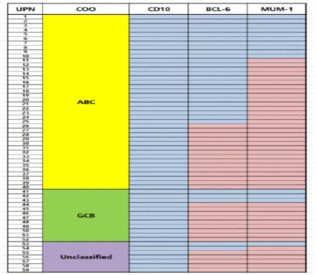
Hans criteria based on the expression levels of CD10, BCL6, and MUM1 were used to define non-GCB type DLBCL by IHC.7,21 For patients whose IHC results were not available in medical records, additional IHC was performed. Cut-off levels of 30% for positive are used for CD10 and MUM-1, and 40% for BCL-6.
Double-expressor lymphoma (DEL) was defined as DLBCLs with MYC and BCL2 protein concurrent expression, as determined by IHC. Cut-off values of 50% for BCL2 and 40% for MYC were used.19
8
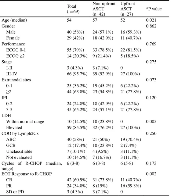
Table 1. Patient characteristics and the upfront ASCT
Total (n=69) Non-upfront ASCT (n=42) Upfront ASCT (n=27) *P value Age (median) 54 57 52 0.021 Gender 0.862 Male 40 (58%) 24 (57.1%) 16 (59.3%) Female 29 (42%) 18 (42.9%) 11 (40.7%) Performance 0.769 ECOG 0-1 55 (79%) 33 (78.5%) 22 (81.5%) ECOG ≥2 14 (20.3%) 9 (21.4%) 5 (18.5%) Stage 0.275 I-II 3 (4.3%) 3 (7.1%) 0 III-IV 66 (95.7%) 39 (92.9%) 27 (100%) Extranodal sites 0.073 0-1 25 (36.2%) 19 (45.2%) 6 (22.2%) ≥2 44 (63.8%) 23 (54.8%) 21 (77.8%) IPI 0.120 0-2 24 (24.8%) 18 (42.9%) 6 (22.2%) 3-5 45 (65.2%) 24 (57.1%) 21 (77.8%) LDH
Within normal range 10 (14.5%) 10 (23.8%) 0 0.005 Elevated 59 (85.5%) 32 (76.2%) 27 (100%) COO by Lymph2Cx 0.250 ABC 40 (58%) 21 (50%) 19 (70.4%) GCB 12 (17.4%) 10 (23.8%) 2 (7.4%) Unclassifiable 7 (10.1%) 4 (9.5%) 3 (11.1%) Not evaluated 10 (14.5%) 7 (16.7%) 3 (11.1%) Cycles of R-CHOP (median,
range)
6 (3-8) 6 (3-8) 6 (5-8) 0.173
EOT Response to R-CHOP 0.002
CR 42 (60.9%) 31 (73.8%) 11 (40.7%) PR 24 (34.8%) 8 (19%) 16 (59.3%) SD or PD 3 (4.3%) 3 (7.1%) 0
Abbreviations : ASCT, autologous stem cell transplantation; ECOG, Eastern Cooperative Oncology Group; IPI, International Prognostic Index; LDH, lactase dehydrogenase; COO, cell of origin; ABC, activated B cell; GCB, germinal center B cell; R-CHOP, rituximab/cyclophosphamide/doxorubicin/vincristine/prednisolone; EOT, end of treatment; CR, complete response; PR, partial response; SD, stable disease; PD, progressive disease.
9
3. RNA extraction and NanoString assay
NanoString assays were performed as described previously.8 Briefly, total RNA was extracted from FFPE slides of tumor tissues. Up to five slides per sample (2 µm sections consisting of >60% of the surface area) were collected for each patient, with similar tumor volumes collected for each patient. The recommended surface area of each slide is 1 cm2, which means if the biopsy surface were 10 mm by 5 mm, then two slides should be used. Slides were stored at room temperature until extraction. Nucleic acids were extracted using the MasterPure Complete DNA and RNA Purification Kit (Epicentre Biotechnologies, Madison, WI, USA). RNA was quantified by spectrophotometry (NanoDrop, Thermo Science, DE, USA). GEP was performed on 200 ng of RNA using NanoString technology (NanoString Technologies, WA, USA). Total RNA was hybridized to the custom code sets at 65°C overnight (15.5 to 22.5 h).
The hybridization reaction was processed on the nCounter Prep Station and then gene expression data were acquired on the nCounter Digital Analyzer at the high-resolution setting. The reference sample consisted of 100 nt oligonucleotides representing the hybridization targets within the genes. Standard quality control methods (Bioanalyzer and nSolver Analysis Software, NanoString Technologies, WA, USA) were employed, with samples validated against the positive spike-in controls, with individual gene ranges limited to 0.3 to 3 times the geometric mean of the total positive spike-in for that cartridge.
4. Analysis of Lymph2Cx and additional ABC-related genes
For COO discrimination, the Lymph2Cx code sets, consisting of 20 genes validated in previous publications, were used.8 In addition, we designed 13 custom code sets to assess tumor-associated genes previously associated with the BCR
10
signaling pathway (Table 2). The research-use-only (RUO) version of the NanoString Lymphoma Subtyping Test (LST) algorithm was used to determine the COO molecular subtype of each sample. The LST algorithm measures the geometric mean of five housekeeping genes (HK geomean) to ensure RNA quality based on a predefined clinical quality control (QC) threshold. Each sample meeting the QC threshold is reported as one of the two molecular subtypes, activated-B-cell (ABC) or germinal center-B-cell (GCB), or unclassified within an equivocal zone. Subtypes were determined using a linear predictor score (LPS), computed by summing the products of 15 weighted gene coefficients and the gene expression measurements and applying predefined thresholds (ABC when LPS ≥ 2433.5; GCB when LPS ≤ 1907.8).
Table 2. Genes included in the Lymph2Cx code sets
Category Genes ABC CCDC50 ABC CREB3L2 ABC CYB5R2 ABC IRF4 ABC LIMD1 ABC PIM2 ABC RAB7L1 ABC THFRSF13B GCB ASB13 GCB ITPKB GCB MAML3 GCB MME GCB MYBL1 GCB S1PR2 GCB SERPINA9 Housekeeping ISY1 Housekeeping R3HDM1 Housekeeping TRIM56 Housekeeping UBXN4 Housekeeping WDR55
11
5. Statistical methods
The primary end point was progression-free survival (PFS) of each COO group, as categorized based on the Lymph2Cx code set. Secondary end points included comparison of COO data between Lymph2Cx and immunohistochemistry, and to investigate the distribution of other clinical risk factors and biomarkers according to the COO by Lymph2Cx and immunohistochemistry. PFS was calculated using the Kaplan-Meier methods with the log rank test. PFS was defined from the date of diagnosis to the date of documented progression of lymphoma or death from any cause. For parametric and nonparametric tests, t-tests and Mann-Whitney U tests were used. For categorical variables, Chi-square tests with a two-sided P value < 0.05 were used. A Cox proportional hazard model was performed to identify prognostic factors in univariate and multivariate analyses. Receiver operating characteristic (ROC) curve analyses were used to investigate cut-off expression levels for candidate biomarker genes, which can discriminate PFS events such as lymphoma relapse, progression, or death. All statistical analyses were done using SPSS statistical software (v.21.0; IBM Corp., Armonk, NY, USA).
III. RESULTS
1. Patient characteristics
A total of 69 patients with high-risk non-GCB type DLBCL were included in this study. A summary of clinical characteristics for each of the patient groups is shown in Table 1. The median age was 54 years (range 23–65) and 40 (58%) patients were male. Patients were designated as high-risk based on advanced stage (66 patients, 95.7%), IPI score ≥ 3 (45 patients, 63.8%), and elevated serum levels
12
of LDH (59 patients, 65.2%). The median number of R-CHOP cycles was six (range 3–8). At the end of all planned R-CHOP treatments, 42 (60.9%) patients achieved complete response (CR) and 24 (34.8%) had achieved a partial response (PR). Three patients (4.3%) had no response to the first-line treatment. All three primary refractory patients underwent salvage chemotherapy.
Among patients who showed a response to first-line chemotherapy, 27 (39.1%) received upfront ASCT. Patients undergoing upfront ASCT were younger (p = 0.021) with higher serum levels of LDH (p = 0.005) and responsive but residual disease (p = 0.002).
2. Cell of origin analyses
All 69 patients included in this analysis were non-GCB type, as determined by IHC. Among them, the COO of 59 patients could be interpreted by GEP with the NanoString assay and the Lymph2Cx algorithm. Based on these analyses, we identified 19 discrepancies in which the molecular subtype was reclassified from non-GCB type to either GCB (12 patients, 17.3%) or unclassifiable (7 patients, 10.1%) based on molecular analyses. COO and IHC results for each case are shown in Figure 1.
3. DEL in high risk and ABC type DLBCL
The frequency of DEL, which is associated with worse prognosis in GCB type DLBCL, is shown in Table 3. DHL was not analyzed due to lack of fluorescent in
situ hybridization data. DEL was frequently observed in high-risk ABC type
DLBCL (27.5%), although its influence on survival outcome was not statistically significant (Table 4).
13
Figure 1. Cell of origin by Hans criteria and Lymph2Cx algorithm
Table 3. Frequency of DEL according to COO
DEL
ABC 11/40 (27.5%)
GCB 1/12 (8.3%)
Unclassifiable 1/7 (14.3%)
Not evaluated 0/10 (0%)
14
Table 4. Univariate analyses of factors affecting OS and PFS: all patient cohort PFS P value OS P value HR (95% CI) HR (95% CI) Age 1.01 (0.97-1.05) 0.524 1.03 (0.98-1.08) 0.311 Gender (male) 0.944 (0.44-2.01) 0.882 1.18 (0.50-2.80) 0.707 ECOG (≥2) 1.92 (0.83-4.48) 0.129 1.19 (0.43-3.26) 0.741 Stage (III-IV) 21.73 (0->100) 0.471 21.63 (0->100) 0.548 Multiple extranodal sites (>1) 1.50 (0.62-3.61) 0.371 1.67 (0.61-4.65) 0.313 IPI (≥3) 2.11 (0.85-5.24) 0.108 3.16 (0.92-10.82) 0.067 LDH (x ULN) 1.16 (1.06-1.26) 0.001 1.26 (1.11-1.42) <0.001 COO (ABC) 0.62 (0.28-1.37) 0.236 0.57 (0.24-1.40) 0.223 DEL 0.27 (0.06-1.14) 0.074 0.51 (0.15-1.75) 0.284 Response to therapy (non-CR) 2.71 (1.23-5.98) 0.014 5.30 (2.05-13.72) 0.001 Upfront ASCT (not done) 3.03 (1.23-7.30) 0.014 3.94 (1.29-12.03) 0.016 Abbreviation : OS, overall survival; PFS, progression-free survival; ECOG, Eastern Cooperative Oncology Group; IPI, International Prognostic Index; LDH, lactase dehydrogenase; COO, cell of origin; ABC, activated B cell; DEL, double expressor lymphoma ; CR, complete response; ASCT, autologous stem cell transplantation; HR, hazard ratio.
4. Clinical outcomes of all patients
The median PFS and OS values across all patients were 59.7 and 686 months, respectively, with a median follow-up time of 52.1 months (range 5.2–113.1; Figure 2). Eighteen patients died, of which 15 were associated with lymphoma progression. COO did not affect CR rate or prolong survival. The ABC subtype was not significantly different in terms of PFS (p = 0.265) or OS (p = 0.128) relative to non-ABC subtypes. CR rate was not different between the COO subgroups (p = 0.325).
15
Figure 2. Survival outcomes of all patients
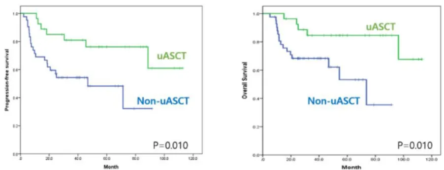
Univariate analyses identified several factors affecting survival outcomes in the all-patient cohort (Table 4). Serum levels of LDH, response to R-CHOP, and upfront ASCT were identified as important clinical factors for predicting PFS and OS. Significant prolongation of PFS (p = 0.010) and OS (p = 0.016) were achieved in patients with high-risk non-GCB DBCL undergoing upfront ASCT (Figure 3). The 5-year PFS rate was significantly better in the upfront ASCT group relative to all other patients (76.2% vs. 48.3%, p = 0.010). Five-year OS rates were 84.6% and 53.2% in the upfront ASCT and non-ASCT groups, respectively (p = 0.010).
Figure 3. Survival outcomes according to upfront autologous stem cell transplantation
16
5. Role of upfront ASCT in subgroups
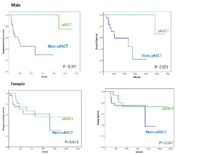
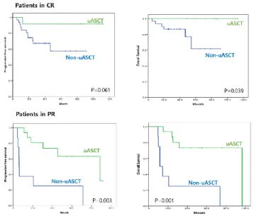
To identify subgroups in whom upfront ASCT is feasible, subgroup analyses were performed. No differences in clinical outcomes were observed based on upfront ASCT status in either the ABC or DEL subgroups. The improvement in survival times associated with upfront ASCT was significantly greater in male patients (Figure 4). Upfront ASCT was beneficial in patients who had response but residual disease (in PR), as well as in patients who achieved CR after R-CHOP (Figure 5). It was also associated with significant OS improvement in patients with higher IPI scores (≥3; p=0.004), but not in those with lower IPI scores (<3; p=0.303).
17
Figure 5. Clinical outcomes according to upfront ASCT status and response to the first line chemotherapy
6. Gene expression profiles and biomarkers associated with survival outcomes
Table 5 shows the expression levels of each gene included in the Lymph2Cx code set, as well as additional genes associated with the BCR signaling pathway, as determined by NanoString. The Lymph2Cx genes specific for the ABC and GCB subtypes were highly expressed in their respective subtypes, as expected. Among the 13 additional genes, there were no differences between the ABC and GCB subtypes, with the exception of IRAK1 (higher in ABC, p = 0.028) and MYC (higher in ABC, p = 0.024).
18
Table 5. Gene expression profiles in patients with non-GCB subtype DLBCL
Category Genes Total (n=59) ABC (n=40) GCB (n=12) P value ABC CCDC50 407±31 479±39 226±37 0.001 ABC CREB3L2 552±47 653±61 270±2 0<001 ABC CYB5R2 422±70 558±96 107±33 <0.001 ABC IRF4 4277±433 4730±3300 3046±2495 0.110 ABC LIMD1 207±21 242±28 103±16 0<001 ABC PIM2 5013±570 6275±764 2005±330 <0.001 ABC RAB7L1 817±86 990±119 397±56 <0.001 ABC TNFRSF13B 578±73 759±96 178±55 0.002 GCB ASB13 90±9 67±6 153±28 0.011 GCB ITPKB 424±38 314±33 641±95 0.006 GCB MAML3 68±8 47±6 130±26 0.009 GCB MME 59±7 51±8 76±17 0.147 GCB MYBL1 61±12 27±5 148±42 0.015 GCB S1PR2 164±32 91±7 360±137 0.076 GCB SERPINA9 128±46 97±60 223±103 0.309 Housekeeping ISY1 151±54 152±8 160±17 0.652 Housekeeping R3HDM1 508±146 522±23 498±38 0.618 Housekeeping TRIM56 644±279 632±35 693±135 0.665 Housekeeping UBXN4 532±129 534±23 527±31 0.880 Housekeeping WDR55 194±76 185±7 175±10 0.461 unknown A20 1361±113 1286±134 1419±258 0.637 unknown Aiolos 1342±73 1316±74 1291±226 0.920 unknown BCL2 726±85 821±118 541±134 0.227 unknown BCL6 954±89 901±120 1057±151 0.510 unknown CRBN 212±18 195±13 196±33 0.961 unknown Ikaros 2170±115 2226±108 1773±201 0.051 unknown IKKbeta 366±16 358±120 395±46 0.408 unknown IL2 23±8 13±4 29±17 0.375 unknown IRAK1 695±36 763±44 566±58 0.028 unknown IRAK4 325±21 310±18 288±27 0.561 unknown IRF7 439±40 396±34 537±128 0.306 unknown MYC 624±41 702±50 462±91 0.024 unknown TP53 270±19 293±29 208±25 0.085 Abbreviations : DLBCL, diffuse large B cell lymphoma; ABC, activated B cell; GCB, germinal center B cell.
19
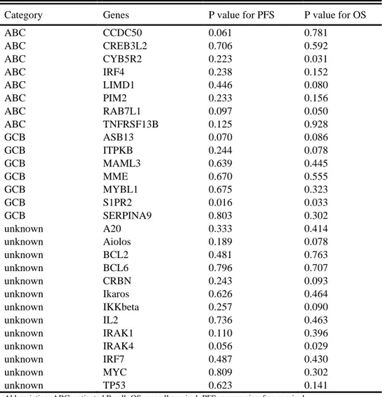
The prognostic value of each of the individual genes was investigated using a Cox proportional hazard model (Table 6). All genes whose expression levels were potentially associated with either PFS or OS (p < 0.1) were included in the analyses, including CCDC50 for PFS; CYB5R2, LIMD1, ITPKB, Aiolos, CRBN, and IKKbeta for OS; and RAB7L1, ASB13, S1PR2, and IRAK4 for both.
Table 6. Univariate analyses of ABC subtype-specific genes associated with survival
Category Genes P value for PFS P value for OS
ABC CCDC50 0.061 0.781 ABC CREB3L2 0.706 0.592 ABC CYB5R2 0.223 0.031 ABC IRF4 0.238 0.152 ABC LIMD1 0.446 0.080 ABC PIM2 0.233 0.156 ABC RAB7L1 0.097 0.050 ABC TNFRSF13B 0.125 0.928 GCB ASB13 0.070 0.086 GCB ITPKB 0.244 0.078 GCB MAML3 0.639 0.445 GCB MME 0.670 0.555 GCB MYBL1 0.675 0.323 GCB S1PR2 0.016 0.033 GCB SERPINA9 0.803 0.302 unknown A20 0.333 0.414 unknown Aiolos 0.189 0.078 unknown BCL2 0.481 0.763 unknown BCL6 0.796 0.707 unknown CRBN 0.243 0.093 unknown Ikaros 0.626 0.464 unknown IKKbeta 0.257 0.090 unknown IL2 0.736 0.463 unknown IRAK1 0.110 0.396 unknown IRAK4 0.056 0.029 unknown IRF7 0.487 0.430 unknown MYC 0.809 0.302 unknown TP53 0.623 0.141
20
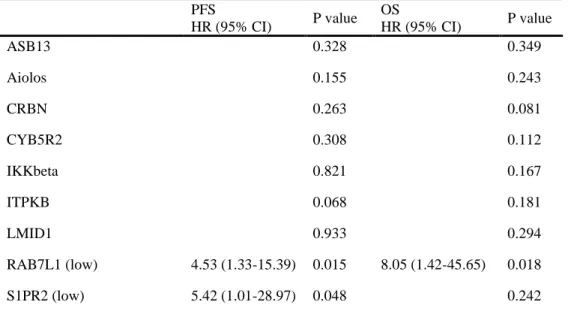
Multivariate analyses of factors affecting survival adjusted for LDH, EOT response to R-CHOP, and upfront ASCT was performed for selected candidate biomarker genes (Table 7). After the adjustment, only RAB7L1 and S1PR2 were revealed as significant prognostic biomarkers for PFS or OS.
Table 7. Multivariate analyses of factors affecting survival adjusted for LDH, EOT response, and upfront ASCT: ABC subgroup
PFS P value OS P value HR (95% CI) HR (95% CI) ASB13 0.328 0.349 Aiolos 0.155 0.243 CRBN 0.263 0.081 CYB5R2 0.308 0.112 IKKbeta 0.821 0.167 ITPKB 0.068 0.181 LMID1 0.933 0.294 RAB7L1 (low) 4.53 (1.33-15.39) 0.015 8.05 (1.42-45.65) 0.018 S1PR2 (low) 5.42 (1.01-28.97) 0.048 0.242 Abbreviations : ASCT, autologous stem cell transplantation; LDH, lactase dehydrogenase; ABC, activated B cell; EOT, end of treatment; PFS, progression-free survival; OS, overall survival; HR, hazard ratio.
The cut-off levels for gene expression that were predictive of lymphoma relapse, progression, or death were calculated using ROC, defining high and low gene expression groups. Low RAB7L1 expression (<641.25) was associated with worse PFS (37.7% vs. 78.0%, p = 0.003) and OS (5-year rate, 45.5% vs. 87.0%, p = 0.004) compared to the high-expression group (Figure 6). Low S1PR2 expression (<78.85) was also associated with worse PFS (5-year rates, 50.0% vs. 68.7%, p = 0.026) and OS (5-year rates, 51.4% vs. 77.3%, p = 0.018; Figure 7). Both ASB13
21
and IRAK4 were associated with PFS and OS in univariate analyses; however, ROC curve analyses revealed a biphasic curve, suggesting that these genes were not appropriate for use as a biomarker.
Figure 6. RAB7L1 expression and survival outcomes in non-GCB type DLBCL
22
Next, we performed subgroup analyses of GEP-confirmed ABC type DLBCL. Low RAB7L1 (p = 0.025 for PFS, p = 0.023 for OS) and S1PR2 (p = 0.008 for PFS, p = 0.021 for OS) expression remained significant prognostic factors for poor outcome in both PFS and for OS; however, the prognostic value of these genes was not significant in non-ABC types.
The results of multivariate analyses of RAB7L1 and S1PR2 expression for survival outcomes are shown in Tables 8 and 9. Low expression of RAB7L1 was an independent biomarker for PFS and OS with a hazard ratio (HR) of 4.53 (95%CI 1.33–15.39, p = 0.015) and 8.05 (95%CI 1.42–45.65, p = 0.018) in high-risk patients with ABC type DLBCL. Low expression of S1PR2 was an independent predictor for PFS with an HR of 5.42 (95% CI 1.01–28.97, p = 0.048), but not OS, in high-risk patients with ABC type DLBCL.
Table 8. Multivariate analyses of factors affecting survival: all patient cohort
PFS P value OS P value HR (95% CI) HR (95% CI) RAB7L1 LDH (x ULN) 1.18 (1.07-1.30) 0.001 1.35 (1.12-1.63) 0.002 Response to therapy (non-CR) 4.74 (1.95-11.53) 0.001 8.19 (2.52-26.57) <0.001 Upfront ASCT (not done) 6.38 (2.34-17.40) <0.001 12.80 (3.37-48.57) <0.001 RAB7L1 (low expression) 2.67 (1.11-6.43) 0.029 0.053 S1PR2
LDH (x ULN) 1.184 0.096
Response to therapy
(non-CR) 4.9 (1.94-12.39) 0.001 9.78 (2.97-32.19) <0.001 Upfront ASCT (not done) 6.83 (2.35-19.78) <0.001 12.94 (3.12-53.64) <0.001
23
Table 9. Multivariate analyses of factors affecting survival: ABC subgroup
PFS P value OS P value HR (95% CI) HR (95% CI) RAB7L1 LDH (x ULN) 1.28 (1.06-1.56) 0.012 1.34 (1.04-1.74) 0.026 Response to therapy (non-CR) 8.98 (1.92-41.94) 0.005 33.84 (4.17-274.88) 0.001 Upfront ASCT (not done) 10.35 (1.97-54.30) 0.006 24.26 (2.97-197.84) 0.003 RAB7L1 (low expression) 4.53 (1.33-15.39) 0.015 8.05 (1.42-45.65) 0.018 S1PR2
LDH (x ULN) 0.158 0.277
Response to therapy
(non-CR) 0.162 8.61 (1.37-54.14) 0.022 Upfront ASCT (not done) 4.67 (1.12-19.37) 0.034 6.11 (1.17-31.94) 0.032 S1PR2 (low expression) 5.42 (1.01-28.97) 0.048 0.242 Abbreviations : ASCT, autologous stem cell transplantation; LDH, lactase dehydrogenase; CR, complete response; PFS, progression-free survival; OS, overall survival; HR, hazard ratio.
IV. DISCUSSION
In this research, I showed that molecular subtyping by digital GEP was possible in most FFPE tissues; however, molecular subtype status was not associated with survival outcomes in the presented patient group.
In terms of the selected patient cohort, we sought to limit our study to a very homogeneous set of patients at high risk for relapse or mortality and who had unmet needs that may be appropriate for risk-adapted therapy. Use of this homogeneous patient group was chosen as a way to minimize confounding factors that could affect survival outcomes and obscure the effect of molecular subtype on survival outcomes.
24
Kim et al. showed that the prognostic impact of non-GCB type DLBCL differed based on the use of upfront ASCT in high-risk DLBCL.21 Patients with non-GCB type DLBCL exhibited worse OS and PFS compared to patients with GCB type disease who did not receive ASCT, with no differences observed in the ASCT group. Although molecular subtyping was limited to IHC only in that study, our results are in line with those findings, showing that COO itself does not have a prognostic impact in patients with high-risk DLBCL who are eligible for ASCT. In Korea, National Health Insurance (NIH) covers upfront ASCT for DLBCL patients with high-risk features at diagnosis, including advanced stage, high serum levels of LDH, and high IPI scores. We adopted criteria similar to that used by the NIH to define high-risk disease, because a physician’s intention to transplant or not might be an important factor in survival analyses.
The benefit of upfront ASCT for aggressive lymphoma has previously been described22 and is now commonly used worldwide. However, upfront ASCT for high-risk DLBCL is still controversial the rituximab era.23, 24 There are still no conclusive data available to guide the use of upfront ASCT for DLBCL after R-CHOP treatment. Although our analyses were limited to a small number of patients, the data do suggest that young, otherwise healthy ABC subtype patients are most likely to benefit from upfront ASCT. The survival gain associated with upfront ASCT was evident not only in patients who achieved a CR after R-CHOP, but also in those who achieved only a PR. Upfront ASCT also overcame higher IPI scores. In addition, upfront ASCT was associated with prolonged survival in male patients, but not in females. This finding that sex is associated with the benefit of upfront ASCT in high-risk DLBCL is consistent with previous publications regarding the unfavorable pharmacokinetics of rituximab in male patients.25, 26 In a prospective randomized phase II trial of different doses and schedules of rituximab with R-CHOP-14 according to sex, Pfreundschuh et al. found that higher rituximab doses for elderly male patients showed similar outcomes and pharmacokinetics to female patients. That clinical trial was limited to elderly
25
DLBCL patients, but this result could explain why upfront ASCT produced more favorable outcomes in male patients. Taken together, these results suggest that inadequate outcomes following R-CHOP therapy in male patients may overcome by using higher-dose chemotherapy during upfront ASCT.
In terms of COO determination, we used both Hans criteria by IHC and Lymph2Cx by digital GEP using NanoString. Microarray GEP approaches are a well-established method for COO typing; however, they are expensive and show poor reproducibility with FFPE containing low-quality RNA samples.27,28 For this reason, GEP-based COO typing has not achieved widespread use in clinical practices.
Most hospitals in Korea have adopted IHC-based methodologies for determining COO due to the availability and cost-effective nature of the assay. The Hans criteria can categorize DLBCLs into GCB and non-GCB types based on the expression of CD10, BCL6, and MUM1 proteins. The concordance rate between Hans criteria and GEP is 80% in the literature.7 Other IHC-based algorithms such as the Choi and the Tally algorithms have been proposed;29, 30 however, such IHC-based methods for COO remain limited due to their poor correlation with GEP, lower accuracy, and limited accuracy in predicting prognosis.7, 30–32
The NanoString assay is less influenced by RNA quality compared to microarray GEP, as it is able to measure mRNA directly using digitally colored code sets attached to gene-specific probes. It has been shown to offer both high sensitivity and reproducibility for mRNA quantification from both frozen and FFPE samples. The NanoString assay can also detect large numbers of genes simultaneously using only a small amount of RNA. Scott et al. used this technology to determine COO and established a COO-interpretation model based on a panel of 20 genes showing the highest reproducibility and predictive power, named Lymph2Cx.8
A validation study performed by a Korean institute showed a discordancy rate of 26.4% between the Hans criteria and Lymph2Cx.33 In our data, we observed a discrepancy of 27.4% for cases categorized as non-GCB by IHC, results that are
26 remarkably similar to that of the previous report.
Improvements in genomic analyses have also enabled us to explore molecular features of DLBCL other than COO. Here, we investigated a set of genes that can predict prognosis in patients with high-risk DLBCL, which were included as part of the NanoString assay. For this analysis, we selected a set of genes that were potentially correlated with survival outcomes, and found that two of them, RAB7L1 and S1PR2, were significant, independent predictive biomarkers for prognosis in high-risk patients with ABC type DLBCL.
Roles in tumorigenesis or prognostic value have not been reported for RAB7L1 in DLBCL to date. RAB7L1 is encoded by the RAB29 gene (1q32.1) and localizes to the lysosomes where it plays an important role in endosomal vascular trafficking.34 It has been actively investigated in Parkinson’s disease.35 Although we found that low expression of the RABL1 gene was correlated with poor survival in high-risk patients with ABC type DLBCL, further validation and mechanism of action studies should be performed.
Little is known about the role of S1PR2 in DLBCL. S1PR2 is a direct repressor of FOXP1, which is transcriptionally regulated by the TGF-beta/TGF-betaR2/SMAD1 axis.36,37 It has been proposed to be a potential regulator of FOXP1 expression in ABC type DLBCL, as its expression is consistently higher than that seen in GCB type DLBCLs. Flori et al. found that DLBCL patients with low S1PR2 and high FOXP1 expression exhibited poorer overall survival compared to the others, although subgroup analyses by COO were not performed.38 Our data are in line with those findings, showing the prognostic value of S1PR2 in DLBCL. In this homogenous study population, we showed the potential prognostic value of S1PR2 as a marker of PFS in ABC type DLBCL compared to other COOs. This result is supported by the data that S1PR2 plays a different role between constitutionally FOXP1-overexpressed ABC type and FOXP1-low GCB type DLBCLs. Therefore, S1PR2 may represent a COO-specific biomarker in ABC type DLBCL. Nonetheless, our findings require further
27
validation due to the limited number of patients and tumor samples included.
V. CONCLUSION
The Lymph2Cx assay effectively discriminate ABC type DLBCLs from non-GCB diseases, however, COO alone was not associated with survival outcomes in transplant-eligible patients with high-risk DLBCL. Upfront ASCT may improve survival in high risk ABC patients in PR as well as CR. Digital GEP identified strong correlations between RAB7L1 and S1PR2 expression levels and disease prognosis, although this requires further validation.
28
REFERENCES
1. Park HJ, Park EH, Jung KW, et al. Statistics of hematologic malignancies in Korea: incidence, prevalence and survival rates from 1999 to 2008. Korean J Hematol 2012;47:28-38.
2. Coiffier B. Rituximab in diffuse large B-cell lymphoma. Clin Adv Hematol Oncol 2004;2:156-157.
3. Kwak JY. Treatment of diffuse large B cell lymphoma. Korean J Intern Med 2012;27:369-377.
4. Ziepert M, Hasenclever D, Kuhnt E, et al. Standard International prognostic index remains a valid predictor of outcome for patients with aggressive CD20+ B-cell lymphoma in the rituximab era. J Clin Oncol 2010;28:2373-2380.
5. Lenz G, Staudt LM. Aggressive lymphomas. N Engl J Med 2010;362:1417-1429.
6. Lenz G, Wright G, Dave SS, et al. Stromal gene signatures in large-B-cell lymphomas. N Engl J Med 2008;359:2313-2323.
7. Hans CP, Weisenburger DD, Greiner TC, et al. Confirmation of the molecular classification of diffuse large B-cell lymphoma by immunohistochemistry using a tissue microarray. Blood 2004;103:275-282.
8. Scott DW, Mottok A, Ennishi D, et al. Prognostic Significance of Diffuse Large B-Cell Lymphoma Cell of Origin Determined by Digital Gene Expression in Formalin-Fixed Paraffin-Embedded Tissue Biopsies. J Clin Oncol 2015;33:2848-2856.
9. Salles G, de Jong D, Xie W, et al. Prognostic significance of immunohistochemical biomarkers in diffuse large B-cell lymphoma: a study from the Lunenburg Lymphoma Biomarker Consortium. Blood 2011;117:7070-7078.
29
10. Horn H, Ziepert M, Becher C, et al. MYC status in concert with BCL2 and BCL6 expression predicts outcome in diffuse large B-cell lymphoma. Blood 2013;121:2253-2263.
11. Johnson NA, Slack GW, Savage KJ, et al. Concurrent expression of MYC and BCL2 in diffuse large B-cell lymphoma treated with rituximab plus cyclophosphamide, doxorubicin, vincristine, and prednisone. J Clin Oncol 2012;30:3452-3459.
12. Wang ML, Rule S, Martin P, et al. Targeting BTK with ibrutinib in relapsed or refractory mantle-cell lymphoma. N Engl J Med 2013;369:507-516.
13. Byrd JC, Furman RR, Coutre SE, et al. Targeting BTK with ibrutinib in relapsed chronic lymphocytic leukemia. N Engl J Med 2013;369:32-42. 14. Davis RE, Ngo VN, Lenz G, et al. Chronic active B-cell-receptor
signalling in diffuse large B-cell lymphoma. Nature 2010;463:88-92. 15. Zheng X, Ding N, Song Y, Feng L, Zhu J. Different sensitivity of
germinal center B cell-like diffuse large B cell lymphoma cells towards ibrutinib treatment. Cancer Cell Int 2014;14:32.
16. Nowakowski GS, LaPlant B, Macon WR, et al. Lenalidomide combined with R-CHOP overcomes negative prognostic impact of non-germinal center B-cell phenotype in newly diagnosed diffuse large B-Cell lymphoma: a phase II study. J Clin Oncol 2015;33:251-257.
17. Yang Y, Shaffer AL, 3rd, Emre NC, et al. Exploiting synthetic lethality for the therapy of ABC diffuse large B cell lymphoma. Cancer Cell 2012;21:723-737.
18. Kronke J, Udeshi ND, Narla A, et al. Lenalidomide causes selective degradation of IKZF1 and IKZF3 in multiple myeloma cells. Science 2014;343:301-305.
19. Lu G, Middleton RE, Sun H, et al. The myeloma drug lenalidomide promotes the cereblon-dependent destruction of Ikaros proteins. Science
30 2014;343:305-309.
20. Wilson WH, Young RM, Schmitz R, et al. Targeting B cell receptor signaling with ibrutinib in diffuse large B cell lymphoma. Nat Med 2015;21:922-926.
21. Kim YR, Kim SJ, Cheong JW et al. The different roles of molecular classification according to upfront autologous stem cell transplantation in advanced-stage diffuse large B cell lymphoma patients with elevated serum lactate dehydrogenase. Ann Hematol 2016;95:1491-1501.
22. Milpied N, Deconinck E, Gaillard F, et al. Initial treatment of aggressive lymphoma with high-dose chemotherapy and autologous stem-cell support. N Engl J Med 2004;350(13):1287–1295.
23. Schmitz N, Nickelsen M, Ziepert M, et al. Conventional chemotherapy (CHOEP-14) with rituximab or high-dose chemotherapy (MegaCHOEP) with rituximab for young, high-risk patients with aggressive B-cell lymphoma: an open-label, randomised, phase 3 trial (DSHNHL 2002-1). Lancet Oncol 2012;13(12):1250–1259.
24. Le Gouill S, Milpied N, Lamy T, et al. First-line rituximab (R) high-dose
therapy (R-HDT) versus R-CHOP 14 for young adults with diffuse large B-cell lymphoma: preliminary results of the GOELAMS 075 prospective multicenter randomized trial. J Clin Oncol 2011;29;15_suppl,8003
25. Muller C, Murawski N, Wiesen, MH, et al. The role of sex and weight on
rituximab clearance and serum elimination halflife in elderly patients with DLBCL. Blood 2012;119, 3276–3284
26. Pfreundschuh M, Murawski N, Zeynalova S, et al. Optimization of
rituximab for the treatment of DLBCL: increasing the dose for elderly
male patients. British J Hematol 2017;179:410-420.
27. Scott DW, Chan FC, Hong F, Rogic S, Tan KL, Meissner B, Ben-Neriah S, Boyle M, Kridel R, Telenius A, Woolcock BW, Farinha P, Fisher RI, et al. Gene expression-based model using formalin-fixed paraffin-embedded
31
biopsies predicts overall survival in advanced-stage classical Hodgkin lymphoma. J Clin Oncol. 2013; 31:692-700.
28. Reis PP, Waldron L, Goswami RS, Xu W, Xuan Y, Perez- Ordonez B, Gullane P, Irish J, Jurisica I, Kamel-Reid S. mRNA transcript quantification in archival samples using multiplexed, color-coded probes. BMC Biotechnol. 2011; 11:46.
29. Rimsza LM, Wright G, Schwartz M, Chan WC, Jaffe ES, Gascoyne RD, Campo E, Rosenwald A, Ott G, Cook JR, Tubbs RR, Braziel RM, Delabie J, et al. Accurate classification of diffuse large B-cell lymphoma into germinal center and activated B-cell subtypes using a nuclease protection assay on formalin-fixed, paraffin-embedded tissues. Clin Cancer Res. 2011; 17:3727-3732.
30. Meyer PN, Fu K, Greiner TC, Smith LM, Delabie J, Gascoyne RD, Ott G, Rosenwald A, Braziel RM, Campo E, Vose JM, Lenz G, Staudt LM, et al. Immunohistochemical methods for predicting cell of origin and survival in patients with diffuse large B-cell lymphoma treated with rituximab. J Clin Oncol. 2011; 29:200-207
31. Fu K, Weisenburger DD, Choi WW, Perry KD, Smith LM, Shi X, Hans CP, Greiner TC, Bierman PJ, Bociek RG, Armitage JO, Chan WC, Vose JM. Addition of rituximab to standard chemotherapy improves the survival of both the germinal center B-cell-like and non-germinal center B-cell-like subtypes of diffuse large B-cell lymphoma. J Clin Oncol. 2008; 26:4587-4594.
32. Gleeson M, Hawkes EA, Cunningham D, Jack A, Linch D. Caution in the Use of Immunohistochemistry for Determination of Cell of Origin in Diffuse Large B-Cell Lymphoma. J Clin Oncol. 2015; 33:3215-3216. 33. Yoon N, Ahn S, Yoo HY et al. Cell-of-Origin of diffuse large B-cell
lymphomas determined by the Lymph2Cx assay: better prognostic indicator than Hans algorithm. Oncotarget, 2017;8(13):22014-22.
32
34. Kuwahara T, Inoue K, D’Agati VD et al. LRRK2 and RAB7L1 coordinately regulate axonal morphology and lysosome integrity in diverse cellular contexts. Sci Rep 2016;6:29945.
35. Chi MM, Che CH, Xu YM. Rab GTPases: The Key Players in the Molecular Pathway of Parkinson's Disease. Front Cell Neurosci 2017;11:81.
36. Stelling A, Hashwah H, Bertram K et al. The tumor suppressive TGF-β/SMAD1/S1PR2 signaling axis is recurrently inactivated in diffuse large B-cell lymphoma. Blood 2018;131(20):2235-46.
37. Bladari CT et al. S1PR2 deficiency in DLBCL: a FOXy connection. Blood 2016;127(11):1380-1.
38. Flori M, Schmid CA, Sumrall ET al. The hematopoietic oncoprotein FOXP1 promotes tumor cell survival in diffuse large B-cell lymphoma by repressing S1PR2 signaling. Blood 2016;127(11):1438-48.
33
ABSTRACT(IN KOREAN)
활성화 B세포 형태의 광범위 큰 B세포 림프종에서 B세포 수용체
신호전달 억제에 대한 예측 생체지표 연구
<지도교수 김 진 석>
연세대학교 대학원 의학과
이 혜 원
연구 배경: 광범위큰B세포림프종(diffuse large B-cell lymphoma,
DLBCL)은 국내 악성림프종 중 가장 흔한 형태이며, 유전자 발현
프로파일링 (gene expression profiling, GEP) 양상에 따라 분자적
세부 아형으로 나뉜다. 이는 세포의 기원 (cell-of-origin, COO)에
기반한 유전자 발현 양상에 따라, 크게 germinal center B cell (GCB)
타입과 activated B-cell (ABC) 타입으로 분류된다. ABC 타입
DBLCL은 특징적인 유전자 발현 과 함께 상대적으로 불량한
예후를 보이는 것으로 알려져 있다. ABC 타입을 구분해내기
위해서 임상 현장에서는 대리 표지자로서 CD10, BCL6, MUM1을
이용한 면역조직화학염색 (immunohistochemistry, IHC)이 널리
사용되고 있으며, 최근에는 보다 정밀하고 활용도가 높은 디지털
GEP가 도입되었다. 본 연구에서는 IHC를 통해 non-GCB로 분류된
고위험군 DLBCL 환자를 대상으로 digital GEP를 수행하여
세포기원의 임상적 중요성을 확인하고자 하였다. 동시에 본
대상군에서 유의한 유전자적 표지자를 탐색해보고자 하였다.
연구 방법: 본 연구에서는 디지털 GEP를 위해 NanoString 및
nCounter 분석법을 활용하였으며, 종양검체는 포르말린 고정
34
파라핀 포매 (FFPE) 조직 블록을 이용하였다. 대상 환자군은
2008년 1월에서 2015년 12월 사이에 세브란스병원에서 DLBCL로
확진된 환자 중, 이식이 가능하고 임상적으로 고위험 요소를 가진
non-GCB
DLBCL
환자로 규정하고 의무기록 검토를 통해
선별하였다. 디지털 GEP에 의한 세포기원 결정은 20개 유전자에
기반하여 개발된 Lymph2Cx 분석 알고리즘에 따라 이루어졌다.
Nanostring 분석을 시행하면서, 세포기원 결정을 위한 20개 유전자
외에 추가적으로 ABC 타입 DLBCL과 관련이 있는 것으로 알려진
유전자 13개의 발현 정도를 함께 측정하였다.
연구 결과: 총 69명의 non-GCB DLBCL 환자가 본 연구를 위해
선별되었으며,
이들은
높은
병기
(95.7%),
3점
이상의
국제예후지표 (International Prognostic Index, IPI) 점수 (63.8%), 높은
젖산탈수소효소 (lactase dehydrogenase, LDH) (65.2%)로 인해
고위험군으로 분류되었다. 관찰기간 중앙값은 52.1개월이었으며,
환자들은 6-8주기의 표준 면역항암화학요법을 시행 받았다.
선제적 자가 조혈모세포이식은 치료반응이 있었던 27명에게
시행되었다. 혈청 LDH, 치료 반응, 선제적 자가 조혈모세포이식은
생존기간에
중요한
요인으로
파악되었으며,
자가
조혈모세포이식을 통한 이득은 여자보다 남자에서 명확하였다.
NanoString 분석은 59건의 검체에서 시행되었고, 그 중 19건
(27.3%)에서 GCB 또는 unclassifiable 타입으로 재분류되어, IHC
기반 non-GCB 분류와 불일치를 보였다. 그러나, 재분류된
세포기원 자체가 생존기간에 영향을 주지는 않았다.
NanoString 분석법으로 측정된 유전자 발현도의 예후인자로서의
가치는
COX
proportional
hazard
model을
이용하여
우선
선별하였으며, p value <0.1 기준으로 하였을 때 무진행생존기간에
대하여 CCDC50. 전체생존기간에 대하여 CYB5R2, LIMD1, ITPKB,
Aiolos, CRBN, IKKbeta, 그리고, 두 가지 생존기간 모두에 대하여
RAB7L1, ASB13, S1PR2, IRAK4가 선택되었다. 이 중, RAB7L1
발현이 낮은 (기준값 641.25) 환자는 발현이 높은 군에 비하여
35