Laparoscopic Ultrasonography–Assisted
Retroperitoneal Lymph Node Sampling
in Patients Evaluated for Stomach
Cancer Recurrence
Honsoul Kim, MD, Woo Jin Hyung, MD, Joon Seok Lim, MD, Mi-Suk Park, MD, Jin Young Choi, MD, Yong Eun Chung, MD, Myeong-Jin Kim, MD, Ki Whang Kim, MD
Objective. The precise localization of target lymph nodes during laparoscopic biopsy can often be technically challenging. The purpose of this series was to report 2 patients who successfully underwent laparoscopic para-aortic lymph node sampling with the assistance of laparoscopic ultrasonography (LUS). Methods. We describe 2 patients previously treated for gastric cancer who later had suspicious retroperitoneal lymphadenopathy detected by computed tomography. They received LUS-assisted lymph node biopsies for histologic evaluation. Results. Laparoscopic ultrasonography precisely localized the target lymph nodes, which measured 1.4 and 1.5 cm, respectively, in both patients. A transperi-toneal laparoscopic approach and target lymph node sampling were successfully performed. No pro-cedure-related technical difficulties or immediate complications occurred. The 2 patients were histologically proven to have nodal tumor recurrence and tuberculous lymphadenopathy, respectively. Conclusions. Precise localization of a small lymph node located deep in the abdomen can be readily performed by intraoperative LUS. Key words: laparoscopic biopsy; laparoscopic surgery; laparoscopic ultrasonography; lymph node recurrence; stomach cancer.
Received March 4, 2008, from the Department of Diagnostic Radiology, Research Institute of Radiological Science (H.K., J.S.L., M.-S.P., J.Y.C., Y.E.C., M.-J.K., K.W.K.), Department of Surgery (W.J.H.), and Institute of Gastroenterology (J.S.L.), Severance Hospital, Yonsei University College of Medicine, Seoul, Korea. Revision requested March 31, 2008. Revised manuscript accepted for publica-tion April 14, 2008.
Address correspondence to Joon Seok Lim, MD, Department of Diagnostic Radiology, Severance Hospital, Seodaemun-ku, Shinchon-dong 134, Seoul 120-752, Korea.
E-mail: jslim1@yuhs.ac Abbreviations
CT, computed tomography; FDG, fluorodeoxyglucose; LUS, laparoscopic ultrasonography; PET, positron emis-sion tomography
aparoscopic biopsy is a safe and effective method that is frequently performed. High diagnostic accuracy and minimal invasiveness have made laparoscopic biopsy an attractive method,1,2 espe-cially for lymph node sampling in patients requiring eval-uation of malignant lymph node metastasis who cannot tolerate invasive laparotomy for diagnostic purposes. Accurate localization of the target lymph nodes for laparoscopic biopsy can entail a challenging and time-consuming procedure, particularly in cases with target nodes buried in the deep compartments of the abdomen. Recently, laparoscopic ultrasonography (LUS) has been used to facilitate the localization of lesions in laparoscop-ic surgeries of various fields.3–6
We report 2 patients with a history of gastric cancer who received LUS-assisted retroperitoneal lymph node sam-pling for histopathologic diagnosis of suspected lymph node tumor recurrence.
Case Descriptions
Case 1
A 57-year-old man was admitted to our institu-tion for evaluainstitu-tion of enlarged retroperitoneal lymph nodes detected by computed tomography (CT). He had undergone a radical total gastrecto-my for stomach cancer 36 months previously, and histopathologic evaluation showed moderately differentiated adenocarcinoma extending to peri-gastric fat tissue. Sixty-six of 127 dissected lymph nodes were reported to have positive findings for metastatic carcinoma (T3N3M0, stage IV). After surgery, he completed 6 cycles of chemotherapy with an irinotecan/cisplatin regimen.
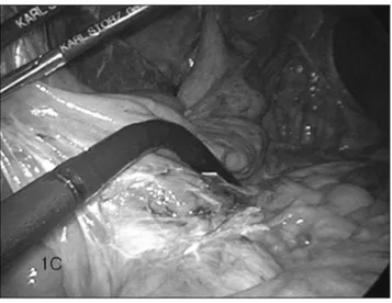
No evidence of tumor recurrence was detected for 3 years after surgery according to abdominal CT performed every 6 months, endoscopy per-formed annually, and positron emission tomog-raphy (PET) performed 2 years after surgery. Routine follow-up CT performed 36 months after surgery showed 2 questionable lymph nodes measuring 1.4 and 0.9 cm, each located in the left para-aortic region inferior to the left renal vein and the aortocaval region posterior to the third portion of the duodenum, respectively. These lymph nodes were thought to be growing because they had measured 1.1 and 0.4 cm on CT performed 6 months previously, further raising the suspicion of tumor recurrence. A faint low-density portion suspicious for necrosis was observed in 1 lymph node, suggestive of malig-nancy (Figure 1A). Subsequent PET and CT showed increased fluorodeoxyglucose (FDG) uptake corresponding to the locations of the enlarged lymph nodes, increasing the suspicion of tumor recurrence (Figure 1B), but no evidence of abnormal FDG uptake was observed else-where. We decided to sample the enlarged left para-aortic lymph node, and laparoscopic lymph node biopsy under LUS guidance was performed (Figure 1, C–E).
Case 2
A 68-year-old man was admitted to our institu-tion for further evaluainstitu-tion of multiple enlarged para-aortic and retroperitoneal lymph nodes. He denied any medical history other than pul-monary tuberculosis, which had been cured by medication. He had undergone a subtotal
gas-trectomy for stomach cancer 29 years previously and then had a second operation (total radical gastrectomy) for locally recurring stomach can-cer (pathologically reported as well-differentiated adenocarcinoma, TisN0M0, stage Ia) 33 months previously.
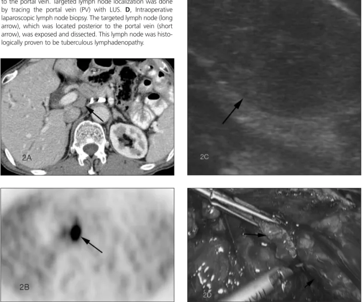
Abdominal CT performed 12 months after the second operation showed multiple enlarged aor-tocaval, para-aortic, and retroperitoneal lymph nodes suspicious for tumor recurrence (Figure 2A). The largest lymph node was located at the posterior part of portal vein, measured 1.5 cm, and had apparently increased in size from 0.9 cm according to a CT scan performed 6 months pre-viously. Subsequent PET (Figure 2B) revealed mul-tiple lesions with intense FDG uptake on the multiple retroperitoneal lymph nodes, lung nod-ules, and hilar lymph nodes, suggesting widespread metastasis. We decided to sample the lymph node located at the posterior part of the portal vein, and laparoscopic lymph node biopsy under LUS guid-ance was performed (Figure 2, C and D).
Procedure
Both patients underwent laparoscopic lymph node biopsy for histologic examination. The surgical procedure was as follows. Under general anesthesia, the patient was placed in the Trendelenburg position with both legs elevated about 15°. The surgeon operated on the patient’s right side; a camera operator was also on the patient’s right side just to the right of the opera-tor; and an assistant surgeon stood on the patient’s left side. After pneumoperitoneum was established by the open technique, 4 ports (12, 10, 5, and 5 mm in diameter) were placed. After the trocars were inserted, a laparoscopic probe with a flexible tip fitted with a 7.5-MHz linear transducer (Aloka Co, Ltd, Tokyo, Japan) was introduced through the right paramedian 12-mm port.
In case 1, first the aorta was visualized and traced by LUS to identify the left renal vein lying next to the aorta (Figure 1C). Then the target lymph node was successfully visualized inferior to the left renal vein as revealed on CT (Figure 1D). In case 2, the portal vein was traced by LUS, and the target lymph node shown on CT was successfully visualized posterior to the portal vein (Figure 2C). Once the location of the
target-Figure 1. Case 1. A, Coronal plane portal venous phase CT shows an enlarged left para-aortic lymph node (long arrow) infe-rior to the left renal vein (arrowhead) measuring 1.4 cm in diam-eter with faint internal low density (short black arrow) suspicious for necrosis. Another lymph node (short white arrow) borderline in size at the aortocaval region is also shown. B, Positron emis-sion tomography shows 2 focal leemis-sions with intense FDG uptake at the locations corresponding to the suspicious lymph nodes (arrows) detected by CT. C, Intraoperative LUS. The ultrasound probe was manipulated to first identify the aorta, the left renal vein, and then the targeted retroperitoneal lymph node located just inferior to the left renal vein. D, Sagittal plane of the target-ed lymph node (arrow) visualiztarget-ed as a hypoechoic nodule by LUS. E, The peritoneal layers were dissected to expose the tar-geted lymph node (long arrow) located posterior to the left renal vein. Note the contour of the aorta (short arrow) beside the tar-geted lymph node. Microscopic examination of this lymph node revealed metastatic adenocarcinoma, which probably originated from the previously treated stomach cancer.
ed retroperitoneal lymph node was marked in each patient, a straightforward transperitoneal approach with minimal tissue dissection was attempted. The dissection was advanced into the retroperitoneal compartment without difficulty, and the targeted retroperitoneal lymph node was successfully retrieved (Figures 1E and 2D). After retrieval of the targeted lymph node was com-pleted, postbiopsy LUS was performed to ensure the adequacy of the procedure. No immediate complications occurred.
Histologic Results
The lymph node sampled from case 1 was histo-logically proven to be metastatic adenocarcino-ma, which was assumed to have originated from the patient’s previous stomach cancer. He resumed chemotherapy. Histologic review of the sampled lymph nodes from case 2 revealed chronic granulomatous inflammation consistent with tuberculosis. The remaining enlarged lymph nodes were found to have decreased in size on CT performed 6 months after the patient received antituberculosis medication.
Figure 2. Case 2. A, Transverse plane portal venous phase CT scan shows an enlarged lymph node (arrow) located at the pos-terior part of the portal vein measuring approximately 1.5 cm. B, Transverse plane PET shows intense FDG uptake (arrow) at the location corresponding to the enlarged lymph node posteri-or to the pposteri-ortal vein detected by CT. C, Transverse plane of an enlarged hypoechoic lymph node (arrows) visualized posterior to the portal vein. Targeted lymph node localization was done by tracing the portal vein (PV) with LUS. D, Intraoperative laparoscopic lymph node biopsy. The targeted lymph node (long arrow), which was located posterior to the portal vein (short arrow), was exposed and dissected. This lymph node was histo-logically proven to be tuberculous lymphadenopathy.
Discussion
Evaluation of lymph node status is essential for postoperative patients treated for any kind of malignancy. It was particularly important for the 2 patients in this study because locoregional lymph nodes are well known as major sites of gastric cancer recurrence.7We generally suspect tumor recurrence in cross-sectional imaging studies if a lymph node has a short-axis diameter of greater than 1 cm, a round shape, or an increase in size during serial studies.8The accuracy in pre-dicting lymph node metastasis solely on the basis of radiologic evaluation is far from satisfac-tory, however, and pathologic evidence support-ing tumor recurrence is indispensable. Recently, PET scanning has been widely applied for eval-uation of metastatic lymph nodes, but positive and -negative results remain issues.8,9 Nontumorous lesions with intense FDG uptake on PET such as tuberculous lymphadenopathy can mimic tumorous conditions; thus, the histo-logic nature of the lesion must be clarified before initiating aggressive treatment.
Percutaneous biopsy guided by CT or ultra-sonography could be a reasonable means of per-forming histologic examinations, producing an acceptable diagnostic yield with low morbidi-ty.1,2At the same time, percutaneous sampling of a small lymph node located in the deep abdomen can be technically difficult and some-times even risky. In such conditions, laparo-scopic biopsy may serve as a reliable, minimally invasive alternative.1,2If the lymph node is large enough and displaces the adjacent organs, no additional guidance for lesion localization would be necessary for laparoscopic biopsy. If the targeted lymph node is small (1.4 and 1.5 cm on preoperative CT in this study), however, or lies in the deep compartments of the abdomen, localization of the node itself can become a challenging task. Wide dissection of normal tissue might be necessary merely to achieve an approach route and expose the tar-geted lymph nodes. In such cases, precise local-ization of the target lymph nodes could instead be accomplished with the assistance of LUS, pre-sumably reducing excessive tissue dissection and visceral manipulation and minimizing tis-sue injury and operation time.
In summary, precise localization of small lymph nodes in the deep compartments of the abdomen can be readily performed by intraop-erative LUS. Precise LUS localization of a target-ed lymph node may facilitate laparoscopic biopsy.
References
1. Mortensen MB, Durup J, Pless T, et al. Initial experience with new dedicated needles for laparoscopic ultrasound-guided fine-needle aspiration and histological biopsies. Endoscopy 2001; 33:585–589.
2. Casaccia M, Torelli P, Cavaliere D, et al. Laparoscopic lymph node biopsy in intra-abdominal lymphoma: high diagnos-tic accuracy achieved with a minimally invasive procedure. Surg Laparosc Endosc Percutan Tech 2007; 17:175–178. 3. Bezzi M, Merlino P, Orsi F, et al. Laparoscopic sonography
during abdominal laparoscopic surgery: technique and imaging findings. AJR Am J Roentgenol 1995; 165:1193– 1198.
4. Hyung WJ, Lim JS, Kim TI, Kim WH, Noh SH. Minimally invasive treatment of obscure gastrointestinal bleeding using laparoscopic ultrasonography. Surg Laparosc Endosc Percutan Tech 2007; 17:325–327.
5. Lee SW, Lim JS, Hyung WJ, et al. Laparoscopic ultrasonog-raphy for localization of a retained appendicolith after appendectomy. J Ultrasound Med 2006; 25:1361–1363. 6. Montorsi M, Santambrogio R, Bianchi P, et al. Laparoscopy
with laparoscopic ultrasound for pretreatment staging of hepatocellular carcinoma: a prospective study. J Gastrointest Surg 2001; 5:312–315.
7. D’Angelica M, Gonen M, Brennan MF, Turnbull AD, Bains M, Karpeh MS. Patterns of initial recurrence in completely resected gastric adenocarcinoma. Ann Surg 2004; 240: 808–816.
8. Torabi M, Aquino SL, Harisinghani MG. Current concepts in lymph node imaging. J Nucl Med 2004; 45:1509–1518. 9. Veit P, Ruehm S, Kuehl H, et al. Lymph node staging with dual-modality PET/CT: enhancing the diagnostic accuracy in oncology. Eur J Radiol 2006; 58:383–389.