Agmatine Improves Cognitive Dysfunction and Prevents Cell
Death in a Streptozotocin-Induced Alzheimer Rat Model
Juhyun Song,
1Bo Eun Hur,
1,2Kiran Kumar Bokara,
1Wonsuk Yang,
1Hyun Jin Cho,
1Kyung Ah Park,
1Won Taek Lee,
1Kyoung Min Lee,
3and Jong Eun Lee
1,2 1Department of Anatomy, Yonsei University College of Medicine, Seoul;2Brain Korea 21 Plus Project for Medical Science, Brain Research Institute, Yonsei University College of Medicine, Seoul; 3Department of Neurology, Seoul National University College of Medicine, Seoul, Korea.
Received: December 18, 2013 Revised: February 18, 2014 Accepted: February 18, 2014
Corresponding author: Dr. Jong Eun Lee, Department of Anatomy,
Yonsei University College of Medicine, Brain Korea 21 Plus Project for Medical Science, Brain Research Institute, Yonsei University College of Medicine, 50-1 Yonsei- ro, Seodaemun-gu, Seoul 120-752, Korea.
Tel: 82-2-2228-1646, Fax: 82-2-365-0700 E-mail: jelee@yuhs.ac
∙ The authors have no financial conflicts of interest.
© Copyright:
Yonsei University College of Medicine 2014
This is an Open Access article distributed under the terms of the Creative Commons Attribution Non-Commercial License (http://creativecommons.org/ licenses/by-nc/3.0) which permits unrestricted non-commercial use, distribution, and reproduction in any medium, provided the original work is properly cited.
Purpose: Alzheimer’s disease (AD) results in memory impairment and neuronal cell death in the brain. Previous studies demonstrated that intracerebroventricular administration of streptozotocin (STZ) induces pathological and behavioral altera-tions similar to those observed in AD. Agmatine (Agm) has been shown to exert neuroprotective effects in central nervous system disorders. In this study, we inves-tigated whether Agm treatment could attenuate apoptosis and improve cognitive decline in a STZ-induced Alzheimer rat model. Materials and Methods: We studied the effect of Agm on AD pathology using a STZ-induced Alzheimer rat model. For each experiment, rats were given anesthesia (chloral hydrate 300 mg/ kg, ip), followed by a single injection of STZ (1.5 mg/kg) bilaterally into each lat-eral ventricle (5 μL/ventricle). Rats were injected with Agm (100 mg/kg) daily up to two weeks from the surgery day. Results: Agm suppressed the accumulation of amyloid beta and enhanced insulin signal transduction in STZ-induced Alzheimer rats [experimetal control (EC) group]. Upon evaluation of cognitive function by Morris water maze testing, significant improvement of learning and memory dys-function in the STZ-Agm group was observed compared with the EC group. West-ern blot results revealed significant attenuation of the protein expressions of cleaved caspase-3 and Bax, as well as increases in the protein expressions of Bcl2, PI3K, Nrf2, and γ-glutamyl cysteine synthetase, in the STZ-Agm group. Conclu-sion: Our results showed that Agm is involved in the activation of antioxidant sig-naling pathways and activation of insulin signal transduction. Accordingly, Agm may be a promising therapeutic agent for improving cognitive decline and attenu-ating apoptosis in AD.
Key Words: Agmatine, streptozotocin, Alzheimer’s disease, cognitive dysfunc-tion, apoptosis, insulin signal transduction
INTRODUCTION
Alzheimer’s disease (AD) is the most common form of dementia and is a neurode-generative disorder characterized by the degeneration of neurons, as well as by the
in improving cognitive dysfunction and preventing cell death in a STZ-induced Alzheimer rat model.
MATERIALS AND METHODS
Animal model
Male Sprague-Dawley rats (n=50, weighing 250-330 g) were used in this study. Rats were maintained under controlled hy-gienic conditions with a 12 hr light/dark reverse cycle at a constant temperature with free access to food and water. All animal experiments were performed in accordance with the Korean Food and Drug Administration guidelines. Protocols were reviewed and approved by the Institutional Animal Care and Use Committee of the Yonsei Laboratory Animal Research Center (Permit #: 10-115).
Drug treatments
For each experiment, rats were given anesthesia [chloral hy-drate 300 mg/kg, intraperitoneal (i.p)], followed by a single injection of STZ (1.5 mg/kg, dissolved in a vehicle consist-ing of 0.05 M citrate buffer) bilaterally into each lateral ven-tricle (5 μL/venven-tricle). Control animals were given an equal volume of intracerebroventricular (icv) vehicle via the same procedure. Agm was purchased from Sigma (Sigma, St. Louis, MO, USA), dissolved in normal saline (pH 7.4), and administered to rats via an intra-peritoneal route. Rats were injected with Agm (100 mg/kg) daily for up to two weeks after surgery. The concentration of Agm (100 mg/kg) was determined based upon the results of our previous study.25
Experimental design
The sham group (n=15) received bilateral icv injection of saline, 5 μL in each rat. The rats in the EC group (n=17) were given injection of STZ-icv (1.5 mg/kg, 1st and 3rd days after surgery) (Sigma, St. Louis, MO, USA) bilaterally.40 Rats in
the STZ-Agm group (n=18) were given 5 uL injection of STZ-icv (1.5 mg/kg, 1st and 3rd days after surgery) (Sig-ma, St. Louis, MO, USA) bilaterally and treated with Agm [100 mg/kg, intraperitoneal (i.p.) daily for two weeks from the 1st day after surgery]. The heads of the rats were posi-tioned in a stereotactic frame (coordinates of 1.5 mm poste-rior to the bregma, 1.5 mm lateral to the sagittal suture, 2.5 mm ventral to the surface of the brain).
Cognition assessment tests
Cognitive evaluation of rats was tested using a Morris wa-progressive decline of cognitive function. AD exhibits the
hallmarks of both senile plaques derived from amyloid beta (Aβ) and neurofibrillary tangles, especially in the hippocam-pus or cerebral cortex, relevant to learning and memory.1,2
When Aβ exists in high concentrations, it forms insoluble and fibrillar Aβ plaques, which activate ion channels in the cell membrane to induce membrane depolarization and de-stabilization of intracellular calcium homeostasis.3-5 In
partic-ular, Aβ oligomers cause intracellular Ca2+ overload, leading
to neuronal death, which can be prevented by N-methyl-D-aspartate (NMDA) receptor antagonists.6,7
Streptozotocin (STZ) is a glucosamine-nitrosourea com-pound that, when metabolized, generates a cytotoxic prod-uct that preferentially destroys pancreatic β cells.8 The
alkyl-ating properties of STZ metabolites generate reactive oxygen species and cause oxidative stress.8 Previously,
in-tracerebroventricular streptozotocin (STZ-icv) administra-tion was shown to induce oxidative stress,9-11 neuronal cell
damage,12,13 and dysfunctions in learning and memory.8,14,15
Accordingly, STZ-icv models have been used to assess the therapeutic potential of various drugs, as well as other non-drug therapeutic strategies.16 Additionally, STZ-induced
learning and memory dysfunction is associated with oxida-tive stress in animal models.17 Therefore, to attenuate the
death of neuronal cells caused by a variety of neurodegener-ative diseases, antioxidants have been spotlighted in poten-tial treatments of neurodegenerative diseases such as AD.17,18
Agmatine (Agm) is an endogenous peptide synthesized by arginine decarboxylase, and is reported to be present in glia and neuronal cells. Several researchers have investigated the potential of Agm to improve cognitive function and neuronal cell death in various animal models.19-23 Agm, a
neurotrans-mitter or neuromodulator, exerts neuroprotective effects in various central nervous system injury models, including neu-rotrauma and neonatal ischemia animal models. 24-28 Agm, as
an NMDA receptor antagonist, plays a crucial role in regu-lating the production of nitric oxide (NO). Since NO can enhance a cell’s survival rate under oxidative stress, Agm also could protect against damage to cells under oxidative stress.29,30 As well, nuclear factor-erythroid 2 related factor
2 (Nrf2) protects the cell against various stresses and regu-lates the expression of antioxidant genes, including super-oxide dismutase, NAD(P)H, and γ-GCS.31-36 Previous
stud-ies demonstrated that Nrf2 is related to cognitive decline.37-39
Accordingly, this study attempted to investigate whether Agm promotes Nrf2 mediated antioxidant signaling. In par-ticular, we aimed to determine the potential benefits of Agm
IRS-1) (1:200, Santa Cruz, CA, USA), 8-oxo-2’-deoxy-guanosine (8-OHdG) (1:200, Chemicon, Billerica, MA, USA), and anti-Aβ (1:200, Millipore, Billerica, MA, USA) were applied to the samples for 24 hours at 4°C, followed by a 90-minute incubation with appropriate florescence sec-ondary antibody (1:100, Invitrogen, Carlsbad, CA, USA) and three washings in PBS for 10 minutes each. After three washes in 0.1% phosphate buffered saline with Tween-20 (PBST), the sections were incubated with rhodamine-con-jugated sheep anti-rabbit or sheep anti-mouse secondary antibody that was diluted to 1:200 with 5% BSA fraction V in 0.1% PBST for 2 h in the dark at room temperature. Af-ter three washing in PBS, all sections were incubated with 1 μg/mL of 4’,6-diamidino-2-phenylindole (Sigma-Aldrich, Sigma, St. Louis, MO, USA) and 2 μg/mL of propidium io-dide (Sigma-Aldrich, USA) for a counter staining. Tissues were then visualized under a confocal microscope (Zeiss LSM 700, Carl Zeiss, Thornwood, NY, USA).
Western blot analysis
For Western blot analysis, rats from all groups were sacri-ficed and their brains were perfused with saline through the heart aorta to rinse away traces of blood. Next, the portion of the brain comprising the hippocampus and cortex was dissected for extraction of proteins and treated with lysis buffer (1% Triton X-100, 0.5% NP-40, 150 mM NaCl, 10 mM Tris (pH 7.4), 1 mM Ethylene-di-amine tetra acetic acid, 1 mM ethylene glycol tetraacetic acid (pH 8.0), 0.2 mM sodium orthovanadate, 0.2 mM phenyl methyl sulfo-nyl fluoride, and protease inhibitor cocktail). Isolated pro-teins were centrifuged at 12000 rpm at 4°C. Equal amounts of protein (20 μg) from the supernatants were separated on a 10% acrylamide gel and proteins were electrophoretically transferred onto nitrocellulose membranes. After blocking with 5% skim milk for 2 hrs, the membranes were incubated with primary antibodies against Bcl2 (1:1000, Santa Cruz, San Jose, CA, USA), Bax (1:1000, Santa Cruz, San Jose, CA, USA), cleaved caspase-3 (1:1000, Cell signaling, Bill-erica, MA, USA), PI3K (1:2000, Millipore, BillBill-erica, MA, USA), Nrf2 (1:200, Santa Cruz, San Jose, CA, USA), γ-GCS (1:500, Thermo Scientific, Bremen, Germany), and β-actin (1:1000, Millipore, Billerica, MA, USA) at 4°C overnight. Later the membranes were washed three times for 5 min each with TBST. The detection of secondary anti-rabbit and anti-mouse antibodies (1:3000, New England Bio labs, USA) was conducted for 1 hour at room temperature. After washing with PBST (0.05% with Tween 20) three times, ter maze.41 Before conducting the Morris water maze test,
we conducted pre-training. For all trials, a single rat was placed in a pool, facing the wall, at a different starting point (NW, NE, SW, or SE), and was then allowed to swim for a maximum of 60 seconds or until it reached the platform. If the rat failed to find the platform during the trial, it was manually guided to the platform by the investigator and placed on top of it for 15 seconds. Next, an apparatus con-sisting of a circular pool (200 cm diameter, 60 cm high) filled with water (depth 30 cm; 24±1°C) was placed in a room with consistently positioned spatial cues. An escape platform (15 cm diameter) was placed in the middle of one of the quadrants, 1.5 cm below the water surface, equidis-tant from the sidewall and the middle of the pool. The time required to reach the platform (escape latency) was mea-sured in each trial. After the acquisition stage, a probe test was performed after removing the platform. We measured the latency up to the point when the rat traversed the loca-tion where the platform used to be as a measure of memory.
Hematoxylin and eosin (H&E) staining
Rats were anesthetized and perfused as previously men-tioned.42 Rat brains were cut into coronal slices of 2 mm
thickness using a rat brain matrix (Ted Pella, Redding, CA, USA). Next, the brain slices were fixed with 4% parafor-maldehyde (pH 7.4) for 3 days and subsequently embedded in a paraffin block. The paraffin blocks containing the hip-pocampus and cortex were deparaffinized and re-hydrated with different concentrations of alcohol and xylene. After hematoxylin and eosin (H&E) staining, stained hippocam-pus and cortex sections were examined under a microscope.
Immunohistochemistry
Five μm thick frozen brain sections were harvested onto clean glass slides (Thermo Scientific, Bremen, Germany), air-dried, and fixed in cold acetone for 10 minutes at -20°C. The slides were washed first in Tris-buffered saline (TBS) and then incubated with 0.3% H2O2 in methanol to quench
endogenous peroxidase activity. Followed by a series of washes (three times with distilled water), the sections were blocked with 10% normal rabbit serum. Frozen brain sec-tions (20 μm) were fixed in ice-cold acetone for 20 minutes. To block specific staining, sections were incubated in 5% bovine serum albumin (BSA) (Sigma-Aldrich, Sigma, St. Louis, MO, USA) diluted in PBS for 30 minutes before ad-dition of primary and secondary antibodies. Primary anti-bodies for phosphorylated insulin receptor substrate-1
(p-Our findings suggest that Agm treatment could improve learning and memory dysfunction in STZ-induced Al-zheimer rats.
Agmatine treatment attenuates histological
abnormalities in hippocampus and cortex regions of STZ-icv rats
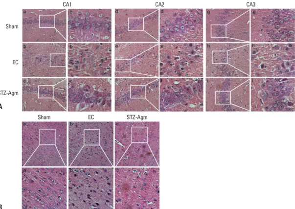
To investigate morphological differences in cells, we con-ducted H&E staining. The H&E staining of Cornu Ammo-nis (CA)1, CA2, and CA3 regions of the hippocampus re-vealed a large number of degenerated cells in the EC group compared with STZ-Agm group (Fig. 2A), which were evi-denced by a decrease in the number of H&E stained cells. The EC group exhibited greater shrinkage of the nuclei of cells than those in the STZ-Agm group. Fig. 2B shows that the number of abnormal cells in the cortical region were higher in the EC group than the STZ-Agm group. Fig. 2 in-dicates that Agm treatment reduces STZ-induced histologi-cal abnormalities in hippocampus and cortex regions, com-pared with the EC group.
Agmatine treatment inhibits the cell death pathway in STZ-icv rats
To confirm apoptotic cell death in the EC group and the STZ-Agm group, western blot analysis was conducted to check expression of apoptotic proteins. Fig. 3A indicates that the presence of cleaved caspase-3, an active form of caspase-3, was attenuated in the STZ-Agm group com-pared with the EC group. Additionally, in the present study, the anti-apoptotic effect of Agm was investigated by check-ing the expression of proteins, such as Bax, Bcl2, and PI3K, using western blotting. Among the quantitative west-ern blot results, the expression of Bax, known as a pro-apoptotic protein, was higher in the EC group than the STZ-Agm group (Fig. 3C). In contrast, Bcl2, known as an anti-apoptotic protein, was expressed more in the Agm group than the EC group (Fig. 3B). Also, the STZ-Agm group showed increased expression of PI3K, known to be related to survival pathways and insulin signal trans-duction, compared with the EC group (Fig. 3D). Fig. 3 sug-gests that Agm treatment inhibits cell death signaling in STZ-induced Alzheimer rats.
Agmatine treatment promotes the Nrf2-mediated antioxidant pathway in STZ-icv rats
To detect the generation of Reactive Oxygen Species (ROS), which causes DNA damage to the cells, we conducted immunoreactive signals were detected by
chemilumines-cence with an ECL detection system (Amersham Life Sci-ence, London, UK) using the LAS 4000 program.
Statistical analysis
Statistical analyses were carried out using SPSS 18.0 soft-ware (IBM Portsmouth, IBM North Harbour, Portsmouth, UK). All data are expressed as means±S.E.M. Statistical significance in intergroup differences was determined by one-way analysis of variance, followed by Scheffe’s post hoc multiple comparison test. Each experiment included at least three replicates per condition. Differences with a p value less than 0.05 were considered statistically signifi-cant.
RESULTS
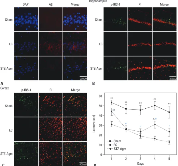
Agmatine treatment attenuates Aβ accumulation and improves cognitive dysfunction in STZ-icv rats
To determine the accumulation of Aβ, on the 21st day fol-lowing STZ injection, Aβ staining was performed in the sham, EC, and STZ-Agm groups. The expression of Aβ was considerably decreased in the STZ-Agm group com-pared to the EC group (Fig. 1A). To confirm phosphoryla-tion of IRS-1 by Agm treatment in STZ-icv rats, we conduct-ed immunohistochemistry using phospho-IRS-1 antibody, because IRS-1 plays a key role in transmitting signals from the insulin receptors to intracellular pathways. In the STZ-Agm group, both hippocampus (Fig. 1B) and cortex re-gions (Fig. 1C) showed an increase in IRS-1 phosphoryla-tion compared to the EC group. These data indicated that Agm treatment could promote IRS-1 phosphorylation in STZ-icv rat models.
In previous studies, Morris water maze tests have been commonly applied for the assessment of cognition and memory functions. To confirm the enhancement of memo-ry function in our STZ-induced Alzheimer rat model upon treatment with Agm, we conducted Morris water maze tests. The swimming times of four trials per day for 5 days in each group are shown in Fig. 1. Escape latency time (days 1-5) (to find a hidden platform) was significantly prolonged in the EC group compared to the sham group (Fig. 1D). Fig. 1D shows that the STZ-Agm group animals presented a significantly lower latency to find the platform than the EC group. The animals of the STZ-Agm group demonstrat-ed improvdemonstrat-ed Morris water maze acquisition performance.
DISCUSSION
AD is one of the most prevalent degenerative diseases in the elderly. AD is characterized by progressive memory loss and cognitive impairment.43 The administration of STZ to the
central nervous system (CNS) generates similar pathology to
AD.44-46 Previously, STZ-icv administration was shown to
in-duce oxidative stress, neuronal cell damage, and dysfunction in learning and memory.8,14,15 Accordingly, STZ-icv models
have been used to assess the therapeutic potential of various drugs. Hence, we employed a STZ-induced Alzheimer rat 8-OHdG staining. The immunohistochemical localization
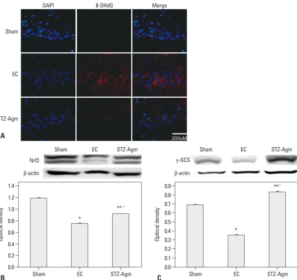
of 8-OHdG revealed a decreased number of 8-OHdG posi-tive cells in the STZ-Agm group compared to the EC group (Fig. 4A). Fig. 4A indicates that ROS generation is de-creased in the STZ-Agm group compared with the EC group. The western blot data for Nrf2 (known as an antioxi-dant transcription factor) (Fig. 4B) and γ-GCS (an impor-tant enzyme in glutathione synthesis) (Fig. 4C) show that Agm treatment promotes Nrf2-mediated antioxidant path-way signaling. Fig. 4 indicates that Agm treatment prevents cell death and promotes Nrf2-mediated antioxidant signal-ing in STZ-induced Alzheimer rats.
Fig. 1. Agmatine attenuated Aβ accumulation and promoted phosphorylation of IRS-1 in the STZ-icv rat model. (A) The expression of Aβ accumulation in the sham group, EC group, and STZ-Agm group. The image was shown at the magnification of 400. Scale bar: 200 μM. (B) The expression of phosphorylated IRS-1 in the sham, EC, and STZ-Agm groups in hippocampus sections. The image was shown at the magnification of 200. Scale bar: 400 μM. (C) The expression of phosphorylated IRS-1 in the sham, EC, and STZ-Agm groups in cortex sec-tions. The image was shown at the magnification of 200. Scale bar: 400 μM. (D) The latency time of Morris water maze was measured in the sham, EC, and STZ-Agm groups. The time required to reach the platform (escape latency) was measured on each day (1-5 days). Data were expressed as mean±SEM, and were analyzed statistically using one-way ANOVA, followed by Scheffe’s post hoc (*p<0.05, **p<0.001 compared to Sham group, †p<0.05, ††p<0.01 compared to EC group with STZ-Agm group). DAPI, 4’,6-diamidino-2-phenylindole; PI, propidium iodide; p-IRS-1, phosphorylated IRS-1; STZ, streptozotocin; EC, experimental control.
DAPI Aβ Merge Hippocampus p-IRS-1 PI Merge
Sham Sham EC EC STZ-Agm STZ-Agm p-IRS-1 PI Merge Cortex Sham EC STZ-Agm C D A B Days 0 10 20 30 40 50 60 La te nc y ( se c) 1 2 3 4 5 Sham EC STZ-Agm ** ** *,† *,†† †† †† † ** ** **
are associated with cognitive function in CNS diseases.56
Insulin dysregulation contributes to AD pathologies by re-ducing brain glucose utilization.57-59 Insulin affects neuronal
cognition and memory by regulating ion channels and neu-rotransmitter receptors in AD brains.60,61 Considering that
insulin signal transduction is important for cognitive func-tion,59,62-64 our data indicated that Agm could induce the
ac-tivation of brain insulin signal transduction and improve learning and cognitive decline in STZ-induced Alzheimer rat model.
In addition, Agm inhibits cell death by regulating the pro-duction of NO,29,30,65 and also attenuates neuronal cell death
in neurodegenerative animal models.19-23,66-68 During the
on-set of AD, there is increased oxidative stress leading to the retardation of cognitive ability.69-71 In the present study,
wa-ter maze test results highlighted significant improvements in test scores in the STZ-Agm group, compared with EC group. These functional outcomes may because Agm, a NMDA an-tagonist, improves memory function in EC rats.72,73
Addition-ally, our H&E staining data indicated that the excessive production and accumulation of ROS by Aβ can cause functional and structural changes in critical macromole-cules leading to lipid peroxidation, protein oxidation, and model to investigate AD like pathologies.
In previous studies, the administration of STZ in the CNS generated similar pathology to AD, mainly accumulation of
Aβ.46,47 In regards to the pathologic status of AD, the hyper
activation of glutamate receptor and continuous Ca2+ influx
by Aβ results in neuronal damage and cognitive dysfunc-tion.48-50 Additionally, a previous study showed that
meman-tine, as a glutamate NMDA receptor channel antagonist, blocks NMDA overstimulation upon excitoxicity; accord-ingly, memantine was suggested to be of potential use in the treatment of AD.51 Several studies reported the
neuro-protective effects of Agm in a variety of neurodegenerative pathologies through possible blockade via NMDA chan-nels.24,25,30,52-54 As shown in our data, Aβ accumulation in
damaged brain regions was decreased in the STZ-Agm group compared to the EC group.
Also, STZ-icv in rats can induce brain insulin system dysfunction and induce progressive deficits in learning, memory, and cognitive behavior like sporadic AD.12,46 As
shown in our data, STZ inhibits phosphorylation of IRS-1 in rats. IRS-1 plays a key role in transmitting signals from insulin receptors to intracellular pathways.55 Accordingly,
several studies demonstrated that IRS-1 signaling pathways
Fig. 2. Histological analysis of the hippocampus and cortex regions in STZ-icv rats. (A) Hippocampus sections from the sham group (a, b,
c), EC group (d, e, f), and STZ-Agm group (g, h, i). CA1 (a, d, g), CA2 (b, e, h), CA3 (c, f, i). Scale bars were indicated. (B) Cortex sections from the sham group (a), EC group (b), and STZ-Agm group (c). All slides were stained by hematoxylin and eosin (H&E). STZ, streptozoto-cin; EC, experimental control.
CA1 CA2 CA3
A B Sham EC STZ-Agm Sham EC STZ-Agm
lated in AD models.83 Bax also plays an essential role in
oligomeric Aβ-induced neuronal cell death.84 In the present
study, Bax and Bcl285,86 were measured. Agm treatment
sig-nificantly decreased the expression of 8-OHdG, cleaved caspase-3, and Bax, and increased the expression of Bcl2 in STZ-induced Alzheimer rats.
PI3K signaling cascade promotes NO generation through the activation of endothelial nitric oxide synthase.63,87 In
ad-dition, the PI3K/Akt signaling pathway plays crucial roles in cell survival, growth, gene expression, apoptosis, metab-olism,88 and also, neuronal survival.89 Liu, et al.64
demon-strated that the insulin-PI3K/Akt signaling pathway is re-duced in AD brain. As IRS-1 plays a key role in transmitting signals from the insulin receptors to intracellular pathways including the PI3K pathway, the phosphorylation of IRS-1 by Agm treatment could improve cognitive decline and protect against cell death by activating the PI3K pathway. DNA cleavage. Earlier studies have suggested that
antioxi-dant treatment could therapeutically cure and prevent neu-rodegenerative diseases, especially sporadic AD.17,18
More-over, previous researchers reported that antioxidants such as melatonin, vitamin E, and selegiline can be used to cure
AD.74-77
Also, STZ-induced learning and memory dysfunction is considerably associated with oxidative stress in animal models.78-80 The levels of molecular markers for DNA
(par-ticularly 8-OHdG) are reported to be elevated in the brains of patients with AD.72 Bcl2 is neuroprotective against
apop-totic cell death caused by Aβ.81 Accordingly,
overexpres-sion of Bcl2 could attenuate the processing of amyloid pre-cursor protein and tau and reduce extracellular deposits of Aβ.82 Bcl2 protects neuronal cells by inhibiting the activation
of caspase-3.81,82 Previously, Bcl-2 expression was shown to
be upregulated, while Bax and caspase-3 were down
regu-Fig. 3. Agmatine treatment decreased the expression of apoptotic proteins in STZ-icv rats. (A) Representative blots showed the protein
levels of cleaved caspase-3 from the total protein extracts prepared from the hippocampus regions of each group. Bars represented the relative protein quantification of active cleaved caspase-3 on the basis of β-actin, respectively. Representative blots showed the protein levels of Bcl2 (B) and Bax (C) from the total protein extracts prepared from the hippocampus regions of each group. (D) Representative blots showed the protein levels of PI3K from the total protein extracts prepared from the hippocampus regions of each group (*p<0.05, **p<0.01 compared to Sham group, †p<0.05, ††p<0.01 compared to EC group). STZ, streptozotocin; EC, experimental control.
0.0 0.0 0.0 0.0 0.2 0.2 0.2 0.2 0.4 0.4 0.4 0.6 0.8 0.6 0.4 0.8 1.0 0.6 0.6 1.0 1.2 0.8 0.8 1.2 1.4 1.0 1.4 1.6 1.0 1.6 1.8 1.2 1.2 Op iti ca l d en sit y Op iti ca l d en sit y Op iti ca l d en sit y Op iti ca l d en sit y Sham Sham Sham Sham EC ** EC EC EC STZ-Agm **,†† STZ-Agm STZ-Agm STZ-Agm A C B D Cleaved caspase 3 Bax Bcl 2 PI3K β-actin β-actin β-actin β-actin * *, † * *, † *
for alleviating neuronal cell apoptosis and cognitive de-cline in AD.
ACKNOWLEDGEMENTS
This research was supported by the Brain Research Program through the National Research Foundation of Korea (NRF) funded by the Ministry of Science, ICT & Future Planning (2012-0005827). This work was supported by the Brain Ko-rea 21 Plus Project for Medical Science, Yonsei University. We would like to thank Jae Ho Seo (Department of Pharma-cology, Yonsei University College of Medicine) for behav-ior test assistance.
Nrf2 plays an important role in regulating cellular oxidative stress and controls the expression of many detoxifying genes such as catalase, superoxide dismutase, heme oxygenase-1 (HO-1), NAD(P)H, and γ-GCS.31-34 Nrf2 activation can
in-duce the antioxidant pathway and protect cells against oxi-dative stress.35,36 In addition, Nrf2 ameliorates cognitive
im-pairment.37-39 Our western blot data for Nrf2 and γ-GCS
indicated that Agm could promote Nrf2-mediated antioxi-dant pathways in STZ-induced Alzheimer rats.
In conclusion, Agm could improve cognitive decline by decreasing accumulation of Aβ and ameliorating insulin signal transduction. Also, Agm could protect against dam-age to cells by activating Nrf2-mediated antioxidant sig-naling. Hence, Agm may be a promising therapeutic agent
Fig. 4. Agmatine treatment decreased the immunoreactivity of 8-OHdG and increased the expression of Nrf2 and γ-GCS in STZ-icv rats. (A) Immunohistochemistry images showed immunostaining of 8-OhdG (red) in the sham group, EC group and STZ-Agm group. The image was shown at the magnification of 400. Scale bar: 200 μM. (B) Western blot showed the amount of Nrf2 protein from the total protein ex-tracts prepared from the hippocampus regions of each group. Bar graph showed the quantification of Nrf2 protein levels in all groups. (C) Western blot showed the expression levels of γ-GCS from the total protein extracts prepared from the hippocampus regions of each group. Bar graph showed the quantification of γ-GCS protein levels (*p<0.05, **p<0.01 compared to the sham group, †p<0.05 compared to the EC group). DAPI, 4’,6-diamidino-2-phenylindole; STZ, streptozotocin; GCS, glutamyl cysteine synthetase; EC, experimental control.
DAPI 8-OHdG Merge
Sham EC STZ-Agm 0.0 0.0 0.2 0.20.1 0.4 0.6 0.3 0.4 0.8 0.5 1.0 0.60.7 1.2 1.4 0.8 0.9 Op iti ca l d en sit y Op iti ca l d en sit y Sham EC Sham * EC STZ-Agm **, † STZ-Agm B A C Nrf2 γ-GCS β-actin β-actin * **,†
18. Ansari MA, Ahmad AS, Ahmad M, Salim S, Yousuf S, Ishrat T, et al. Selenium protects cerebral ischemia in rat brain mitochondria. Biol Trace Elem Res 2004;101:73-86.
19. Arteni NS, Lavinsky D, Rodrigues AL, Frison VB, Netto CA. Ag-matine facilitates memory of an inhibitory avoidance task in adult rats. Neurobiol Learn Mem 2002;78:465-9.
20. Liu P, Collie ND. Behavioral effects of agmatine in naive rats are task- and delay-dependent. Neuroscience 2009;163:82-96. 21. Lu W, Dong HJ, Gong ZH, Su RB, Li J. Agmatine inhibits
mor-phine-induced memory impairment in the mouse step-down inhibi-tory avoidance task. Pharmacol Biochem Behav 2010;97:256-61. 22. McKay BE, Lado WE, Martin LJ, Galic MA, Fournier NM.
Learning and memory in agmatine-treated rats. Pharmacol Bio-chem Behav 2002;72:551-7.
23. Zarifkar A, Choopani S, Ghasemi R, Naghdi N, Maghsoudi AH, Maghsoudi N, et al. Agmatine prevents LPS-induced spatial memory impairment and hippocampal apoptosis. Eur J Pharmacol 2010;634:84-8.
24. Feng Y, Piletz JE, Leblanc MH. Agmatine suppresses nitric oxide production and attenuates hypoxic-ischemic brain injury in neona-tal rats. Pediatr Res 2002;52:606-11.
25. Kim JH, Yenari MA, Giffard RG, Cho SW, Park KA, Lee JE. Ag-matine reduces infarct area in a mouse model of transient focal ce-rebral ischemia and protects cultured neurons from ischemia-like injury. Exp Neurol 2004;189:122-30.
26. Regunathan S, Dozier D, Takkalapalli R, Phillips WJ. Agmatine levels in the cerebrospinal fluid of normal human volunteers. J Pain Palliat Care Pharmacother 2009;23:35-9.
27. Reis DJ, Regunathan S. Agmatine: a novel neurotransmitter? Adv Pharmacol 1998;42:645-9.
28. Regunathan S, Youngson C, Raasch W, Wang H, Reis DJ. Imidaz-oline receptors and agmatine in blood vessels: a novel system in-hibiting vascular smooth muscle proliferation. J Pharmacol Exp Ther 1996;276:1272-82.
29. Halaris A, Plietz J. Agmatine: metabolic pathway and spectrum of activity in brain. CNS Drugs 2007;21:885-900.
30. Reis DJ, Regunathan S. Is agmatine a novel neurotransmitter in brain? Trends Pharmacol Sci 2000;21:187-93.
31. Alam J, Stewart D, Touchard C, Boinapally S, Choi AM, Cook JL. Nrf2, a Cap’n’Collar transcription factor, regulates induction of the heme oxygenase-1 gene. J Biol Chem 1999;274:26071-8. 32. Motohashi H, Yamamoto M. Nrf2-Keap1 defines a
physiological-ly important stress response mechanism. Trends Mol Med 2004; 10:549-57.
33. Ramprasath T, Selvam GS. Potential impact of genetic variants in Nrf2 regulated antioxidant genes and risk prediction of diabetes and associated cardiac complications. Curr Med Chem 2013;20: 4680-93.
34. Wild AC, Gipp JJ, Mulcahy T. Overlapping antioxidant response element and PMA response element sequences mediate basal and beta-naphthoflavone-induced expression of the human gamma-glutamylcysteine synthetase catalytic subunit gene. Biochem J 1998;332(Pt 2):373-81.
35. Tomobe K, Shinozuka T, Kuroiwa M, Nomura Y. Age-related changes of Nrf2 and phosphorylated GSK-3β in a mouse model of accelerated aging (SAMP8). Arch Gerontol Geriatr 2012;54:e1-7. 36. Venugopal R, Jaiswal AK. Nrf1 and Nrf2 positively and c-Fos and
Fra1 negatively regulate the human antioxidant response element-mediated expression of NAD(P)H:quinone oxidoreductase1 gene. Proc Natl Acad Sci U S A 1996;93:14960-5.
REFERENCES
1. Hyman BT, Damasio H, Damasio AR, Van Hoesen GW. Alzheim-er’s disease. Annu Rev Public Health 1989;10:115-40.
2. Van Hoesen GW, Augustinack JC, Dierking J, Redman SJ, Than-gavel R. The parahippocampal gyrus in Alzheimer’s disease. Clin-ical and preclinClin-ical neuroanatomClin-ical correlates. Ann N Y Acad Sci 2000;911:254-74.
3. Arispe N, Rojas E, Pollard HB. Alzheimer disease amyloid beta protein forms calcium channels in bilayer membranes: blockade by tromethamine and aluminum. Proc Natl Acad Sci U S A 1993; 90:567-71.
4. Furukawa K, Abe Y, Akaike N. Amyloid beta protein-induced ir-reversible current in rat cortical neurones. Neuroreport 1994;5: 2016-8.
5. Mattson MP, Barger SW, Cheng B, Lieberburg I, Smith-Swin-tosky VL, Rydel RE. beta-Amyloid precursor protein metabolites and loss of neuronal Ca2+ homeostasis in Alzheimer’s disease. Trends Neurosci 1993;16:409-14.
6. Alberdi E, Sánchez-Gómez MV, Cavaliere F, Pérez-Samartín A, Zugaza JL, Trullas R, et al. Amyloid beta oligomers induce Ca2+ dysregulation and neuronal death through activation of ionotropic glutamate receptors. Cell Calcium 2010;47:264-72.
7. Kelly BL, Ferreira A. beta-Amyloid-induced dynamin 1 degrada-tion is mediated by N-methyl-D-aspartate receptors in hippocam-pal neurons. J Biol Chem 2006;281:28079-89.
8. Lester-Coll N, Rivera EJ, Soscia SJ, Doiron K, Wands JR, de la Monte SM. Intracerebral streptozotocin model of type 3 diabetes: relevance to sporadic Alzheimer’s disease. J Alzheimers Dis 2006;9:13-33.
9. Butterfield DA. Proteomics: a new approach to investigate oxida-tive stress in Alzheimer’s disease brain. Brain Res 2004;1000:1-7. 10. Lannert H, Hoyer S. Intracerebroventricular administration of
streptozotocin causes long-term diminutions in learning and mem-ory abilities and in cerebral energy metabolism in adult rats. Be-hav Neurosci 1998;112:1199-208.
11. Gibson GE, Huang HM. Oxidative stress in Alzheimer’s disease. Neurobiol Aging 2005;26:575-8.
12. Grünblatt E, Salkovic-Petrisic M, Osmanovic J, Riederer P, Hoyer S. Brain insulin system dysfunction in streptozotocin intracerebro-ventricularly treated rats generates hyperphosphorylated tau pro-tein. J Neurochem 2007;101:757-70.
13. Weinstock M, Shoham S. Rat models of dementia based on reduc-tions in regional glucose metabolism, cerebral blood flow and cy-tochrome oxidase activity. J Neural Transm 2004;111:347-66. 14. Dröge W, Kinscherf R. Aberrant insulin receptor signaling and
amino acid homeostasis as a major cause of oxidative stress in ag-ing. Antioxid Redox Signal 2008;10:661-78.
15. Hoyer S, Lannert H. Inhibition of the neuronal insulin receptor causes Alzheimer-like disturbances in oxidative/energy brain me-tabolism and in behavior in adult rats. Ann N Y Acad Sci 1999; 893:301-3.
16. Hoyer S. Glucose metabolism and insulin receptor signal trans-duction in Alzheimer disease. Eur J Pharmacol 2004;490:115-25. 17. Ahmad M, Saleem S, Ahmad AS, Yousuf S, Ansari MA, Khan
MB, et al. Ginkgo biloba affords dose-dependent protection against 6-hydroxydopamine-induced parkinsonism in rats: neu-robehavioural, neurochemical and immunohistochemical evidenc-es. J Neurochem 2005;93:94-104.
Alzheimer’s disease. Biochem Soc Trans 2012;40:721-7. 56. Freude S, Hettich MM, Schumann C, Stöhr O, Koch L, Köhler C,
et al. Neuronal IGF-1 resistance reduces Abeta accumulation and protects against premature death in a model of Alzheimer’s dis-ease. FASEB J 2009;23:3315-24.
57. Craft S, Asthana S, Newcomer JW, Wilkinson CW, Matos IT, Baker LD, et al. Enhancement of memory in Alzheimer disease with insulin and somatostatin, but not glucose. Arch Gen Psychia-try 1999;56:1135-40.
58. Park CR, Seeley RJ, Craft S, Woods SC. Intracerebroventricular insulin enhances memory in a passive-avoidance task. Physiol Be-hav 2000;68:509-14.
59. van der Heide LP, Ramakers GM, Smidt MP. Insulin signaling in the central nervous system: learning to survive. Prog Neurobiol 2006;79:205-21.
60. Wang YT, Salter MW. Regulation of NMDA receptors by tyrosine kinases and phosphatases. Nature 1994;369:233-5.
61. van der Heide LP, Kamal A, Artola A, Gispen WH, Ramakers GM. Insulin modulates hippocampal activity-dependent synaptic plastic-ity in a N-methyl-d-aspartate receptor and phosphatidyl-inositol-3-kinase-dependent manner. J Neurochem 2005;94:1158-66. 62. Zhao WQ, Chen H, Quon MJ, Alkon DL. Insulin and the insulin
receptor in experimental models of learning and memory. Eur J Pharmacol 2004;490:71-81.
63. Park CR. Cognitive effects of insulin in the central nervous sys-tem. Neurosci Biobehav Rev 2001;25:311-23.
64. Liu Y, Liu F, Grundke-Iqbal I, Iqbal K, Gong CX. Deficient brain insulin signalling pathway in Alzheimer’s disease and diabetes. J Pathol 2011;225:54-62.
65. Ahn SK, Hong S, Park YM, Choi JY, Lee WT, Park KA, et al. Protective effects of agmatine on lipopolysaccharide-injured mi-croglia and inducible nitric oxide synthase activity. Life Sci 2012; 91:1345-50.
66. Bokara KK, Kwon KH, Nho Y, Lee WT, Park KA, Lee JE. Retro-viral expression of arginine decarboxylase attenuates oxidative burden in mouse cortical neural stem cells. Stem Cells Dev 2011; 20:527-37.
67. Seo SK, Yang W, Park YM, Lee WT, Park KA, Lee JE. Overex-pression of human arginine decarboxylase rescues human mesen-chymal stem cells against H2O2 toxicity through cell survival pro-tein activation. J Korean Med Sci 2013;28:366-73.
68. Liu P, Bergin DH. Differential effects of i.c.v. microinfusion of agmatine on spatial working and reference memory in the rat. Neuroscience 2009;159:951-61.
69. Frölich L, Riederer P. Free radical mechanisms in dementia of Al-zheimer type and the potential for antioxidative treatment. Arz-neimittelforschung 1995;45:443-6.
70. Launer LJ, Kalmijn S. Anti-oxidants and cognitive function: a re-view of clinical and epidemiologic studies. J Neural Transm Suppl 1998;53:1-8.
71. Mikati MA, Abi-Habib RJ, El Sabban ME, Dbaibo GS, Kurdi RM, Kobeissi M, et al. Hippocampal programmed cell death after status epilepticus: evidence for NMDA-receptor and ceramide-mediated mechanisms. Epilepsia 2003;44:282-91.
72. Brouillette J. The Effects of Soluble Aβ Oligomers on Neurode-generation in Alzheimer’s Disease. Curr Pharm Des 2013. [Epub ahead of print]
73. Texidó L, Martín-Satué M, Alberdi E, Solsona C, Matute C. Amy-loid β peptide oligomers directly activate NMDA receptors. Cell Calcium 2011;49:184-90.
37. Kanninen K, White AR, Koistinaho J, Malm T. Targeting Glyco-gen Synthase Kinase-3β for Therapeutic Benefit against Oxidative Stress in Alzheimer’s Disease: Involvement of the Nrf2-ARE Pathway. Int J Alzheimers Dis 2011;2011:985085.
38. Li XH, Li CY, Xiang ZG, Hu JJ, Lu JM, Tian RB, et al. Allicin ameliorates cardiac hypertrophy and fibrosis through enhancing of Nrf2 antioxidant signaling pathways. Cardiovasc Drugs Ther 2012;26:457-65.
39. Yang Y, Zhang J, Liu H, Zhang L. Change of Nrf2 expression in rat hippocampus in a model of chronic cerebral hypoperfusion. Int J Neurosci 2013. [Epub ahead of print]
40. Rodrigues L, Dutra MF, Ilha J, Biasibetti R, Quincozes-Santos A, Leite MC, et al. Treadmill training restores spatial cognitive defi-cits and neurochemical alterations in the hippocampus of rats sub-mitted to an intracerebroventricular administration of streptozoto-cin. J Neural Transm 2010;117:1295-305.
41. Morris R. Developments of a water-maze procedure for studying spatial learning in the rat. J Neurosci Methods 1984;11:47-60. 42. Paul CA, Beltz B, Berger-Sweeney J. Perfusion of brain tissues
with fixative. CSH Protoc 2008;2008:pdb.prot4802.
43. Flynn BL. Pharmacologic management of Alzheimer disease, Part I: Hormonal and emerging investigational drug therapies. Ann Pharmacother 1999;33:178-87.
44. Sharma M, Gupta YK. Chronic treatment with trans resveratrol prevents intracerebroventricular streptozotocin induced cognitive impairment and oxidative stress in rats. Life Sci 2002;71:2489-98. 45. Selkoe DJ. Alzheimer’s disease results from the cerebral accumu-lation and cytotoxicity of amyloid beta-protein. J Alzheimers Dis 2001;3:75-80.
46. Salkovic-Petrisic M, Hoyer S. Central insulin resistance as a trig-ger for sporadic Alzheimer-like pathology: an experimental ap-proach. J Neural Transm Suppl 2007:217-33.
47. Maftei M, Thurm F, Schnack C, Tumani H, Otto M, Elbert T, et al. Increased levels of antigen-bound β-amyloid autoantibodies in serum and cerebrospinal fluid of Alzheimer’s disease patients. PLoS One 2013;8:e68996.
48. Selkoe DJ. Alzheimer’s disease: a central role for amyloid. J Neu-ropathol Exp Neurol 1994;53:438-47.
49. Varadarajan S, Yatin S, Aksenova M, Butterfield DA. Review: Al-zheimer’s amyloid beta-peptide-associated free radical oxidative stress and neurotoxicity. J Struct Biol 2000;130:184-208. 50. Varadarajan S, Kanski J, Aksenova M, Lauderback C, Butterfield
DA. Different mechanisms of oxidative stress and neurotoxicity for Alzheimer’s A beta(1--42) and A beta(25--35). J Am Chem Soc 2001;123:5625-31.
51. Kornhuber J, Bormann J, Retz W, Hübers M, Riederer P. Meman-tine displaces [3H]MK-801 at therapeutic concentrations in post-mortem human frontal cortex. Eur J Pharmacol 1989;166:589-90. 52. Piletz JE, Aricioglu F, Cheng JT, Fairbanks CA, Gilad VH,
Hae-nisch B, et al. Agmatine: clinical applications after 100 years in translation. Drug Discov Today 2013;18:880-93.
53. Salloway S, Mintzer J, Weiner MF, Cummings JL. Disease-modi-fying therapies in Alzheimer’s disease. Alzheimers Dement 2008;4:65-79.
54. Wang WP, Iyo AH, Miguel-Hidalgo J, Regunathan S, Zhu MY. Agmatine protects against cell damage induced by NMDA and glutamate in cultured hippocampal neurons. Brain Res 2006;1084: 210-6.
55. O’Neill C, Kiely AP, Coakley MF, Manning S, Long-Smith CM. Insulin and IGF-1 signalling: longevity, protein homoeostasis and
overexpression protects against amyloid-beta and prion toxicity in GT1-7 neural cells. J Alzheimers Dis 2007;12:223-8.
82. Rohn TT, Vyas V, Hernandez-Estrada T, Nichol KE, Christie LA, Head E. Lack of pathology in a triple transgenic mouse model of Alzheimer’s disease after overexpression of the anti-apoptotic protein Bcl-2. J Neurosci 2008;28:3051-9.
83. Kong J, Ren G, Jia N, Wang Y, Zhang H, Zhang W, et al. Effects of nicorandil in neuroprotective activation of PI3K/AKT pathways in a cellular model of Alzheimer’s disease. Eur Neurol 2013;70: 233-41.
84. Kudo W, Lee HP, Smith MA, Zhu X, Matsuyama S, Lee HG. In-hibition of Bax protects neuronal cells from oligomeric Aβ neuro-toxicity. Cell Death Dis 2012;3:e309.
85. Lin MT, Beal MF. Mitochondrial dysfunction and oxidative stress in neurodegenerative diseases. Nature 2006;443:787-95.
86. Slee EA, Adrain C, Martin SJ. Serial killers: ordering caspase acti-vation events in apoptosis. Cell Death Differ 1999;6:1067-74. 87. Montagnani M, Chen H, Barr VA, Quon MJ. Insulin-stimulated
activation of eNOS is independent of Ca2+ but requires phosphor-ylation by Akt at Ser(1179). J Biol Chem 2001;276:30392-8. 88. Scheid MP, Woodgett JR. PKB/AKT: functional insights from
ge-netic models. Nat Rev Mol Cell Biol 2001;2:760-8.
89. Brunet A, Datta SR, Greenberg ME. Transcription-dependent and -independent control of neuronal survival by the PI3K-Akt signal-ing pathway. Curr Opin Neurobiol 2001;11:297-305.
74. Feng Z, Zhang JT. Protective effect of melatonin on beta-amyloid-induced apoptosis in rat astroglioma C6 cells and its mechanism. Free Radic Biol Med 2004;37:1790-801.
75. Montiel T, Quiroz-Baez R, Massieu L, Arias C. Role of oxidative stress on beta-amyloid neurotoxicity elicited during impairment of energy metabolism in the hippocampus: protection by antioxi-dants. Exp Neurol 2006;200:496-508.
76. Pavlik VN, Doody RS, Rountree SD, Darby EJ. Vitamin E use is associated with improved survival in an Alzheimer’s disease co-hort. Dement Geriatr Cogn Disord 2009;28:536-40.
77. Sano M, Ernesto C, Thomas RG, Klauber MR, Schafer K, Grund-man M, et al. A controlled trial of selegiline, alpha-tocopherol, or both as treatment for Alzheimer’s disease. The Alzheimer’s Dis-ease Cooperative Study. N Engl J Med 1997;336:1216-22. 78. Ishrat T, Khan MB, Hoda MN, Yousuf S, Ahmad M, Ansari MA,
et al. Coenzyme Q10 modulates cognitive impairment against in-tracerebroventricular injection of streptozotocin in rats. Behav Brain Res 2006;171:9-16.
79. Pathan AR, Viswanad B, Sonkusare SK, Ramarao P. Chronic ad-ministration of pioglitazone attenuates intracerebroventricular streptozotocin induced-memory impairment in rats. Life Sci 2006;79:2209-16.
80. Sharma M, Gupta YK. Intracerebroventricular injection of strep-tozotocin in rats produces both oxidative stress in the brain and cognitive impairment. Life Sci 2001;68:1021-9.