Review
Correspondence to: Won Ho Kim
Department of Medicine and Institute of Gastroenterology, Yonsei University College of Medicine, 134, Shinchon-dong, Seodaemun- gu, Seoul 120-752, Korea
Tel: +82-2-2228-1950, Fax: +82-2-393-6884, E-mail: kimwonho@yuhs.ac Received on October 1, 2009. Accepted on October 4, 2009.
DOI: 10.5009/gnl.2010.4.1.1
Inflammatory Bowel Disease in Korea: Epidemiological, Genomic,
Clinical, and Therapeutic Characteristics
Eun Soo Kim and Won Ho Kim
Department of Internal Medicine and Institute of Gastroenterology, Yonsei University College of Medicine, Seoul, Korea
Inflammatory bowel disease (IBD) describes chronic inflammatory disease of the intestines and has a vari-able course; Crohn’s disease and ulcerative colitis comprise the two main forms of the condition. Although IBD occurs worldwide, its epidemiologic and clinical characteristics vary depending upon the geo-graphic location and the ethnicity of the population. Identifying the characteristic features of IBD in pop-ulations living in different geographical locations and with different ethnicities may provide significant clues about its etiology and pathophysiology, which in turn may be helpful in the development of more appro-priate treatment strategies for IBD for these different populations. Therefore, it is important for each country and region to evaluate critically the epidemiology, ge-nomics, and clinical characteristics of IBD among its own population. We have performed a critical review of the recent data in Korea, and describe herein the current epidemiologic and genotypic status, as well as the clinical manifestations and therapeutic responses of IBD that are unique to Korean patients. (Gut Liver
2010;4:1-14)
Key Words: Inflammatory bowel diseases; Crohn’s
disease; Ulcerative colitis; Korea
INTRODUCTION
Inflammatory bowel disease (IBD) is a chronic relapsing inflammatory disorder of the bowel, consisting mainly of Crohn’s disease (CD) and ulcerative colitis (UC). The in-cidence of IBD has been rising not only in Western coun-tries, but also in Asia, including Korea.1 Intriguingly, the characteristics of Western and Asian IBD patients differ
in epidemiology, phenotype and genetic susceptibility.2-7 Evaluating these geographically unique features may pro-vide substantial clues for identifying the pathophysiology and the etiology of IBD. However, such efforts have been challenging in most Asian countries, where there are lack of population-based registries, limited access to health care facilities, limited availability of diagnostic devices, and the occurrence of infectious diseases that mimic IBD, which hindered the accurate assessment of the incidence and the prevalence, the risk factors and the clinical char-acteristics of IBD.8 Until recently, only Japan has been able to report the reliable data, as it has the accessible and efficient health care system and maintains a national IBD registry under the Ministry of Health, Labor and Welfare.3 In Korea, the Korean Association for the Study of Intestinal Diseases (KASID) has recently taken a lead-ing role in establishlead-ing the organizational structural bases for investigating IBD and has performed a series of na-tionwide studies.1,9,10 Korea has a relatively small nation of territory and the population is ethnically homogeneous. Members of KASID from each province of the nation par-ticipate to perform multicenter studies and patients with IBD have been registered by members of KASID.1,9 This review will describe the updated data on Korean IBD pa-tients based on the recent publications.1,8,11-20
EPIDEMIOLOGY
Despite the advances in scientific technologies used to study IBD, its etiology and pathogenesis remain unclear. Traditionally, it has generally been thought that IBD oc-curs predominantly in the industrialized Western
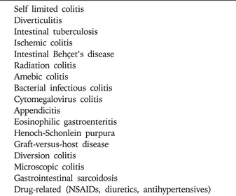
coun-Table 1. Differential Diagnosis of Intestinal Inflammation
Self limited colitis Diverticulitis
Intestinal tuberculosis Ischemic colitis
Intestinal Behçet’s disease Radiation colitis
Amebic colitis
Bacterial infectious colitis Cytomegalovirus colitis Appendicitis Eosinophilic gastroenteritis Henoch-Schonlein purpura Graft-versus-host disease Diversion colitis Microscopic colitis Gastrointestinal sarcoidosis
Drug-related (NSAIDs, diuretics, antihypertensives)
tries. However, as many other parts of the world includ-ing Asia, South America and Eastern Europe became dustrialized since the middle of the last century, the cidence and the prevalence of IBD in these areas in-creased dramatically, suggesting that urbanization may be a risk factor for IBD.2,3,8,21-27 On the other hand, in Western Europe and North America, the incidence and the prevalence of IBD have stabilized or only slightly in-creased over time.28-31
In Korea, the incidence and prevalence of CD and UC are low compared to those of Western countries, but are increasing rapidly. According to a recent update on the descriptive epidemiology of IBD in Korea1 based on the population-based data from an urban district in Seoul, the mean annual incidence rates of CD and UC increased from 0.05 to 0.34 per 100,000 inhabitants in 1986-1990, and from 1.34 to 3.08 per 100,000 inhabitants in 2001-2005, respectively. The adjusted prevalence rates of CD and UC per 100,000 inhabitants were 11.24 (95% CI, 9.29-13.18) and 30.87 (95% CI, 27.47-34.27), respecti-vely. In addition, it was observed that the ratio of the cidence rates of UC to CD was decreasing in Korea, dicating that CD showed a trend of more accelerated in-cidence rate compared to UC. This finding is similar to that observed in many Western countries.28,32,33 Male pre-dominance in the prevalence rate of CD was observed in the Korean population, a finding similar to those reported in the recent studies from China2 and Japan34 while the incidence of CD in females is predominant or equal to that in males in Western countries.28,32
DIAGNOSIS
Regarding the diagnosis of IBD, two factors should be considered: first, distinguishing IBD patients from alter-native inflammatory disease patients and secondly, cor-rectly differentiating between UC and CD.35 Before estab-lishing a diagnosis of IBD, other various forms of in-testinal inflammation with identifiable causes should be excluded using relevant history, careful physical examina-tion, prudent laboratory tests, and thorough review of the radiographic, endoscopic and pathologic data. The differ-ential diagnosis of intestinal inflammation is outlined in Table 1. In some Asian countries including Korea, where intestinal tuberculosis is still prevalent, the distinction between intestinal tuberculosis and CD is extremely diffi-cult because of their clinical similarities. Studies from Korea have shown that the proportion of patients receiv-ing anti-tuberculosis treatment before confirmation of CD was nearly 50% of all CD patients.36 In a study conducted in Korea, four endoscopic characteristics were identified
for each disease: anorectal lesions, longitudinal ulcers, aphthous ulcers, and a cobblestone appearance for CD, and involvement of fewer than four segments, a patulous ileocecal valve, transverse ulcers, and scars or pseudopo-lyps for intestinal tuberculosis. Using these endoscopic parameters, the diagnosis of either intestinal tuberculosis or CD was correctly made in 87.5% of patients.18 In Asia, if a patient has an ulceration in the ileocecal region, in-testinal Behçet’s disease (BD) should be included as a dif-ferential diagnosis.35 In an effort to differentiate intestinal BD and CD, a Korean study reported that round ulcer-ations, ≤5 in number, focal distributions, and absence of aphthous and cobblestone lesions were dominant features in intestinal BD.12 The authors stated that these charac-teristics enabled the correct diagnosis of intestinal BD or CD in 92% of patients with ulcers on colonoscopy. As the two main subforms of IBD, UC and CD have similar clinical manifestations, it is very challenging for the clinicians to correctly diagnose IBD. While there are several diagnostic criteria available for IBD including Mendeloff’s criteria,37 Lennard-Jones criteria,38 the inter-national multicentre scoring system of the Organization Mondiale de Gastroenterologie (OMGE),39 and the diag-nostic criteria of the Japanese Research Society on IBD,3 there is no single gold standard for the diagnosis of IBD. All diagnostic criteria use an integrating assessment of the clinical presentation, endoscopic, radiographic, and pathologic findings for diagnosis. More recently, serologic assays, utilizing antineutrophil cytoplasmic autoantibody (ANCA) and anti-Saccharomyces cerevisiae antibody (ASCA) have been added as diagnostic tools with an adjunctive role in differentiating UC and CD.40,41 A Korean study demonstrated that the sensitivity (49.4%) and the
specif-Table 2. Differences in Clinical, Endoscopic, and Histological Features between Ulcerative Colitis and Crohn’s Disease
Ulcerative colitis Crohn’s disease Hematochezia Common Rarely
Palpable abdominal mass Rarely Occasionally in right lower abdomen
UGI involvement No Yes
Distribution Diffuse and continuous Focal and asymmetric with skip segments Fistulas and abscesses Rarely Common
Strictures Rarely Common Perianal lesion Rarely Common
Mucosal appearance Diffuse friability, large ulcers Aphthous ulcers, linear or serpiginous, cobblestone ulcers Granulomas Absent 15-60% present
Crypt abscesses Common May be present
Fissures Rarely Common
Transmural mucosal inflammation No Yes
ANCA Common Rarely
ASCA Rarely Common
UGI, upper gastrointestinal; ANCA, anti-neutrophil cytoplasmic antibodies; ASCA, anti-saccharomyces cerevisiae antibodies.
icity (79.2%) of ASCA for CD are comparable to those seen in Western countries and that the sensitivity (44.2%) and the specificity (95.8%) of the pANCA for UC are also similar to those previously reported in Western countries.42 However, in a recent Korean study, Lee et al. reported that the prevalence of pANCA in Korean patients with UC was relatively low compared to patients in Western countries (22% vs 59-84%).17 The major distinctive clinical, morphological, structural and pathological features between the two subforms of IBD are listed in Table 2.
As genome-wide association studies have been in-troduced in an effort to identify the etiologic factors of IBD, many genetic polymorphisms related to an increased risk of IBD have been discovered.43-52 In the near future, it is expected that a more specific genetic map will be identified to improve the diagnosis of IBD. Combined with serologic assays, genetic testing is promising for a number of reasons. First, genetic testing is non-invasive. Second, genetic testing can be used to predict the pheno-typic features of disease.53 Serologic tests serve as effec-tive means to predict a complicated course of the disease by showing high associations with disease activity and the sites of involvement in IBD.54,55 A judicious combined uti-lization of genetic and serologic tests may be more reli-able for predicting complicated behavior than the use of either test alone and may be helpful in developing a risk-stratified approach for the selection of medical therapy.56 Moreover, genetic evidence could allow clini-cians to identify IBD patients before they become sympto-matic and to choose appropriate early preventive measures.
Genotypic features and clinical characteristics of Korean IBD are somewhat different from those of Western
countries. In Korea, a recent population based study showed that the most common location of the involve-ment CD is in both small bowel and colon (66.7%) fol-lowed by isolated segments of either small bowel (21.0%) or colon (12.3%).1 This finding is similar to that reported by a Japanese study.57 This contrasts with recent Euro-pean studies that reported isolated colonic disease as the most common type of CD at diagnosis.29,58 In a genetic polymorphism study with a large number of Northern European and Korean patients with CD and normal con-trols, CARD15 (NOD2) mutations were not found to be associated with susceptibility to CD in Korean patients, although they were in European populations.59 This find-ing was confirmed by another study also conducted in Korea.15 On the other hand, recent studies showed that genetic variants in IL23R, tumor necrosis factor superfamily member 15 (TNFSF15), and tumor necrosis factor-α (TNF-α) gene but not autophagy-related 16-like 1 (ATG16L1), which are known to be associated with CD in European populations also contribute to CD suscepti-bility in Koreans.60-62 In Japan63,64 and Korea65 studies, the HLA-DRB1*1502 allele was shown to be positively asso-ciated with UC, whereas the results showed discord about this association in Western populations.66-68
The clinical features of UC at diagnosis are reported to be similar in Koreans and Westerners.20 For instance, there are no differences in the extent and the severity of disease at diagnosis or in the male to female ratio of pa-tients with UC between Korea and Western countries.
EVALUATION OF CLINICAL ACTIVITY
To develop an optimal treatment strategy for IBD, it is essential to fully understand the disease’s activity and
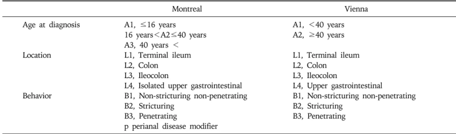
ex-Table 3. The Vienna and Montreal Classifications of Crohn’s Disease
Montreal Vienna
Age at diagnosis A1, ≤16 years A1, <40 years 16 years<A2≤40 years A2, ≥40 years A3, 40 years <
Location L1, Terminal ileum L1, Terminal ileum L2, Colon L2, Colon
L3, Ileocolon L3, Ileocolon
L4, Isolated upper gastrointestinal L4, Upper gastrointestinal
Behavior B1, Non-stricturing non-penetrating B1, Non-stricturing non-penetrating B2, Stricturing B2, Stricturing
B3, Penetrating B3, Penetrating p perianal disease modifier
tension of disease. The Truelove and Witts severity index is one of the most commonly used disease activity in-dexes for UC, and comprises six items: number of stools per day, presence of blood in stools, temperature, pulse, hemoglobin and erythrocyte sedimentation rate (ESR).69 Another popular activity index is the Mayo Score, which is composed of four variables: frequency of bowel move-ments, rectal bleeding, findings of sigmoidoscopy and Physician Global Assessment.70 Generally, the anatomic extent of UC is divided into “distal” when the lesions are not found beyond the splenic flexure and “extensive” when the mucosal inflammation is extended proximally to the splenic flexure.71 In a Korean study of 304 UC pa-tients, 44.1% of patients were diagnosed with proctitis, 22.7% with left side colitis and 33.2% with extensive colitis. The clinical severity was mild in 49.0% of pa-tients, moderate in 41.1% and severe in 8.6%.20 As sero-logic markers for the clinical activity of UC, pANCA and triggering receptors expressed on myeloid cells-1 (TREM-1) have been reported to be useful. The UC activity index was found to be higher in pANCA-positive patients than in pANCA-negative Korean patients.17 The serum TREM-1 level correlated better with disease activity than ESR or CRP, irrespective of disease extent in Korean patients with UC.19
Since CD affects various sites of the intestine and man-ifests as different endoscopic appearances of involvement, it is suggested that CD is a heterogeneous entity and that different genetic backgrounds may explain the different clinical patterns observed for the disease.72,73 It is, there-fore, important to develop a simple classification of CD based on stable and reproducible clinical variables in or-der to study its genetic or environmental bases. In 1998, the World Gastroenterology Meeting in Vienna proposed a new classification based on three clinical variables in-cluding age at diagnosis, location and disease behavior (Table 3).74 According to the Vienna classification,
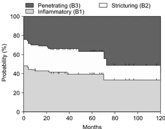
pa-tients are categorized into the A1 group if the diagnosis is made before 40 years of age and A2 if it is made thereafter. Patients are regarded as L1 when the disease is located in the terminal ileum, L2 when it is located in the colon, L3 when it is located in the ileocolon, and L4 when it involves the upper gastrointestinal tract. The B1 group includes patients with non-stricturing non-penetrat-ing disease, the B2 group includes patients with strictur-ing disease, and the B3 group includes patients with pen-etrating disease. Among these variables, disease location appears to remain stable, while significant sustained changes are noted in disease behavior over time.75 Recently, the Vienna classification was revised and a new Montreal classification was proposed.76 Regarding age-of- onset, the Montreal revision categorizes early onset of disease as an A1 category for those with an age of diag-nosis of 16 years or younger. With respect to disease lo-cation, the Montreal classification allows upper gastro-intestinal disease to coexist with more distal disease. Lastly, in terms of disease behavior, perianal disease is recognized as a separate subclassification. The data from Severance Hospital in Korea following the Vienna classi-fication showed that patients in groups A1 and A2 made up 88.9% and 11.1%, respectively, of the entire patient population, and that L1, L2, L3, and L4 made up 19.5%, 13.9%, 45.8%, and 20.8%, respectively.77 Notably, the most common location was L3, which is different from the pattern normally seen in Western countries. During the follow-up period, the proportion of B1 and B2 dimin-ished from 48.6% and 27.8% to 37.5% and 25%, re-spectively, while that of B3 increased from 23.6% up to 37.5% (Fig. 1). The Crohn’s disease activity index (CDAI), the most widely applied activity index for CD, consists of eight variables: the number of liquid stools, the extent of abdominal pain, general well-being, the oc-currence of extraintestinal symptoms, the need for an-ti-diarrheal drugs, the presence of abdominal masses,
Fig. 1. Changes in disease behavior in Korean CD patients
according to the Vienna classification.
hematocrit and body weight.78 The range of scores is 0 to 600 and in clinical trials, active CD is indicated when the CDAI score is over 150. As the CDAI has been frequently used in clinical trials over two decades and vigorously va-lidated, this index is considered the gold standard for evaluation of disease activity.79 The Harvey Bradshaw dex is another convenient and commonly used activity in-dex for CD.80
MANAGEMENT OF IBD
IBD is characterized by its chronicity, with cycles of re-mission and relapse that occur over the patient’s lifetime. The goals of therapy for IBD, therefore, are not only the induction but also the maintenance of remission of symp-toms without the use of corticosteroids.
CROHN’S DISEASE
1. Medical management 1) Induction of remissionClinical trials have documented that sulfasalazine, bu-desonide, and conventional corticosteroids are effective at inducing remission in mild to moderately active CD. Although oral aminosalicylates have been widely used in clinical practice to treat active CD, their use is con-troversial evidence. In the case of mild to moderate CD, clinical trials demonstrate that sulfasalazine administered at 2-6 g/day is more effective than placebo for inducing remission.81,82 This effect is most notable in patients with active colonic or ileocolonic disease.83,84 In contrast, mesa-lazine has failed to show consistent results in various controlled studies.85-87 Some patients are intolerant of the
adverse effects of sulfasalazine, including headache, nau-sea, skin rash, fever, hepatitis, autoimmune hemolysis, aplastic anemia, leucopenia, folate deficiency, pancreatitis and male infertility. Most of these effects are related to the sulfapyridine moiety of sulfasalazine. Considering that the intestinal bacterial flora probably plays a role in the pathogenesis of CD, antibiotics have been used as ther-apeutic agents. Although metronidazole and ciprofloxacin are often used for the treatment of CD, controlled trials have failed to show convincing evidence of their efficacy for the induction of remission in active CD.88-91 Budesonide undergoes extensive first-pass hepatic metab-olism, which reduces the side effects associated with sys-temic corticosteroids. Well controlled studies have re-vealed that budesonide is significantly more effective than placebo and 5-ASA, and is about as effective as conven-tional corticosteroids for inducing remission in active CD.92-94 Therefore, budesonide has been recommended as a first-line therapy for mild to moderate CD of the ileum and proximal colon in an American Gastroenterological Association position statement and in the European Crohn’s and Colitis Organization Consensus.95,96
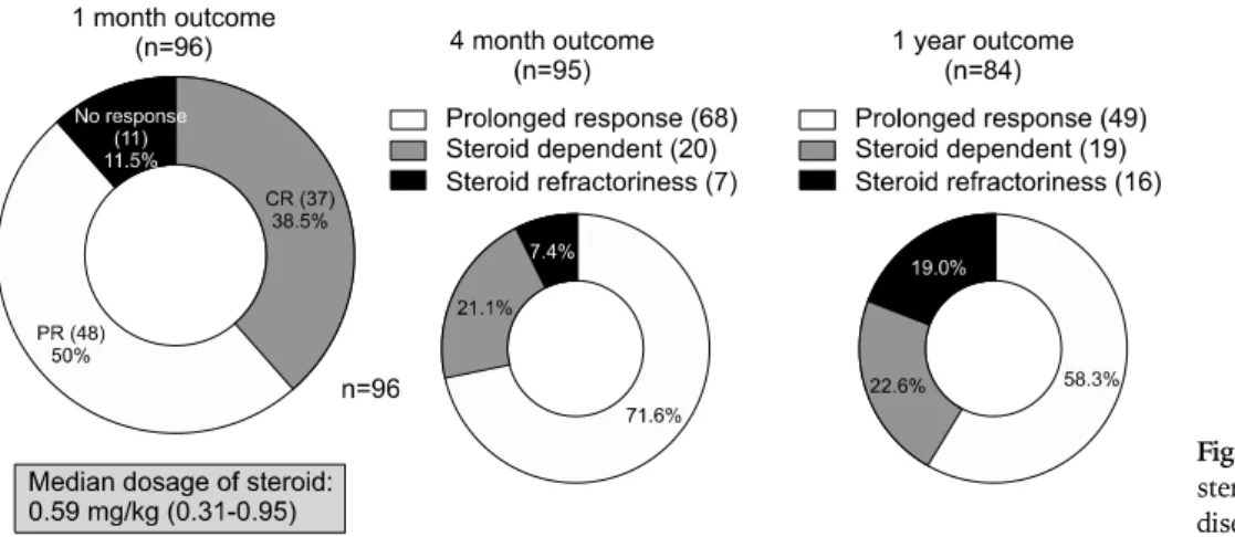
For those patients who do not respond to the first-line therapy described above and for those with moderate to severe CD, oral prednisone 40-60 mg/day should be given. In the case of fulminant and refractory in-flammatory CD, remission may be induced with intra-venous corticosteroids, such as methylprednisolone at 1-1.5 mg/kg per day. A Korean study showed that serum albumin level could be a significant predictive factor for response to initial steroid therapy in active CD patients.97 Compared to steroid responders, a steroid dependent group was characterized by lower serum albumin levels. According to the data from a Korean study, when a me-dian dosage of oral steroids at 0.59 mg/kg was ad-ministered, the overall response rates to the steroid at one month and one year were as high as 88.5% and 58.3%, respectively.98 These rates were comparable to the best response rates from Western studies (Fig. 2). Patients with moderate to severe disease who fail to re-spond to 5-ASA, budesonide and conventional cortico-steroids, and patients intolerant of corticocortico-steroids, may be treated with anti-TNF-α antibody agents including in-fliximab (intravenous, 5 mg/kg at 0, 2, and 6 weeks) and adalimumab (subcutaneous, 160 mg at 0 and 80 mg at 2 weeks). The results of a nationwide survey conducted in Korea,99 showed that the clinical response rate for in-fliximab in luminal CD was 51%, and that the clinical re-mission rate was 36%. Thirteen percent of patients did not respond to infliximab treatment. In patients with fist-ular disease, the clinical response rate was 45%, clinical
Fig. 2. Overall response rates to
steroids in Korean Crohn’s disease patients.
remission 31%, and no response 24%. These results are similar to the clinical response rates reported in Western studies. Interestingly, the most common reason for in-fliximab cessation in Korea was the limitation of the standard national health insurance reimbursement, as it previously covered only one year of infliximab treatment for active CD patients. However, the national health in-surance coverage for infliximab has been extended up to 28 injections, regardless of period in a recent amend-ment.100
2) Maintenance therapy
Despite the widespread use of 5-ASA in clinical practice for the maintenance of medically induced remission in CD patients, this application is not evidence-based. Clinical studies have revealed that sulfasalazine and mesa-lazine are not actually effective for maintaining medically induced remission in CD patients. In general, cortico-steroids are not recommended for maintaining remission due to related systemic adverse effects and lack of efficacy. The results of a Korean study showed that pre-operative corticosteroid treatment increased the risk of postoperative infections in patients with CD.101 Treatment with budesonide at 6 mg/day, however, has been shown to prolong the time to relapse and to be effective at maintaining remission for at least six months, but less than one year.102
Immunosuppressive agents such as azathioprine (AZA) and 6-mercaptopurine (6-MP) have been reported to be effective as maintenance therapy and to be advantageous for reducing or discontinuing steroid usage in ste-roid-dependent CD patients.103-105 However, due to slow onset of action, these drugs are not used as induction therapy in patients with active CD.103 Because azathio-prine or mercaptopurine are frequently continued indef-initely as maintenance therapy,106 their long term side ef-fects should be kept in mind. These agents, therefore, are
currently reserved for steroid-dependent or refractory patients. Myelotoxicity is one of the significant adverse effects of thiopurine drugs and is reported to be asso-ciated with decreased thiopurine methyltransferase (TPMT) and deficiency of inosine triphosphate py-rophosphatase (ITPA).107 Korean patients with IBD who are treated with AZA/6-MP experience myelotoxicity more frequently than similarly treated Europeans. Among 133 IBD patients treated with azathioprine in Korea, leu-copenia occurred in 75 cases (56.4%), which is a higher incidence than that reported in Western studies.108 Another Korean study revealed that there is no correla-tion between TPMT genotype and AZA toxicity.109 Methotrexate is another effective immunosuppressive agent for maintaining remission of CD.
For patients who are unable to maintain remission or who are dependent on steroids in spite of treatment with AZA or 6-MP, infliximab can be used for maintenance of remission, steroid-sparing and mucosal healing (5 mg/kg every eight weeks).110 Alternatively, adalimumab can be given subcutaneously for maintenance therapy at 40 mg every other week. With advances in the understanding of the pathophysiology of CD, a range of new therapeutic agents apart from anti-TNF drugs are under investigation. Among these innovative drugs are the T-cell blocking agents,111 anti-adhesion molecules,112 anti-vascular adhe-sion protein-1 monoclonal antibody113 and avidity multi-mers (avimulti-mers)114 with high ligand affinity and specificity that allows binding to many targets at the same time.
2. Medical management of fistulizing CD
One of the distinct characteristics of Korean CD pa-tients is the high incidence of perianal fistulas. The in-cidence rate of perianal fistulas in Korea is 48.5%, which is higher than that reported in other countries (13-38%).13 Because the incidence of CD in Korea has been increasing substantially, the possibility of CD should
be considered in Korean patients with perianal fistulas. The first-line therapy for fistulas in patients with CD is antibiotic treatment with metronidazole or cipro-floxacin,96,104,115,116 with AZA or 6-MP being the typical second-line treatment. In cases in which active fistulizing disease fails to respond to treatment with antibiotics and immunosuppressive agents, infliximab or adalimumab can be administered.
3. Surgical management
A significant proportion of CD patients will need surgi-cal management an average of 20 years after diagnosis. Failure of medical therapy remains the most common in-dication for surgical treatment of CD.117,118 The occur-rence of complications despite medical treatment is an additional indication for surgical management.
Worsening obstruction is a frequent complication of CD, especially with small bowel involvement. Whereas acute obstructions are often resolved with medical treat-ment, chronic obstructions are usually caused by fi-brostenotic lesions and require surgical management.119 Not all fistulas require surgery. Although intestinal fis-tulas occur in one-third of CD patients, only a few pa-tients require surgical management.120 Generally, fistulas associated with abscesses or strictures, that are connected to the genitourinary tract, or that cause intestinal mal-absorption are indications for surgical management. The lifetime risk for intraabdominal abscess in CD pa-tients is approximately 25%.121 Almost all abscesses re-quire drainage either surgically or percutaneously. Although there is controversy as to whether or not intra-abdominal CD abscess should be managed surgically, sur-gical resection is required when an abscess has enteric contents.122 Korean data have shown that nonsurgical treatment including percutaneous catheter drainage can be considered an initial therapy for a CD-related abscess. The overall success rate and cumulative recurrence rate at seven months were 66.7% and 12.5%, respectively.16 A recent Korean study reported that the clinical efficacy of nonsurgical treatment is comparable to that of surgical treatment.123 Other indications for surgical management in CD include free perforation, bleeding and cancer. In CD, 70-90% of patients will require surgical manage-ment during the disease course. Another study reported that the cumulative operation rate was 44%, 61%, and 71% at one, five, and 10 years after diagnosis, respectively.124 Unlike the above Western studies, the rate was much lower in the results of a Korean study with 11.5%, 13.1%, and 18.2% at one, three, and six years af-ter diagnosis, respectively.36 The rate reported in another Korean study was also lower than that reported for
Western countries.125 This disparity could be attributed to the different perspectives of different backgrounds toward surgical intervention in CD patients. Another possible reason could be that the Western study was conducted early, when aggressive surgery for CD was prevalent and Korean studies are more recent, when surgical treatment is rarely performed due to the advances in medical treatment.
The rate of complications with surgical management for CD in Korea is 13.6%, which is comparable to that of other countries.126
ULCERATIVE COLITIS
1. Medical managementUC can be treated according to anatomical extent of the disease. In distal UC, in which disease involvement is limited to below the splenic flexure, a topical agent can be effectively applied. Similar to CD, the management of UC is focused on both induction and maintenance of dis-ease remission. In addition, the therapeutic decision de-pends mainly on clinical severity rather than on histologic severity of inflammation.
1) Induction of remission
In mild to moderate active UC patients, the first line regimen is 5-ASA, including oral and rectal formulations. Effective oral doses for each drug are as follows; sulfasa-lazine 4-6 g/day, mesalamine 2-4 g/day, balsalazide 6.75 g/day, olsalazine 1.5-3 g/day. The effects of these drugs are usually observed within 2-4 weeks in 40-80% of patients.127,128 Left side colitis can also be managed with topical agents such as rectal mesalazine or rectal corticosteroid. Controlled trials show that topical mesal-amine is superior to oral 5-ASA or topical corticosteroid alone in inducing clinical improvement in patients with mild to moderate distal colitis.110,129,130 Furthermore, the combination of oral and topical 5-ASA may produce a better response in the management of mild to moderate distal colitis than administration of either oral or topical formulations alone.131
If the patients fail to respond to 5-ASA agents, they should be managed with oral prednisone 40-60 mg daily for one to two weeks. At one year after starting cortico-steroids, a prolonged response is observed in 49% of pa-tients, and 22% of patients become steroid dependent.132 Patients with severe active UC showing aggravated symptoms despite treatment with oral corticosteroids should be hospitalized for intravenous steroid therapy. Further failure to respond to intravenous steroid might require the use of cyclosporine, tacrolimus and a biologic
Table 4. Characteristics of Korean Patients with CD Compared to Those of Western Countries
Differences Similarities Epidemiology • Incidence and prevalence of CD in Korea are low, but are
increasing rapidly whereas those of Western countries have stabilized or only slightly increased over time1,28-31 • Male is predominant in the prevalence rate of CD in
Korean population while female is predominant or equal to male in Western countries1,28,32
• CD shows a trend of more accelerated incidence rate compared to UC1
Genomics • CARD15 (NOD2) and ATG16L1 mutations were not found to be associated with susceptibility to CD15,59,62
• Genetic variants in IL23R, TNFSF15, and TNF-α gene were associated with suscep-tibility to CD60-62
Diagnosis • Intestinal tuberculosis and intestinal Behcet’s disease should be included as a differential diagnosis because of their relatively high prevalence in Korean population35,36
• Sensitivity and specificity of ASCA for CD are 49.4% and 79.2%, respectively42
Clinical features • The most common location of CD is both small bowel and colon type in Korean population while isolated colonic disease is the most common type in Western countries1
• The incidence rate of perianal fistulas in Korea is higher than Western countries (48.5% vs 13-38%)13
Therapeutic efficacy • The most common reason for infliximab cessation was the limitation of the standard national health insurance reimbursement99
• Korean patients who are treated with AZA/6-MP experience myelotoxicity more frequently than similarly treated Westerners108
• The cumulative operation rate of Korean CD patients is much lower than that of Western CD patients36,125
• The clinical response rate and clinical remission rate for infliximab in Korean luminal CD and fistular disease are similar with those of Western countries99
• The rate of complications with surgical management for CD in Korea is comparable to that of Western countries126
agent such as infliximab. If patients do not respond to maximal medical treatment, superimposed infection with cytomegalovirus or C. difficile should be considered as a potential cause of aggravation.
Although there have been many studies in Western countries indicating the usefulness of cyclosporine in the treatment of severe UC, the efficacy of cyclosporine has been disappointing in Korea, with a reported response rate of only 14%.133 One Korean study was conducted to evaluate the efficacy of cyclosporine and corticosteroid combination therapy in patients with severe UC that was refractory to an initial intravenous corticosteroid treat-ment. Unlike Western studies, the Korean study con-cluded that this combination failed to present any addi-tional benefit over corticosteroid therapy alone in terms of short-term response.134 These results suggest that there might be differences in the metabolism of this agent or differences in phenotypes, in terms of responsiveness, based on differences in genetic backgrounds.
Korean UC patients seem to show a better clinical re-sponse to medical treatment than their Western counterparts. The overall remission rate in the first attack was 90% in Western population-based studies, whereas the rate in the Korean study with 311 first diagnosed UC
patients was 97.4%.20
2) Maintenance management
5-ASA agents are the mainstay of effective treatment for relapse-prevention of UC. Topical 5-ASA reduces the relapse rates in proctitis or left sided colitis, but topical corticosteroids have not shown effectiveness as a main-tenance treatment in distal colitis.127,135,136 AZA or 6-MP may be another choice for relapse-prevention in patients who do not respond to 5-ASA or who are dependent on oral steroids. Patients, who have frequent relapses or are steroid-dependent, despite treatment with 5-ASA and im-munomodulators, can be treated effectively with inflixi-mab. Recent studies have shown that 45% of infliximab responders remain in the non-relapsed state, compared to 20% of initial responders in the placebo group after 54 weeks of maintenance therapy with infliximab.137 These promising results with infliximab encourage further trials for confirmation of the role of infliximab as an effective management of UC.
2. Surgical management
A surgical approach is required for treatment of UC when there are life-threatening complications such as
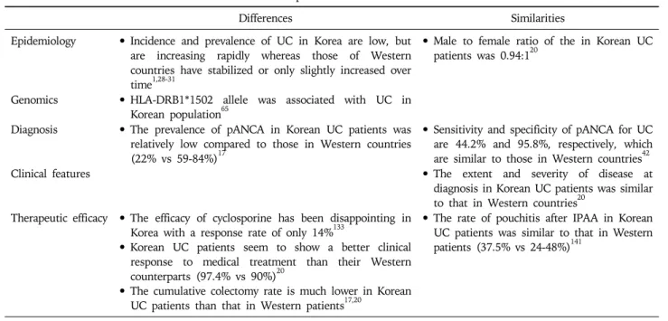
Table 5. Characteristics of Korean Patients with UC Compared to Those of Western Countries
Differences Similarities Epidemiology • Incidence and prevalence of UC in Korea are low, but
are increasing rapidly whereas those of Western countries have stabilized or only slightly increased over time1,28-31
• Male to female ratio of the in Korean UC patients was 0.94:120
Genomics • HLA-DRB1*1502 allele was associated with UC in Korean population65
Diagnosis • The prevalence of pANCA in Korean UC patients was relatively low compared to those in Western countries (22% vs 59-84%)17
• Sensitivity and specificity of pANCA for UC are 44.2% and 95.8%, respectively, which are similar to those in Western countries42 Clinical features • The extent and severity of disease at
diagnosis in Korean UC patients was similar to that in Western countries20
Therapeutic efficacy • The efficacy of cyclosporine has been disappointing in Korea with a response rate of only 14%133
• Korean UC patients seem to show a better clinical response to medical treatment than their Western counterparts (97.4% vs 90%)20
• The cumulative colectomy rate is much lower in Korean UC patients than that in Western patients17,20
• The rate of pouchitis after IPAA in Korean UC patients was similar to that in Western patients (37.5% vs 24-48%)141
hemorrhage or perforation, and documented dysplasia or cancer. Other indications for surgery include fulminant colitis with or without toxic megacolon refractory to max-imal medical management or intolerance to immuno-suppression or medical therapy. Growth failure due to in-tractable disease course in pediatric UC patients can also be an indication for surgery.138 Since 20-24% of strictures in UC are malignant139 and biopsies of the stricture site are inadequate for excluding malignancy, strictures should be managed with surgery.140
Total proctocolectomy with ileal J-pouch anal anasto-mosis (IPAA) is the most commonly used surgical meth-od for treatment of UC. Large cohort studies have de-scribed this method as relatively safe and durable with a low perioperative mortality rate of 0.2-1.0%. In a recent study of 20 Korean UC patients treated with IPAA, post-operative functional outcomes including sexual and uri-nary function were found to be tolerable. Despite a rela-tively high postoperative complication rate (35%), most cases were managed conservatively.14
Pouchitis is the most common long-term complication of IPAA. It develops in 24-48% of IPAA patients and can be controlled with antibiotics including ciprofloxacin or metronidazole. Similar to data from other countries, 37.5% of Korean UC patients undergoing total proctoco-lectomy with IPAA have pouchitis and most of these cas-es occur within one year after surgery.141
One of the unique characteristics is that the cumulative probability of colectomy is much lower in Korean UC pa-tients than in Western papa-tients. In Western countries, the
cumulative colectomy rate is shown to be 3-10% after one year,142-144 8-20% after five years,144,145 and 24-34% after 10 years.146,147 In contrast, studies from Korea reveal that it is 2.0-3.0% after one year and 3.3% after five to 15 years.17,20 Furthermore, the author showed that there were no deaths associated with UC in that study, sug-gesting that Korean UC patients tend to have a milder form of the disease.
CONCLUSION
Although IBD is a worldwide disease, its incidence rates and clinical features vary depending on geographic area and population origin. The incidence and the preva-lence rates of IBD have been increasing dramatically in Korea. There are some clinical and genotypic character-istics that are unique to Korean IBD patients (Tables 4 and 5). Most of all, the clinical course of IBD in Korean patients seems milder than that in Western countries, as indicated by the lower rates of surgical intervention and better responses to medical management in Korean patients. These differing characteristics may provide sig-nificant clues about the etiology and the pathophysiology of IBD. Further comprehensive population-based studies are required to clarify these differences.
Although biological therapies have shown a promising role and have altered the management of IBD, they must be used judiciously based on the existing evidence of effi-cacy and safety. The recent emergence of several promis-ing biologic agents suggests that these novel biologic
agents may replace or be used in combination with TNF antagonists in the near future.
REFERENCES
1. Yang SK, Yun S, Kim JH, et al. Epidemiology of in-flammatory bowel disease in the Songpa-Kangdong district, Seoul, Korea, 1986-2005: a KASID study. Inflamm Bowel Dis 2008;14:542-549.
2. Leong RW, Lau JY, Sung JJ. The epidemiology and pheno-type of Crohn's disease in the Chinese population. Inflamm Bowel Dis 2004;10:646-651.
3. Morita N, Toki S, Hirohashi T, et al. Incidence and preva-lence of inflammatory bowel disease in Japan: nationwide epidemiological survey during the year 1991. J Gastroen-terol 1995;30 Suppl 8:1-4.
4. Yang SK, Hong WS, Min YI, et al. Incidence and preva-lence of ulcerative colitis in the Songpa-Kangdong District, Seoul, Korea, 1986-1997. J Gastroenterol Hepatol 2000;15: 1037-1042.
5. Ling KL, Ooi CJ, Luman W, Cheong WK, Choen FS, Ng HS. Clinical characteristics of ulcerative colitis in Singa-pore, a multiracial city-state. J Clin Gastroenterol 2002;35: 144-148.
6. Iida M, Yao T, Okada M. Long-term follow-up study of Crohn's disease in Japan: the Research Committee of Inflammatory Bowel Disease in Japan. J Gastroenterol 1995;30 Suppl 8:17-19.
7. Inoue N, Tamura K, Kinouchi Y, et al. Lack of common NOD2 variants in Japanese patients with Crohn's disease. Gastroenterology 2002;123:86-91.
8. Yang SK, Loftus EV Jr, Sandborn WJ. Epidemiology of in-flammatory bowel disease in Asia. Inflamm Bowel Dis 2001;7:260-270.
9. Kim BJ, Yang SK, Kim JS, et al. Trends of ulcerative col-itis-associated colorectal cancer in Korea: a KASID study. J Gastroenterol Hepatol 2009;24:667-671.
10. Chang DK, Kim YH, Byeon JS, et al. The current status of ulcerative colitis-associated colorectal cancer in Korea: a KASID study. Korean J Gastroenterol 2005;46:276-282. 11. Cheon JH, Kim JH, Kim BY, et al. Allele frequency of
thio-purine methyltransferase and inosine triphosphate py-rophosphatase gene polymorphisms in Korean patients with inflammatory bowel diseases. Hepatogastroenterology 2009;56:421-423.
12. Lee SK, Kim BK, Kim TI, Kim WH. Differential diagnosis of intestinal Behcet's disease and Crohn's disease by colo-noscopic findings. Endoscopy 2009;41:9-16.
13. Kim JY, Yang SK, Byeon JS. The incidence and natural his-tory of perianal fistulas in Korean patients with Crohn's disease. Intest Res 2006;4:22-31.
14. Ko YT, Kim NK, Min BS, et al. Twenty cases of restorative proctocolectomy for ulcerative colitis of Asian patients: analysis of operative safety and functional outcomes in sin-gle institution experience. Int J Colorectal Dis 2008;23: 131-132.
15. Lee GH, Kim CG, Kim JS, Jung HC, Song IS. Frequency analysis of NOD2 gene mutations in Korean patients with Crohn's disease. Korean J Gastroenterol 2005;45:162-168. 16. Lee H, Kim YH, Kim JH, et al. Nonsurgical treatment of
abdominal or pelvic abscess in consecutive patients with Crohn's disease. Dig Liver Dis 2006;38:659-664.
17. Lee JH, Cheon JH, Kim ES, et al. The prevalence and clin-ical significance of perinuclear anti-neutrophil cytoplasmic antibody in Korean patients with ulcerative colitis. Dig Dis Sci. Forthcoming. DOI: 10.1007/s10620-009-0847-8. 18. Lee YJ, Yang SK, Byeon JS, et al. Analysis of colonoscopic
findings in the differential diagnosis between intestinal tu-berculosis and Crohn's disease. Endoscopy 2006;38:592-597. 19. Park JJ, Cheon JH, Kim BY, et al. Correlation of se-rum-soluble triggering receptor expressed on myeloid cells-1 with clinical disease activity in inflammatory bowel disease. Dig Dis Sci 2009;54:1525-1531.
20. Park SH, Kim YM, Yang SK, et al. Clinical features and natural history of ulcerative colitis in Korea. Inflamm Bowel Dis 2007;13:278-283.
21. Russel MG. Changes in the incidence of inflammatory bowel disease: what does it mean? Eur J Intern Med 2000; 11:191-196.
22. Cashman KD, Shanahan F. Is nutrition an aetiological fac-tor for inflammafac-tory bowel disease? Eur J Gastroenterol Hepatol 2003;15:607-613.
23. Jovanovic Z. Epidemiology of Crohn's disease in the Rijeka-Istra region. Lijec Vjesn 1999;121:8-13.
24. Appleyard CB, Hernandez G, Rios-Bedoya CF. Basic epi-demiology of inflammatory bowel disease in Puerto Rico. Inflamm Bowel Dis 2004;10:106-111.
25. Linares de la Cal JA, Canton C, Hermida C, Perez-Miranda M, Mate-Jimenez J. Estimated incidence of inflammatory bowel disease in Argentina and Panama (1987-1993). Rev Esp Enferm Dig 1999;91:277-286.
26. Lakatos L, Mester G, Erdelyi Z, et al. Striking elevation in incidence and prevalence of inflammatory bowel disease in a province of western Hungary between 1977-2001. World J Gastroenterol 2004;10:404-409.
27. Sincic BM, Vucelic B, Persic M, et al. Incidence of in-flammatory bowel disease in Primorsko-goranska County, Croatia, 2000-2004: a prospective population-based study. Scand J Gastroenterol 2006;41:437-444.
28. Molinie F, Gower-Rousseau C, Yzet T, et al. Opposite evo-lution in incidence of Crohn's disease and ulcerative colitis in Northern France (1988-1999). Gut 2004;53:843-848. 29. Lapidus A. Crohn's disease in Stockholm County during
1990-2001: an epidemiological update. World J Gastroen-terol 2006;12:75-81.
30. Loftus EV Jr, Silverstein MD, Sandborn WJ, Tremaine WJ, Harmsen WS, Zinsmeister AR. Crohn's disease in Olmsted County, Minnesota, 1940-1993: incidence, prevalence, and survival. Gastroenterology 1998;114:1161-1168.
31. Loftus EV Jr, Silverstein MD, Sandborn WJ, Tremaine WJ, Harmsen WS, Zinsmeister AR. Ulcerative colitis in Olmsted County, Minnesota, 1940-1993: incidence, preva-lence, and survival. Gut 2000;46:336-343.
32. Bernstein CN, Wajda A, Svenson LW, et al. The epidemiol-ogy of inflammatory bowel disease in Canada: a popula-tion-based study. Am J Gastroenterol 2006;101:1559-1568. 33. Gearry RB, Richardson A, Frampton CM, et al. High
in-cidence of Crohn's disease in Canterbury, New Zealand: results of an epidemiologic study. Inflamm Bowel Dis 2006;12:936-943.
diagnostic criteria and epidemiology. Dis Colon Rectum 2000;43(10 Suppl):S85-S93.
35. Sands BE. From symptom to diagnosis: clinical distinctions among various forms of intestinal inflammation. Gastroen-terology 2004;126:1518-1532.
36. Park JB, Yang SK, Myung SJ, et al. Clinical characteristics at diagnosis and course of Korean patients with Crohn's disease. Korean J Gastroenterol 2004;43:8-17.
37. Calkins BM, Lilienfeld AM, Garland CF, Mendeloff AI. Trends in incidence rates of ulcerative colitis and Crohn's disease. Dig Dis Sci 1984;29:913-920.
38. Lennard-Jones JE. Classification of inflammatory bowel disease. Scand J Gastroenterol Suppl 1989;170:2-6.
39. Myren J, Bouchier IA, Watkinson G, Softley A, Clamp SE, de Dombal FT. The OMGE multinational inflammatory bowel disease survey 1976-1986: a further report on 3175 cases. Scand J Gastroenterol Suppl 1988;144:11-19. 40. Saxon A, Shanahan F, Landers C, Ganz T, Targan S. A
dis-tinct subset of antineutrophil cytoplasmic antibodies is as-sociated with inflammatory bowel disease. J Allergy Clin Immunol 1990;86:202-210.
41. Vermeire S, Joossens S, Peeters M, et al. Comparative study of ASCA (Anti-Saccharomyces cerevisiae antibody) assays in inflammatory bowel disease. Gastroenterology 2001;120:827-833.
42. Kim BG, Kim YS, Kim JS, Jung HC, Song IS. Diagnostic role of anti-Saccharomyces cerevisiae mannan antibodies combined with antineutrophil cytoplasmic antibodies in pa-tients with inflammatory bowel disease. Dis Colon Rectum 2002;45:1062-1069.
43. Hugot JP, Chamaillard M, Zouali H, et al. Association of NOD2 leucine-rich repeat variants with susceptibility to Crohn's disease. Nature 2001;411:599-603.
44. Ogura Y, Bonen DK, Inohara N, et al. A frameshift muta-tion in NOD2 associated with susceptibility to Crohn's disease. Nature 2001;411:603-606.
45. Hampe J, Cuthbert A, Croucher PJ, et al. Association be-tween insertion mutation in NOD2 gene and Crohn's dis-ease in German and British populations. Lancet 2001;357: 1925-1928.
46. Stoll M, Corneliussen B, Costello CM, et al. Genetic varia-tion in DLG5 is associated with inflammatory bowel disease. Nat Genet 2004;36:476-480.
47. Till A, Rosenstiel P, Krippner-Heidenreich A, et al. The Met-196 → Arg variation of human tumor necrosis factor receptor 2 (TNFR2) affects TNF-alpha-induced apoptosis by impaired NF-kappaB signaling and target gene ex-pression. J Biol Chem 2005;280:5994-6004.
48. Duerr RH, Taylor KD, Brant SR, et al. A genome-wide as-sociation study identifies IL23R as an inflammatory bowel disease gene. Science 2006;314:1461-1463.
49. Hampe J, Franke A, Rosenstiel P, et al. A genome-wide as-sociation scan of nonsynonymous SNPs identifies a sus-ceptibility variant for Crohn disease in ATG16L1. Nat Genet 2007;39:207-211.
50. Rioux JD, Xavier RJ, Taylor KD, et al. Genome-wide asso-ciation study identifies new susceptibility loci for Crohn disease and implicates autophagy in disease pathogenesis. Nat Genet 2007;39:596-604.
51. Parkes M, Barrett JC, Prescott NJ, et al. Sequence variants in the autophagy gene IRGM and multiple other
replicat-ing loci contribute to Crohn's disease susceptibility. Nat Genet 2007;39:830-832.
52. Raelson JV, Little RD, Ruether A, et al. Genome-wide as-sociation study for Crohn's disease in the Quebec Founder Population identifies multiple validated disease loci. Proc Natl Acad Sci U S A 2007;104:14747-14752.
53. Riis L, Vind I, Vermeire S, et al. The prevalence of genetic and serologic markers in an unselected European pop-ulation-based cohort of IBD patients. Inflamm Bowel Dis 2007;13:24-32.
54. Targan SR, Landers CJ, Yang H, et al. Antibodies to CBir1 flagellin define a unique response that is associated in-dependently with complicated Crohn's disease. Gastroen-terology 2005;128:2020-2028.
55. Braun J, Targan SR. Multiparameter analysis of im-munogenetic mechanisms in clinical diagnosis and manage-ment of inflammatory bowel disease. Adv Exp Med Biol 2006;579:209-218.
56. Nikolaus S, Schreiber S. Diagnostics of inflammatory bow-el disease. Gastroenterology 2007;133:1670-1689.
57. Oriuchi T, Hiwatashi N, Kinouchi Y, et al. Clinical course and longterm prognosis of Japanese patients with Crohn's disease: predictive factors, rates of operation, and mortali-ty. J Gastroenterol 2003;38:942-953.
58. Vind I, Riis L, Jess T, et al. Increasing incidences of in-flammatory bowel disease and decreasing surgery rates in Copenhagen City and County, 2003-2005: a population- based study from the Danish Crohn colitis database. Am J Gastroenterol 2006;101:1274-1282.
59. Croucher PJ, Mascheretti S, Hampe J, et al. Haplotype structure and association to Crohn's disease of CARD15 mutations in two ethnically divergent populations. Eur J Hum Genet 2003;11:6-16.
60. Yang SK, Lee SG, Cho YK, Lim J, Lee I, Song K. Associa-tion of TNF-alpha/LTA polymorphisms with Crohn's dis-ease in Koreans. Cytokine 2006;35:13-20.
61. Yang SK, Lim J, Chang HS, et al. Association of TNFSF15 with Crohn's disease in Koreans. Am J Gastroenterol 2008;103:1437-1442.
62. Yang SK, Park M, Lim J, et al. Contribution of IL23R but not ATG16L1 to Crohn's disease susceptibility in Koreans. Inflamm Bowel Dis 2009;15:1385-1390.
63. Yoshitake S, Kimura A, Okada M, Yao T, Sasazuki T. HLA class II alleles in Japanese patients with inflammatory bow-el disease. Tissue Antigens 1999;53:350-358.
64. Masuda H, Nakamura Y, Tanaka T, Hayakawa S. Distinct relationship between HLA-DR genes and intractability of ulcerative colitis. Am J Gastroenterol 1994;89:1957-1962. 65. Myung SJ, Yang SK, Jung HY, et al. HLA-DRB1*1502
con-fers susceptibility to ulcerative colitis, but is negatively as-sociated with its intractability: a Korean study. Int J Colorectal Dis 2002;17:233-237.
66. Satsangi J, Welsh KI, Bunce M, et al. Contribution of genes of the major histocompatibility complex to suscepti-bility and disease phenotype in inflammatory bowel disease. Lancet 1996;347:1212-1217.
67. De La Concha EG, Fernandez-Arquero M, Santa-Cruz S, et al. Positive and negative associations of distinct HLA-DR2 subtypes with ulcerative colitis (UC). Clin Exp Immunol 1997;108:392-395.
Ulcerative colitis and HLA phenotype. Gut 1985;26:952- 954.
69. Truelove SC, Witts LJ. Cortisone in ulcerative colitis; final report on a therapeutic trial. Br Med J 1955;2:1041-1048. 70. Schroeder KW, Tremaine WJ, Ilstrup DM. Coated oral
5-aminosalicylic acid therapy for mildly to moderately ac-tive ulceraac-tive colitis. A randomized study. N Engl J Med 1987;317:1625-1629.
71. Shivananda S, Lennard-Jones J, Logan R, et al. Incidence of inflammatory bowel disease across Europe: is there a dif-ference between north and south? Results of the European Collaborative Study on Inflammatory Bowel Disease (EC-IBD). Gut 1996;39:690-697.
72. Modigliani R, Mary JY, Simon JF, et al. Clinical, biological, and endoscopic picture of attacks of Crohn's disease. Evolution on prednisolone. Groupe d'Etude Therapeutique des Affections Inflammatoires Digestives. Gastroenterology 1990;98:811-818.
73. Fiocchi C. Inflammatory bowel disease: etiology and patho-genesis. Gastroenterology 1998;115:182-205.
74. Gasche C, Scholmerich J, Brynskov J, et al. A simple classi-fication of Crohn's disease: report of the Working Party for the World Congresses of Gastroenterology, Vienna 1998. Inflamm Bowel Dis 2000;6:8-15.
75. Louis E, Collard A, Oger AF, Degroote E, Aboul Nasr El Yafi FA, Belaiche J. Behaviour of Crohn's disease according to the Vienna classification: changing pattern over the course of the disease. Gut 2001;49:777-782.
76. Satsangi J, Silverberg MS, Vermeire S, Colombel JF. The Montreal classification of inflammatory bowel disease: con-troversies, consensus, and implications. Gut 2006;55:749- 753.
77. Han JY. Clinical outcome of Crohn's disease. Korean J Gastroenterol 2002;40(Suppl 5):187A.
78. Best WR, Becktel JM, Singleton JW. Rederived values of the eight coefficients of the Crohn's Disease Activity Index (CDAI). Gastroenterology 1979;77:843-846.
79. Sandborn WJ, Feagan BG, Hanauer SB, et al. A review of activity indices and efficacy endpoints for clinical trials of medical therapy in adults with Crohn's disease. Gastroen-terology 2002;122:512-530.
80. Harvey RF, Bradshaw JM. A simple index of Crohn's-dis-ease activity. Lancet 1980;1:514.
81. Van Hees PA, Van Lier HJ, Van Elteren PH, et al. Effect of sulphasalazine in patients with active Crohn's disease: a controlled double-blind study. Gut 1981;22:404-409. 82. Anthonisen P, Barany F, Folkenborg O, et al. The clinical
effect of salazosulphapyridine (Salazopyrin r) in Crohn's disease. A controlled double-blind study. Scand J Gastroen-terol 1974;9:549-554.
83. Summers RW, Switz DM, Sessions JT Jr, et al. National Cooperative Crohn's Disease Study: results of drug treat-ment. Gastroenterology 1979;77:847-869.
84. Malchow H, Ewe K, Brandes JW, et al. European Coopera-tive Crohn's Disease Study (ECCDS): results of drug treat-ment. Gastroenterology 1984;86:249-266.
85. Tremaine WJ, Schroeder KW, Harrison JM, Zinsmeister AR. A randomized, double-blind, placebo-controlled trial of the oral mesalamine (5-ASA) preparation, Asacol, in the treatment of symptomatic Crohn's colitis and ileocolitis. J Clin Gastroenterol 1994;19:278-282.
86. Singleton JW, Hanauer SB, Gitnick GL, et al. Mesalamine capsules for the treatment of active Crohn's disease: re-sults of a 16-week trial. Pentasa Crohn's Disease Study Group. Gastroenterology 1993;104:1293-1301.
87. Hanauer SB, Stromberg U. Oral Pentasa in the treatment of active Crohn's disease: A meta-analysis of double-blind, placebo-controlled trials. Clin Gastroenterol Hepatol 2004; 2:379-388.
88. Sutherland L, Singleton J, Sessions J, et al. Double blind, placebo controlled trial of metronidazole in Crohn's disease. Gut 1991;32:1071-1075.
89. Ambrose NS, Allan RN, Keighley MR, et al. Antibiotic therapy for treatment in relapse of intestinal Crohn's dis-ease: a prospective randomized study. Dis Colon Rectum 1985;28:81-85.
90. Blichfeldt P, Blomhoff JP, Myhre E, Gjone E. Metronida-zole in Crohn's disease: a double blind cross-over clinical trial. Scand J Gastroenterol 1978;13:123-127.
91. Steinhart AH, Feagan BG, Wong CJ, et al. Combined bu-desonide and antibiotic therapy for active Crohn's disease: a randomized controlled trial. Gastroenterology 2002;123: 33-40.
92. Greenberg GR, Feagan BG, Martin F, et al. Oral budeso-nide for active Crohn's disease: Canadian Inflammatory Bowel Disease Study Group. N Engl J Med 1994;331:836- 841.
93. Tremaine WJ, Hanauer SB, Katz S, et al. Budesonide CIR capsules (once or twice daily divided-dose) in active Crohn's disease: a randomized placebo-controlled study in the United States. Am J Gastroenterol 2002;97:1748- 1754.
94. Kane SV, Schoenfeld P, Sandborn WJ, Tremaine W, Hofer T, Feagan BG. The effectiveness of budesonide therapy for Crohn's disease. Aliment Pharmacol Ther 2002;16: 1509-1517.
95. Lichtenstein GR, Abreu MT, Cohen R, Tremaine W. American Gastroenterological Association Institute medi-cal position statement on corticosteroids, immunomo-dulators, and infliximab in inflammatory bowel disease. Gastroenterology 2006;130:935-939.
96. Travis SP, Stange EF, Lemann M, et al. European evi-dence based consensus on the diagnosis and management of Crohn's disease: current management. Gut 2006; 55(Suppl 1):i16-i35.
97. Hyun JG, Kim JJ, Kim YH, et al. Analysis of clinical, bio-chemical and pathologic factors according to the response to initial steroid therapy in active Crohn's disease. Korean J Gastrointest Endosc 2001;22:406-410.
98. Kim DH. Response rate to oral steroid therapy in pa-tients with Crohn's disease and its clinical predictive fac-tors [master’s thesis]. Seoul: Yonsei University, 2009. 99. Lee KM, Kim JS, Ye BD, et al. Infliximab treatment in
Crohn's disease: a nation-wide survey in Korea. Paper presented at: The 3rd Japan-Korea IBD symposium; 2008 Sep 20; Seoul, Korea.
100. Health Insurance Review and Assessment Service [Internet]. Seoul: Health Insurance Review and Assess-ment Service, c2009. Available from: http://www.hira. or.kr/.
101. Ahn HS, Lee SK, Kim HJ, et al. Risk of postoperative in-fection in patients with inflammatory bowel disease.
Korean J Gastroenterol 2006;48:306-312.
102. Sandborn WJ, Lofberg R, Feagan BG, Hanauer SB, Campieri M, Greenberg GR. Budesonide for maintenance of remission in patients with Crohn's disease in medically induced remission: a predetermined pooled analysis of four randomized, double-blind, placebo-controlled trials. Am J Gastroenterol 2005;100:1780-1787.
103. Present DH, Korelitz BI, Wisch N, Glass JL, Sachar DB, Pasternack BS. Treatment of Crohn's disease with 6-mer-captopurine: a long-term, randomized, double-blind study. N Engl J Med 1980;302:981-987.
104. Hanauer SB, Sandborn W. Management of Crohn's dis-ease in adults. Am J Gastroenterol 2001;96:635-643. 105. Candy S, Wright J, Gerber M, Adams G, Gerig M,
Goodman R. A controlled double blind study of azathio-prine in the management of Crohn's disease. Gut 1995;37:674-678.
106. Lemann M, Mary JY, Colombel JF, et al. A randomized, double-blind, controlled withdrawal trial in Crohn's dis-ease patients in long-term remission on azathioprine. Gastroenterology 2005;128:1812-1818.
107. Gearry RB, Barclay ML. Azathioprine and 6-mercaptopur-ine pharmacogenetics and metabolite monitoring in in-flammatory bowel disease. J Gastroenterol Hepatol 2005;20:1149-1157.
108. Kim JH, Cheon JH, Kim WH. The frequency and the course of the adverse effects of azathioprine/6-mercapto-purine treatment in patients with inflammatory bowel disease. Korean J Gastroenterol 2008;51:291-297.
109. Jun JB, Cho DY, Kang C, Bae SC. Thiopurine S-methyl-transferase polymorphisms and the relationship between the mutant alleles and the adverse effects in systemic lu-pus erythematosus patients taking azathioprine. Clin Exp Rheumatol 2005;23:873-876.
110. Baumgart DC, Sandborn WJ. Inflammatory bowel disease: clinical aspects and established and evolving therapies. Lancet 2007;369:1641-1657.
111. Smolen JS, Aletaha D, Koeller M, Weisman MH, Emery P. New therapies for treatment of rheumatoid arthritis. Lancet 2007;370:1861-1874.
112. Olaussen RW, Karlsson MR, Lundin KE, Jahnsen J, Brandtzaeg P, Farstad IN. Reduced chemokine receptor 9 on intraepithelial lymphocytes in celiac disease suggests persistent epithelial activation. Gastroenterology 2007; 132:2371-2382.
113. Salmi M, Jalkanen S. Developmental regulation of the ad-hesive and enzymatic activity of vascular adhesion pro-tein-1 (VAP-1) in humans. Blood 2006;108:1555-1561. 114. Silverman J, Liu Q, Bakker A, et al. Multivalent avimer
proteins evolved by exon shuffling of a family of human receptor domains. Nat Biotechnol 2005;23:1556-1561. 115. Carter MJ, Lobo AJ, Travis SP. Guidelines for the
man-agement of inflammatory bowel disease in adults. Gut 2004;53 Suppl 5:V1-V16.
116. Sandborn WJ, Fazio VW, Feagan BG, Hanauer SB. AGA technical review on perianal Crohn's disease. Gastroente-rology 2003;125:1508-1530.
117. Hurst RD, Molinari M, Chung TP, Rubin M, Michelassi F. Prospective study of the features, indications, and sur-gical treatment in 513 consecutive patients affected by Crohn's disease. Surgery 1997;122:661-667.
118. Michelassi F, Block GE. Surgical management of Crohn's disease. Adv Surg 1993;26:307-322.
119. Prantera C. Indications for surgery in Crohn's disease. Am J Gastroenterol 1990;85:900-901.
120. Broe PJ, Bayless TM, Cameron JL. Crohn's disease: are enteroenteral fistulas an indication for surgery? Surgery 1982;91:249-253.
121. Ribeiro MB, Greenstein AJ, Yamazaki Y, Aufses AH Jr. Intra-abdominal abscess in regional enteritis. Ann Surg 1991;213:32-36.
122. Gardiner KR, Dasari BV. Operative management of small bowel Crohn's disease. Surg Clin North Am 2007;87: 587-610.
123. Kim DH, Cheon JH, Moon CM, et al. Clinical efficacy of nonsurgical treatment of Crohn's disease-related intra-abdominal abscess. Korean J Gastroenterol 2009;53:29-35. 124. Bernell O, Lapidus A, Hellers G. Risk factors for surgery
and postoperative recurrence in Crohn's disease. Ann Surg 2000;231:38-45.
125. Kim CG, Kim JW, Kim HD, et al. Clinical features of Crohn's disease in Korea. Korean J Gastroenterol 2002; 40:173-180.
126. Kim HA, Chung SS, Kim KH, Lee RA. The change of the clinical features that Crohn's disease treated by surgery. J Korean Surg Soc 2005;69:135-138.
127. Cohen RD, Woseth DM, Thisted RA, Hanauer SB. A meta-analysis and overview of the literature on treatment options for left-sided ulcerative colitis and ulcerative proctitis. Am J Gastroenterol 2000;95:1263-1276.
128. Stein RB, Hanauer SB. Medical therapy for inflammatory bowel disease. Gastroenterol Clin North Am 1999;28:297- 321.
129. Campieri M, Lanfranchi GA, Bazzocchi G, et al. Treat-ment of ulcerative colitis with high-dose 5-aminosalicylic acid enemas. Lancet 1981;2:270-271.
130. Marshall JK, Irvine EJ. Rectal corticosteroids versus alter-native treatments in ulcerative colitis: a meta-analysis. Gut 1997;40:775-781.
131. Safdi M, DeMicco M, Sninsky C, et al. A double-blind comparison of oral versus rectal mesalamine versus com-bination therapy in the treatment of distal ulcerative colitis. Am J Gastroenterol 1997;92:1867-1871.
132. Faubion WA Jr, Loftus EV Jr, Harmsen WS, Zinsmeister AR, Sandborn WJ. The natural history of corticosteroid therapy for inflammatory bowel disease: a population- based study. Gastroenterology 2001;121:255-260.
133. Lee DH, Lee JH, Park DY, et al. Experience of cyclo-sporine A therapy in severe ulcerative colitis refractory to steroid treatment. Korean J Gastroenterol 1997;30:58-65. 134. Chung GE, Cheon JH, Lee JY, et al. Efficacy of combina-tion of intravenous cyclosporin A and steroid therapy ver-sus prolonged intravenous steroid therapy alone in pa-tients with severe ulcerative colitis refractory to initial in-travenous steroid therapy. Korean J Gastroenterol 2006; 48:263-268.
135. Hanauer S, Good LI, Goodman MW, et al. Long-term use of mesalamine (Rowasa) suppositories in remission main-tenance of ulcerative proctitis. Am J Gastroenterol 2000; 95:1749-1754.
136. d'Albasio G, Paoluzi P, Campieri M, et al. Maintenance treatment of ulcerative proctitis with mesalazine
supposi-tories: a double-blind placebo-controlled trial. The Italian IBD Study Group. Am J Gastroenterol 1998;93:799-803. 137. Rutgeerts P, Sandborn WJ, Feagan BG, et al. Infliximab
for induction and maintenance therapy for ulcerative colitis. N Engl J Med 2005;353:2462-2476.
138. Berger M, Gribetz D, Korelitz BI. Growth retardation in children with ulcerative colitis: the effect of medical and surgical therapy. Pediatrics 1975;55:459-467.
139. Gumaste V, Sachar DB, Greenstein AJ. Benign and malig-nant colorectal strictures in ulcerative colitis. Gut 1992; 33:938-941.
140. Cohen JL, Strong SA, Hyman NH, et al. Practice parame-ters for the surgical treatment of ulcerative colitis. Dis Colon Rectum 2005;48:1997-2009.
141. Park IJ, Yu CS, Kim HC, et al. Analysis of pouchitis after restorative proctocolectomy. Korean J Gastroenterol 2005; 46:99-104.
142. Moum B, Ekbom A, Vatn MH, et al. Clinical course dur-ing the 1st year after diagnosis in ulcerative colitis and Crohn's disease: results of a large, prospective
pop-ulation-based study in southeastern Norway, 1990-93. Scand J Gastroenterol 1997;32:1005-1012.
143. Leijonmarck CE, Persson PG, Hellers G. Factors affecting colectomy rate in ulcerative colitis: an epidemiologic study. Gut 1990;31:329-333.
144. Langholz E, Munkholm P, Davidsen M, Binder V. Course of ulcerative colitis: analysis of changes in disease activity over years. Gastroenterology 1994;107:3-11.
145. Sinclair TS, Brunt PW, Mowat NA. Nonspecific proctoco-litis in northeastern Scotland: a community study. Gastro-enterology 1983;85:1-11.
146. Farmer RG, Easley KA, Rankin GB. Clinical patterns, nat-ural history, and progression of ulcerative colitis: a long-term follow-up of 1116 patients. Dig Dis Sci 1993; 38:1137-1146.
147. Hendriksen C, Kreiner S, Binder V. Long term prognosis in ulcerative colitis: based on results from a regional pa-tient group from the county of Copenhagen. Gut 1985; 26:158-163.