and Min Goo Lee
Orlowski, Eunjoon Kim, Kyung Hwan Kim
Kim, Hyun Woo Park, Sergio Grinstein, John
Jung-Soo Lee, Young Mee Lee, Joo Young
Protein-Protein Interaction
through a Shank2-mediated
Exchanger 3
+
/H
+
Pix Up-regulates Na
β
doi: 10.1074/jbc.M109.055079 originally published online January 14, 2010 2010, 285:8104-8113.
J. Biol. Chem.
10.1074/jbc.M109.055079 Access the most updated version of this article at doi:
. JBC Affinity Sites Find articles, minireviews, Reflections and Classics on similar topics on the
Alerts:
When a correction for this article is posted •
When this article is cited •
to choose from all of JBC's e-mail alerts Click here
Supplemental material:
http://www.jbc.org/content/suppl/2010/01/14/M109.055079.DC1.html http://www.jbc.org/content/285/11/8104.full.html#ref-list-1This article cites 42 references, 26 of which can be accessed free at
at YONSEI UNIVERSITY on January 7, 2014
http://www.jbc.org/
Downloaded from
at YONSEI UNIVERSITY on January 7, 2014
http://www.jbc.org/
Pix Up-regulates Na
ⴙ
/H
ⴙ
Exchanger 3 through a
Shank2-mediated Protein-Protein Interaction
*
□SReceived for publication, August 11, 2009, and in revised form, December 29, 2009 Published, JBC Papers in Press, January 15, 2010, DOI 10.1074/jbc.M109.055079
Jung-Soo Lee‡, Young Mee Lee‡, Joo Young Kim‡, Hyun Woo Park‡, Sergio Grinstein§, John Orlowski¶, Eunjoon Kim储, Kyung Hwan Kim‡, and Min Goo Lee‡1
From the‡Department of Pharmacology and Brain Korea 21 Project for Medical Science, Institute of Gastroenterology, Yonsei University College of Medicine, Seoul 120-752, Korea, the§Program in Cell Biology, The Hospital for Sick Children,
Toronto, Ontario M5G 1X8, Canada, the¶Department of Physiology, McGill University, Montreal, Quebec H3G 1Y6, Canada, and 储Creative Research for Synaptogenesis and the Department of Biological Sciences, Korea Advanced Institute of Science and Technology, Daejeon 305-701, Korea
Naⴙ/Hⴙexchanger 3 (NHE3) plays an important role in neu-tral Naⴙtransport in mammalian epithelial cells. The Rho fam-ily of small GTPases and the PDZ (PSD-95/discs large/ZO-1) domain-based adaptor Shank2 are known to regulate the mem-brane expression and activity of NHE3. In this study we exam-ined the role ofPix, a guanine nucleotide exchange factor for the Rho GTPase and a strong binding partner to Shank2, in NHE3 regulation using integrated molecular and physiological approaches. Immunoprecipitation and pulldown assays re-vealed that NHE3, Shank2, andPix form a macromolecular complex when expressed heterologously in mammalian cells as well as endogenously in rat colon, kidney, and pancreas. In addi-tion, these proteins co-segregated at the apical surface of rat colonic epithelial cells, as detected by immunofluorescence staining. When expressed in PS120/NHE3 cells,Pix increased membrane expression and basal activity of NHE3. Interestingly, the effects ofPix on NHE3 were abolished by cotransfection with dominant-negative Shank2 mutants and by treatment with
Clostridium difficile toxin B, a Rho GTPase inhibitor, indicating
that Shank2 and Rho GTPases are involved inPix-mediated NHE3 regulation. Knockdown of endogenous Pix by RNA interference decreased Shank2-induced increase of NHE3 membrane expression in HEK 293T cells. These results indicate thatPix up-regulates NHE3 membrane expression and activity by Shank2-mediated protein-protein interaction and by activat-ing Rho GTPases in the apical regions of epithelial cells.
Members of the Na⫹/H⫹exchanger (NHE)2family are inte-gral membrane proteins that catalyze the extrusion of
intracel-lular proton (H⫹) ions in exchange for extracellular sodium (Na⫹) ions and play vital roles in the regulation of cellular pH as well as transepithelial ion and water transport (1, 2). To date, eleven mammalian NHE proteins, including nine NHE isoforms belonged to the SLC9A family and two Na⫹/H⫹ antiporter proteins (3, 4), have been identified with unique tissue distribution and functional properties. As one of the bet-ter characbet-terized isoforms, Na⫹/H⫹ exchanger 3 (NHE3, or SLC9A3) is known to be expressed in the apical membrane of epithelial cells of the renal proximal tubules and gastrointesti-nal tract where it plays a major role in acid-base and systemic fluid volume homeostasis (5). NHE3 knock-out mice have chronic diarrhea and altered salt and water homeostasis (6). NHE3 is known to be regulated by many hormones, neuro-transmitters, and associated signaling systems such as cAMP, cGMP, and elevated intracellular calcium, but the underlying mechanisms are still only partially understood (5).
It has been shown that adaptor proteins with PDZ (PSD-95/ discs large/ZO-1) domains play an important role in the mem-brane expression and acute regulation of NHE3 activity in polarized epithelia. For example, the NHERF family of adaptor proteins, which have two or four PDZ domains, are linked to cAMP-dependent inhibition of NHE3 in colon and kidney epi-thelia (7, 8). In addition, recent studies have indicated that another PDZ-based adaptor, Shank2, participates in the regu-lation of transepithelial salt and water transport by affecting NHE3 expression and activity (5, 9).
The Shank family of proteins was initially known for mak-ing molecular scaffolds in neuronal cells, where they serve as central coordinators of membrane and cytoplasmic protein complexes in the postsynaptic density (PSD) (10, 11). Shank polypeptides contain multiple sites for specific protein-protein interactions, including ankyrin repeats, an SH3 domain, a PDZ domain, a long proline-rich region, and a sterile␣ motif (SAM) (10). Currently there are three known members of the Shank fam-ily: Shank1, Shank2, and Shank3. Among them, Shank2 has been shown to be localized to the apical poles of pancreatic, colonic,
*This work was supported in part by Grants R11-2007-040-01001-0 and R01-2007-000-20710-0 from the Korea Science and Engineering Foundation, Ministry of Education, Science and Technology, Korea, and by Grant A070001 from the Korea Healthcare Technology R&D Project, Ministry for Health, Welfare and Family Affairs, Republic of Korea.
□S The on-line version of this article (available at http://www.jbc.org) contains
supplemental Figs. 1– 4.
1Supported by a faculty research program of Yonsei University College of Medicine. To whom correspondence should be addressed: Dept. of Phar-macology, Yonsei University College of Medicine, 134 Sinchon-Dong, Seoul 120-752, Korea. Tel.: 82-2-2228-1737; Fax: 82-2-313-1894; E-mail: mlee@yuhs.ac.
2The abbreviations used are: NHE, Na⫹/H⫹exchanger; GEF, guanine nucleo-tide exchange factor; PDZ, PSD-95/Dlg/ZO-1; PSD, postsynaptic density; PAK,p21-activatedkinase;Pix,PAK-interactingexchangefactor;PKA,cAMP-dependent protein kinase; SAM, sterile␣ motif; SH3, Src homology-3; HA,
hemagglutinin; GFP, green fluorescent protein; BCECF, acetoxymethyl ester of 2⬘,7⬘-bis(carboxyethyl)-5(6)-carboxyfluorescein; TxB, toxin B; DSP, dithiobis(succinimidyl propionate); CMV, cytomegalovirus; MDCK, Madin-Darby canine kidney cell; siRNA, small interference RNA; GST, glutathione
S-transferase; HEK, human embryonic kidney; pHi, intracellular pH; CFTR, cystic fibrosis transmembrane conductance regulator.
THE JOURNAL OF BIOLOGICAL CHEMISTRY VOL. 285, NO. 11, pp. 8104 –8113, March 12, 2010 © 2010 by The American Society for Biochemistry and Molecular Biology, Inc. Printed in the U.S.A.
at YONSEI UNIVERSITY on January 7, 2014
http://www.jbc.org/
hepatic, and renal epithelia and to modulate the activity of specific membrane transport proteins, such as the cystic fibrosis trans-membrane conductance regulator (CFTR) and the type IIa sodium phosphate cotransporter (9, 12–14). In addition, Shank2 associ-ates with NHE3 and up-regulassoci-ates the membrane expression and basal activity of NHE3 in epithelial cells (5). However, the under-lying mechanisms of these functions are still unclear.
Shank proteins interact withPix and promote synaptic accumulation ofPix-associated signaling molecules at the PSD of excitatory synapses (15). It is believed that these asso-ciations may contribute to Shank-dependent organization of the PSD and to the regulation of dendritic spine dynamics (15). Pix (PAK-interacting exchange factor)/Cool (cloned out of library) is a family of guanine nucleotide exchange factors (GEFs) for the Rho family of small GTPases (16, 17). A very interesting feature is that Rho GTPases are critical for the retention and targeting of NHE3 in the apical membrane of epithelial cells (18, 19). Therefore, it is conceivable thatPix also associates with the Shank2-NHE3 complex and partici-pates in NHE3 regulation by activating Rho GTPases near the apical pole in epithelial cells. In this study, we investigated the role ofPix in NHE3 regulation using integrated molecular and physiological approaches.
EXPERIMENTAL PROCEDURES
Materials and Cell Culture—Polyclonal antibodies against NHE3 (#1568 and #1314) (20, 21), Shank2 (#1136 and #3856) (11), andPix (#1254 and #1257) (15) have been described pre-viously. The anti-HA epitope monoclonal antibody (Cell Sig-naling Technology, Danvers, MA) and anti-GFP and-actin polyclonal antibodies (Santa Cruz Biotechnology, Santa Cruz, CA) were purchased from commercial sources. The acetoxym-ethyl ester of 2⬘,7⬘-bis(carboxyethyl)-5(6)-carboxyfluorescein (BCECF) and Clostridium difficile toxin B (TxB) were pur-chased from Molecular Probes (Eugene, OR) and TechLab (Blacksburg, VA), respectively. Dithiobis(succinimidyl propio-nate) (DSP, cross-linking reagent), sulfo-NHS-SS-biotin, and NeutrAvidin were obtained from Pierce. All other chemicals, including nigericin, were purchased from Sigma. The pcDNA3-HA-rShank2 and pcDNA3.1-rShank2/CortBP1 plas-mids (5, 9) containing wild-type Shank2/CortBP1 cDNA and the pcDNA3.1-rShank2(H109A) plasmid harboring the H109A mutation in the PDZ domain of Shank2 (14) (Fig. 1A) have been described previously. To generate pcDNA3.1-rShank2(⌬SAM), the SAM domain-deleted rShank2 (amino acids 1–1163 and amino acids⌬1164–1253) was PCR-amplified and subcloned into pcDNA3.1 using BamHI and NotI restriction sites. The pCMV-rNHE3 (22), pCMV-rNHE3⬘38HA3(18, 23), and pEGFP-Pix (15) constructs have been described previously.
PS120 (NHE-deficient hamster fibroblast) and HEK 293T (human embryonic kidney) cells were maintained in Dulbecco’s modified Eagle’s medium-high glucose (Invitrogen) supple-mented with 10% fetal bovine serum and penicillin (50 IU/ml)/ streptomycin (50g/ml). Plasmids expressing NHE3 were sta-bly transfected into PS120 cells using Lipofectamine Plus Reagent (Invitrogen). NHE3 stable transfectants were selected by resistance to the antibiotic Geneticin (G418, Invitrogen) and by an H⫹-killing method (24). Madin-Darby canine kidney
(MDCK)-NHE3⬘38HA3-stable cells, described previously (18), were maintained in the 1:1 Dulbecco’s modified Eagle’s medi-um/nutrient mixture F-12 with 5% fetal bovine serum and with G418 selection (500g/ml). To knock down endogenous Pix expression in HEK 293T cells, 25-bp double-stranded RNA oligonucleotides specific forPix were synthesized (Invitro-gen) and transfected into cells using Lipofectamine 2000 (Invitrogen). The target small interfering RNA (siRNA) se-quence was 5⬘-GGAGGATTATCATACAGATAGACAA-3⬘. A negative control RNA (StealthTM RNAi Negative Control Duplexes, catalog no. 12935-300, Invitrogen) was used in ap-propriate control experiments. Two days after transfection, cells were harvested in lysis buffer for immunoblotting.
Immunohistochemistry—Immunostaining of frozen sections was performed as reported previously (14). Briefly, colon tissue from Sprague-Dawley rats was embedded in OCT (Miles, Elkhart, IN), frozen in liquid N2, and cut into 4-m sections. The sections were fixed and permeabilized by incubation in cold methanol for 10 min. Nonspecific binding sites were blocked by incubation for 1 h at room temperature with 0.1 ml of phosphate-buffered saline containing 5% goat serum, 1% bovine serum albumin, and 0.1% gelatin (blocking medium). After blocking, the sections were stained by incubating them with Shank2 (#3856), NHE3 (#1314), and/or anti-Pix (#1257) antibodies and then treated with fluorophore-tagged secondary antibodies. To cover the surface of the first primary antibody for double labeling using primary antibodies from the same host species, sections were incubated with unconjugated AffiniPure Fab Fragment Goat Anti-Rabbit IgG (H⫹L) (Jackson ImmunoResearch, West Grove, PA) overnight at 4 °C. Images were obtained with a Zeiss LSM 510 confocal microscope.
Immunoprecipitation and Immunoblotting—For immuno-precipitation, precleared colon, kidney, pancreas, or PS120 lysates (500g of protein) were mixed with the appropriate antibodies and incubated overnight at 4 °C in lysis buffer. The PS120 cells were treated with the cross-linking agent DSP (2 mM) for 30 min at room temperature before extraction.
Immune complexes were collected by binding to protein G beads, which were subsequently washed four times with lysis buffer prior to electrophoresis. The immunoprecipitates or lysates (50g of protein) were suspended in SDS sample buffer and separated by SDS-PAGE. The separated proteins were transferred to nitrocellulose membranes, and the membranes were blocked by 1-h incubation at room temperature in block-ing solution containblock-ing 5% nonfat dry milk. The membranes were then incubated with the appropriate primary and second-ary antibodies, and protein bands were detected with enhanced chemiluminescence solutions (Amersham Biosciences).
Pulldown Assay—cDNA containing full-length Pix was generated by PCR amplification and subcloned into the gluta-thione S-transferase (GST) fusion vector pGEX4T-1 using SalI and NotI restriction sites. The GST-Pix fusion protein was expressed in Escherichia coli (strain B/BL21-DE3) and purified with glutathione-Sepharose 4B (Amersham Biosciences). For pulldown experiments, HEK 293T cells were transfected with pcDNA3-HA-Shank2 and pCMV-rNHE3. Two days after transfection, HEK 293T cells were lysed on ice in a 1% Triton
at YONSEI UNIVERSITY on January 7, 2014
http://www.jbc.org/
X-100 buffer containing 200 mM
NaCl, 2 mMMgCl2, 2 mMCaCl2, 10 mMHEPES (pH 7.4), and proteinase
inhibitors (CompleteMini, Roche Applied Science). After centrifuga-tion, the supernatant was incubated with 50g of GST fusion protein for overnight at 4 °C, followed by pre-cipitation with Sepha-rose 4B resin. The glutathione-Sepharose resin was pelleted and washed with wash buffer (3⫻ 5 min, 4 °C) prior to resuspension in SDS sample buffer and immunoblotting. Cell-surface Biotinylation Assay— Cell-surface biotinylation of NHE3 was performed as described pre-viously (5). Briefly, PS120/NHE3, MDCK/NHE3⬘38HA3, and HEK 293T cells were washed with ice-cold phosphate-buffered saline containing 0.1 mMCaCl2and 1 mM MgCl2, and the plasma membrane proteins were then biotinylated by gently shaking the cells in phos-phate-buffered saline containing sulfo-NHS-SS-biotin (Pierce) for 30 min at 4 °C. After biotinylation, the cells were washed extensively with quenching buffer and phos-phate-buffered saline to remove excess biotin. The cells were then lysed, and NeutrAvidin solution (UltraLink Immobilized NeutrAvidin Beads 10%, Pierce) was added to the supernatant, and the mixture was incubated at 4 °C overnight. Avidin-bound complexes were pel-leted (13,000 rpm) and washed three times. Biotinylated proteins were eluted in SDS sample buffer, resolved by SDS-PAGE, electrotransferred, and immunoblotted with the anti-NHE3 (#1568) antibody.
Measurement of Na⫹/H⫹ Ex-change Activity—Na⫹/H⫹ exchange activity was measured using a stand-ard protocol with some modifica-tions (25). Briefly, cells grown on glass coverslips were loaded with a pH-sensitive fluorescent dye, BCECF, and intracellular pH (pHi) changes
were measured. When Shank2 and Pix constructs were transiently expressed, a GFP-expressing plas-mid was cotransfected, and pHi mea-surements were performed with cells expressing high levels of GFP as pre-FIGURE 1. Interaction ofPix, Shank2, and NHE3 in PS120/NHE3ⴕ38HA3cells. A, a diagram depicting the
domain structure of Shank2. B, immunoprecipitation (IP) was performed in PS120/NHE3⬘38HA3cells that stably
express HA epitope-tagged NHE3 (pCMV-rNHE3⬘38HA3). PS120/NHE3⬘38HA3cells were cotransfected with the
plasmids expressing GFP-Pix (pEGFP-Pix) and Shank2 (pcDNA3.1-rShank2). In some experiments, the Shank2 PDZ domain mutant rShank2/H109A) and SAM domain-deleted mutant (pcDNA3.1-rShank2/⌬SAM) were transfected to identify the role of these domains in protein complex formation. The PS120 cells were treated with the cross-linking agent DSP (2 mM) for 30 min at room temperature before harvesting. Protein samples were precipitated with anti-Pix (#1254) and anti-GFP antibodies and immuno-blotting was carried out using monoclonal anti-HA, polyclonal anti-GFP, and polyclonal anti-Shank2 (#1136) antibodies. In immunoblotting of cell lysates, 50g of protein was loaded into each lane. Immunoprecipitation was performed using a total of 500g of cell lysate. C, pulldown assay. The GST-Pix fusion protein was expressed in E. coli and purified with glutathione-Sepharose 4B. HEK 293T cells were transfected with mock, pcDNA3-HA-Shank2, or pCMV-rNHE3 plasmids, and protein samples were incubated with 50g of GST alone or GST-Pix fusion protein. The pelleted protein by glutathione-Sepharose resin was immunoblotted with anti-HA or anti-NHE3 (#1568) antibodies. Left-hand image shows a Ponceau S stain of immunoblot (mock transfected), and right-hand images represent Shank2 and NHE3 immunoblots. GST-Pix showed a direct interaction with Shank2, but not with NHE3. PRCs, proline-rich clusters; ppI, proline-rich SH3 binding motif; and
SAM, sterile␣ motif.
Pix, Shank2, and NHE3
at YONSEI UNIVERSITY on January 7, 2014
http://www.jbc.org/
viously reported (26). As shown insupplemental Fig. 1, BCECF fluorescence showed at least 10-fold higher intensity than GFP fluorescence, and the background GFP fluorescence did not
affect pHimeasurements. The cells were acidified by an NH4⫹
(20 mM) pulse and subsequent perfusion with a Na⫹-free
solu-tion. The maximal Na⫹-dependent pHirecovery was measured
in cells acidified to a pH of 6.4 – 6.5. The standard perfusion
solution contained (mM): 140 NaCl, 5 KCl, 1 MgCl2, 1 CaCl2, 10
glucose, and 10 HEPES (pH 7.4 adjusted with NaOH). Na⫹-free
solutions were prepared by replacing Na⫹with N-methyl-D
-glucamine⫹. The osmolarity of all solutions was adjusted to 310
mMwith the major salt. The 490/440 nm ratios were calibrated
intracellularly by perfusing the cells with solutions containing
145 mMKCl, 10 mMTris, 5Mnigericin with pH adjusted to
6.2–7.8, as described previously (5, 27). In each experiment, the
intrinsic buffer capacity (i) was calculated by measuring pHiin
response to 5–20 mMNH4Cl pulses (5). However, any gene
modulation did not significantly changei. Therefore, all of the
NHE activity values are expressed as⌬pH/min, and this value
was directly analyzed without compensating fori.
Statistical Analysis—The results of multiple experiments
are presented as the means⫾ S.E. Statistical analysis was
performed with analysis of variance followed by a Tukey
mul-tiple comparison test. p ⬍ 0.05 was considered statistically
significant. RESULTS
Shank2 Mediates Associations between NHE3 andPix—
To investigate a putative relationship between NHE3,Pix,
and Shank2, PS120 cells that stably express a triple HA
epitope-tagged form of NHE3 (NHE3⬘38HA3) were
cotrans-fected with expression plasmids containing GFP-Pix singly
or in combination with wild-type and dominant-negative forms of Shank2. Following 48 h of transfection, direct and indirect interactions among these proteins were examined by immunoprecipitation using the cross-linking agent DSP. As
FIGURE 2. Effects ofPix expression on NHE3 surface expression and activity in PS120/NHE3 cells. PS120/NHE3 cells that stably express non-HA epitope-tagged NHE3 (pCMV-rNHE3) were cotransfected with pEGFP-Pix, pcDNA3.1-rShank2, and each mock plasmid. A, surface-biotinylated proteins and whole cell lysates were immunoblotted with anti-NHE3 (#1568), anti-GFP, and anti-Shank2 (#1136) antibodies. A low level of endogenous Shank2 expression is observed in lanes 1 and 2. Three separate experiments showed similar results. B, PS120/NHE3 cells were transfected with each plasmid, and NHE activities were measured as detailed under “Experimental Procedures.” The cells were kept in serum-supplemented conditions. C, a summary of multiple NHE activity measurements. *, p⬍ 0.05; **, p ⬍ 0.01; difference from lane 1.
at YONSEI UNIVERSITY on January 7, 2014
http://www.jbc.org/
shown in Fig. 1B, an association between NHE3 andPix was evident when wild-type Shank2 was coexpressed in the PS120/ NHE3⬘38HA3cells, whereas very little NHE3 immunoprecipi-tate was detected in cells transfected withPix alone. This lat-ter weak signal presumably reflects a complex of exogenous NHE3⬘38HA3andPIX with low levels of endogenous Shank2 that are present in PS120 cells (Fig. 1B). Although a weak asso-ciation betweenPix and NHE3 can be detected without using the cross-linking agent DSP, treatment with DSP evoked a much stronger interaction in immunoprecipitation (
supple-mental Fig. 2), implying that the association betweenPix and
NHE3 is mediated by an indirect interaction. The direct
inter-action ofPix with Shank2, but not with NHE3, was further
confirmed by the pulldown assay using GST-Pix fusion
pro-tein (Fig. 1C).
The PDZ domain of Shank2 has been shown to mediate
Shank2-NHE3 and Shank2-Pix interactions (5, 15). In general,
the first histidine residue of the second␣-helix of the PDZ
domain (position␣B1, His-109 in rShank2) plays an important
role in class I PDZ interaction by forming a strong hydrogen bond between its N-3 nitrogen and the hydroxyl group of the ⫺2 serine/threonine residue of the ligand (14). Interestingly, the dominant negative Shank2 PDZ (H109A) mutant
com-pletely abolished the association between NHE3 andPix (Fig.
1B), indicating that the PDZ domain of Shank2 is critically
involved in the NHE3-Pix association. Because Shank2
con-tains only one PDZ domain, it is unlikely that a Shank2
mole-cule can bind simultaneously to both NHE3 andPix. Instead,
Shank proteins can multimerize via the SAM domain, a domain known to mediate oligomerization (28). Thus, we examined the
FIGURE 3. Role of Shank2 in thePix-induced up-regulation of NHE3 in PS120/NHE3 cells. PS120/NHE3 cells were cotransfected with pEGFP-Pix and/or pcDNA3.1-rShank2/H109A, and surface biotinylation and NHE activity measurements were performed. A, surface-biotinylated proteins and whole cell lysates were immunoblotted with anti-NHE3 (#1568), anti-GFP, and anti-Shank2 (#1136) antibodies. Four separate experiments showed similar results. B, PS120/NHE3 cells were transfected with each plasmid, and NHE activities were measured. C, a summary of multiple NHE activity measurements. Summarized results from cells transfected with pcDNA3.1-rShank2/⌬SAM (Shank2/⌬SAM) instead of pcDNA3.1-rShank2/H109A (Shank2/H109A) are also illustrated in the last two
columns. Note that the dominant negative Shank2 PDZ domain mutant (H109A) and SAM domain-deleted mutant (⌬SAM) completely blocked the
Pix-induced up-regulation of NHE3. **, p⬍ 0.01; difference from lane 2.
Pix, Shank2, and NHE3
at YONSEI UNIVERSITY on January 7, 2014
http://www.jbc.org/
role of Shank2 multimerization in the NHE3-Pix association by using the SAM domain-deleted Shank2 (⌬SAM) construct. Notably, deletion of Shank2 SAM domain also completely abol-ished the NHE3-Pix interaction (Fig. 1B). Collectively, these results imply that oligomerization of Shank2-NHE3 and Shank2-Pix creates a large protein complex, resulting in asso-ciation between NHE3 andPix.
Pix Increases Surface Expression and Basal Activity of NHE3 in PS120 Cells in a Shank2-dependent Manner—To investigate the functional role ofPix, the surface distribution and activity of NHE3 were investigated in PS120/NHE3 cells. In these experiments, we used non-HA-tagged NHE3 to better approx-imate its native structure. Plasma membrane expression of NHE3 was examined using a surface biotinylation assay. NHE activity was measured as the Na⫹-dependent increase in pHi
after intracellular acidification induced by an NH4⫹ pulse as detailed under “Experimental Procedures.” Some studies mea-suring NHE3 kinetics in PS120 cells have been done under serum-deprived conditions, because serum deprivation for 18 h increases the surface expression of NHE3 (25). However, we used serum-supplemented conditions to preserve the innate regulation of NHE3 in PS120 cells. In addition, it has been shown that molecular scaffold-induced effects were better observed in the serum-supplemented condition (5). Notably, Pix increased the surface expression and activity of NHE3 (Fig. 2). The basal NHE activity of PS120/NHE3 cells was 0.106⫾ 0.016⌬pH/min, and this value was increased to 0.382 ⫾ 0.036 ⌬pH/min byPix expression (Fig. 2C). As reported previously (5), Shank2 also up-regulated the surface expression and basal activity of NHE3. However, neither the surface expression nor the basal FIGURE 4. Effect ofPix knockdown on Shank2-induced up-regulation of NHE3 in HEK 293T cells. HEK 293T cells expressing NHE3 were cotransfected with pcDNA3.1-rShank2 or mock plasmids, and were treated with scrambled RNA or siRNAs against humanPix 1 day after plasmid transfection as detailed under “Experimental Procedures.” Forty-eight hours after siRNA treatment, cell-surface biotinylation and NHE measurements were carried out. A, surface-bio-tinylated proteins and whole cell lysates were immunoblotted with anti-NHE3 (#1568), anti-Pix (#1254), anti-Shank2 (#1136), and anti--actin antibodies. Three separate experiments showed similar results. B, NHE activities were measured in HEK 293T cells transfected with each plasmid and treated with siRNAs. Ethyl-isopropyl-amiloride (EIPA, 5M) was administered during NHE activity measurements to block endogenous NHE1 activity in HEK 293T cells. C, a summary of multiple NHE activity measurements. Note thatPix siRNA completely blocked the Shank2-induced up-regulation of NHE3. Scr: a negative control (scram-bled) RNA (StealthTMRNAi Negative Control Duplexes, Invitrogen); **, p⬍ 0.01; difference from lane 3.
at YONSEI UNIVERSITY on January 7, 2014
http://www.jbc.org/
activity of NHE3 was further increased by coexpression ofPix and Shank2 (Fig. 2), suggesting thatPix and Shank2 share a com-mon pathway in up-regulating NHE3.
Next, we explored the role of Shank2 in the Pix-induced up-regulation of NHE3. In agreement with the results of our immunopre-cipitation experiment (Fig. 1B), the Shank2 PDZ (H109A) mutant com-pletely nullified the effects ofPix on NHE3 surface expression and activity (Fig. 3, A–C). Similar effects were observed with the Shank2 SAM domain-deleted (⌬SAM) mu-tant (Fig. 3C). These results indicate that the PDZ domain-mediated protein-protein interaction and multimerization of Shank2 mole-cules are required for the Pix-in-duced up-regulation of NHE3.
Pix Is Required for Shank2-induced Up-regulation of NHE3— To determine whether Pix is involved in Shank2-induced up-regulation of NHE3, we used RNA interference to knock downPix expression in HEK 293T cells that endogenously express human Pix. Compared with scrambled siRNA control, treatment with Pix siRNA induced a pronounced reduction in Pix protein expres-sion (⫺82.9 ⫾ 6.9%, p ⬍ 0.01, n ⫽ 4 as determined by densitometry) and a partial decrease in Shank2 expres-sion (⫺44.9 ⫾ 4.6%, p ⬍ 0.01, n ⫽ 4) and cytosolic expression of NHE3 (⫺28.0 ⫾ 6.5%, p ⬍ 0.05, n ⫽ 4) without affecting -actin expres-sion (0.6⫾ 4.4%, p ⫽ 0.90, n ⫽ 4). Interestingly, knock down ofPix was paralleled by the loss of NHE3 surface expression (⫺76.6 ⫾ 6.7%, p⬍ 0.01, n ⫽ 4, Fig. 4A). Although partial decrease in Shank2 (⫺45%) might contribute, the profound loss ofPix (⫺83%) would give a better explanation for the compa-rable decrease in NHE3 surface expression (⫺77%). Importantly, knock down of Pix completely abolished the Shank2-induced up-regulation of NHE3 surface expression. Comparable results were also observed in the NHE3 activity measurements (Fig. 4, B and C). These results suggest that associa-tion of Pix with the Shank2-NHE3 complex is an important underlying mechanism for the Shank2-induced NHE3 up-regulation reported previ-ously (5).
FIGURE 5. Effect of Rho GTPase inhibition onPix-induced up-regulation of NHE3 in MDCK cells. MDCK cells stably expressing NHE3⬘38HA3were transfected with pEGFP-Pix or mock plasmids. Forty-eight hours after
transfection, cells were kept with or without the Rho GTPase inhibitor C. difficile TxB (4g/ml) for 4 h at 37 °C.
A, surface NHE3 was visualized using surface biotinylation assay as described under “Experimental
Proce-dures.” B, a summary of densitometric analysis from four separate experiments (relative density to NHE3 alone).
FIGURE 6. Formation of an NHE3,Pix, and Shank2 complex in vivo. A, coimmunoprecipitation of NHE3 with Pix and Shank2 in rat colon, kidney, and pancreas. Detergent extracts of each rat tissue fraction were immunopre-cipitated with control (nonimmune IgG), anti-Shank2 (#3856), orPix(#1257) antibodies and characterized by immunoblotting with anti-NHE3 (#1568), anti-Pix (#1254), and anti-Shank2 (#1136) antibodies. B and C, rat colon slices were immunofluorescently stained with anti-NHE3 (#1314),Pix (#1257), and Shank2 (#3856) antibodies. To perform a double labeling experiment using primary antibodies from the same host species, sections were incu-bated with unconjugated AffiniPure Fab Fragment Goat Anti-Rabbit IgG (H⫹L) after the first staining of NHE3 as described under “Experimental Procedures.” Then, the second labeling forPixorShank2wasconducted.Notethat NHE3,Pix, and Shank2 are colocalized in the apical regions of colonic epithelial cells.
Pix, Shank2, and NHE3
at YONSEI UNIVERSITY on January 7, 2014
http://www.jbc.org/
Rho GTPases Are Involved inPix-induced Up-regulation of NHE3—NHE3 requires an intact cytoskeleton for its optimal function. In most cells, small GTP-binding proteins of the Rho family are major regulators of the actin cytoskeleton (29, 30). In fact, Rho GTPases have been shown to play a critical role in the surface retention of NHE3 in MDCK epithelial cells (18). BecausePix works as an activating factor for Rho GTPases (16, 17), we considered the possibility that Rho GTPases are involved in thePix-induced up-regulation of NHE3. Because the role of Rho proteins in the surface expression of NHE3 was best studied in MDCK cells (18), we analyzed surface expres-sion of NHE3 in MDCK-NHE3⬘38HA3cells after treatment with the Rho GTPase inhibitor C. difficile TxB. As depicted in Fig. 5, treatment with TxB resulted in a 66.2⫾ 9.2% reduction in the surface expression of NHE3. More importantly, TxB com-pletely eliminated thePix-induced increase in NHE3 surface expression. These findings are consistent with the notion that Rho GTPases are involved inPix-Shank2 complex-mediated regulation of NHE3.
NHE3,Pix, and Shank2 Associate in Vivo—The relationship of NHE3,Pix, and Shank2 was examined next in epithelial tissues to explore its physiological relevance. Expression of Shank2 protein was initially confirmed in rat pancreas, ileum, colon, and kidney by immunoblotting (supplemental Fig. 3). As reported previously, kidney tissues express only the long iso-form of Shank2 (Shank2E) (13), whereas colon and pancreas express both the short (CortBP1) and the long (Shank2E) forms
of Shank2 (supplemental Fig. 3and Fig. 6A). Importantly,
coim-munoprecipitation results showed that NHE3 associates with
bothPix and Shank2 in rat tissues, indicating that a protein
complex of NHE3-Pix-Shank2
exists in rat epithelial tissues (Fig. 6A). Lastly, the localization of
NHE3,Pix, and Shank2 was
deter-mined immunohistochemically in rat colon (Fig. 6B). NHE3 was prin-cipally expressed in the apical mem-brane of colonic epithelial cells.
Although small fractions of Pix
and Shank2 were observed in the basolateral area, these two proteins were highly concentrated in the
api-cal pole. Consequently, NHE3,Pix,
and Shank2 were all located near the apical membrane in colonic epithe-lial cells.
DISCUSSION
In the present study, we describe a novel mechanism of NHE3
regula-tion byPix. NHE3 is expressed on
the plasma membranes of many gastrointestinal organs and contrib-utes to the maintenance of intracel-lular pH and volume, transcelintracel-lular
absorption of NaCl and NaHCO3,
and fluid balance as well as regula-tion of systemic pH (31). NHE3 is both rapidly stimulated and inhibited as part of normal diges-tive physiology, and it contributes to multiple pathophysiolog-ical states when it is down-regulated for a prolonged period (7, 31–33). PDZ-based adapter molecules are important mediators of NHE3 regulation, participating in apical targeting, surface retention, and the acute control of NHE3 activity in epithelial cells (7). Shank2 is a PDZ-based adaptor enriched in the apical region of gastrointestinal and kidney epithelia and has been shown to directly bind and regulate NHE3 (5, 12). This associ-ation increases the membrane expression and basal activity of NHE3, but prevents the cAMP-dependent acute inhibition of NHE3 (5). It remains unknown how Shank2 up-regulates the membrane expression and activity of NHE3. Here, we show
thatPix forms a protein complex with Shank2 and increases
the membrane expression and activity of NHE3 in a Rho GTPase-dependent manner. Prevention of the cAMP-GTPase-dependent inhi-bition of NHE3 might be mediated via the binding between the proline-rich domain of Shank2 and the N-terminal regions of phosphodiesterases that cleave cAMP, as has been demon-strated in CFTR regulation at the apical membrane of epithelial
cells (9).Pix does not seem to be involved in the
cAMP-de-pendent regulation of NHE3, because expression ofPix did
not alter the cAMP effects on NHE3 activity (supplemental Fig. 4).
Pix proteins constitute a family of GEF proteins for the Rho GTPases (16, 17, 34). Pix was first cloned as p85SPR (SH3 domain-containing proline-rich protein), and the Pix family
contains two members,␣Pix and Pix (22). These GEFs
acti-vate Rho small G-proteins by facilitating a switch from an inac-tive GDP-bound to an acinac-tive GTP-bound state. The activation
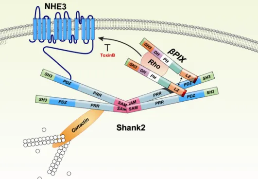
FIGURE 7. A model for the regulation of NHE3 through interaction withPix and Shank2. The molecular machinery implicated in the regulation of NHE3 at the apical membrane of epithelial cells is illustrated. Both NHE3 andPix form protein complexes through the PDZ domain of Shank2. The multimerization of Shank2 via its SAM domain recruits many regulatory and structural proteins, including NHE3 andPix to the complex. Also, the dimerization ofPix facilitates the activation of Rho GTPases. Consequently, NHE3 is directed to the cell surface by its interaction withPix and Shank2. SH3, Src homology3; SAM, sterile alpha motif; PRR, proline-rich region; DH, Dbl homology; PH, pleckstrin homology; and LZ, leucine zipper.
at YONSEI UNIVERSITY on January 7, 2014
http://www.jbc.org/
of Rho proteins influences vesicle movement, impacting endo-cytosis and exoendo-cytosis of integral plasma membrane proteins (35, 36). These encompass a variety of membrane transporters, including Na⫹ channels, K⫹ channels, nonselective cation channels, and CFTR (36 –38). In addition, Rho GTPases play a pivotal role in apical retention and targeting of NHE3 in epithe-lial cells (18). As a major activator of Rho GTPases, GEF pro-teins also play important roles in cytoskeleton rearrangement, membrane trafficking, and transporter regulation (39, 40). However, the molecular nature of the GEF responsible for NHE3 regulation remains obscure. Our data indicate thatPix forms a protein complex with NHE3 in rat epithelial tissues such as colon, kidney, and pancreas (Fig. 6). In addition,Pix up-regulates NHE3 surface expression and activity (Fig. 2), an effect that is blocked by the Rho GTPase inhibitor TxB (Fig. 5). These findings strongly suggest thatPix is one of the major GEFs responsible for NHE3 regulation at the apical membrane of epithelial cells.
Expression ofPix protein increased the membrane expres-sion and basal activity of NHE3, resembling the effects of Shank2 expression. Evidence presented in this study indicates that Shank2 is involved in thePix-induced up-regulation of NHE3 and vice versa. For example,Pix-induced up-regulation of NHE3 was abolished by the Shank2 PDZ- and SAM-domain mutants (Fig. 3). Protein complex formation betweenPix and NHE3 was also abolished by these dominant negative Shank2 mutants (Fig. 1). The PDZ domain of Shank2 mediates binding to NHE3 (5). It is also known that PDZ domain of Shank binds to the C-terminal PDZ-binding motif of Pix. Interestingly, Shank2 has only one PDZ domain. This raises the possibility thatPix and NHE3 may compete with each other to bind to Shank2, rather than form a protein complex. However, coim-munoprecipitation results showed that Shank2 actually medi-ates the association betweenPix and NHE3 (Fig. 1). The C terminus of Shank2 contains a SAM domain, which is known to mediate oligomerization (28). SAM domains are small protein modules that are present in many different proteins in diverse cellular compartments and are involved in wide-ranging func-tions, including scaffolding, signal transduction, and transcrip-tional regulation (41). Unlike other common protein modules in Shank2, such as proline-rich and SH3 domains, they can bind to other SAM domains and self-associate, which suggests that Shank proteins can multimerize in a tail-to-tail manner. The finding that SAM domain-deleted Shank2 mutants abolished the association between NHE3 andPix suggests that Shank2 exists as an oligomer, cross-linking multiple sets of protein complexes with NHE3 andPix.
An interesting finding in this study is that association betweenPix and NHE3 was readily detectable in rat epithelial tissues (Fig. 6), whereas the interaction was faint in the heter-ologous expression system without using the cross-linking agent DSP (supplemental Fig. 2). This raises a possibility that a
new protein may be involved in the stabilization of
Pix-Shank2-NHE3 complex in epithelial tissues. A further study
that identifies the complete binding partners of Pix and
Shank2 in epithelial cells will elucidate this question.
The regulatory mechanisms resulting from the association
of NHE3 with Shank2 andPix are summarized in Fig. 7.
Oligomerization of Shank2 in the apical cytoskeleton forms a large protein complex. This recruits many regulatory and
struc-tural proteins, including NHE3 andPix. In addition, Shank2
oligomerization may facilitate dimerization ofPix, which is
required for activation of Rho GTPases (42). Consequently, activation of Rho GTPases up-regulates membrane expression and activity of NHE3 at the apical membrane of epithelial cells.
Identification ofPix as a regulator of NHE3 will not only shed
new light on electroneutral sodium and hydrogen transport in the gastrointestinal and renal epithelia, but also provide another avenue for the correction of disease states caused by fluid electrolyte imbalance.
Acknowledgments—We thank Dr. Orson Moe (University of Texas Southwestern Medical Center, Dallas, TX) for kindly providing NHE3 antibodies. We also thank Dong-Su Jang for editorial assistance.
REFERENCES
1. Brett, C. L., Donowitz, M., and Rao, R. (2005) Am. J. Physiol. Cell Physiol.
288,C223–C239
2. Orlowski, J., and Grinstein, S. (2004) Pflugers Arch. 447, 549 –565 3. Fuster, D. G., Zhang, J., Shi, M., Bobulescu, I. A., Andersson, S., and Moe,
O. W. (2008) J. Am. Soc. Nephrol. 19, 1547–1556
4. Xiang, M., Feng, M., Muend, S., and Rao, R. (2007) Proc. Natl. Acad. Sci.
U.S.A. 104,18677–18681
5. Han, W., Kim, K. H., Jo, M. J., Lee, J. H., Yang, J., Doctor, R. B., Moe, O. W., Lee, J., Kim, E., and Lee, M. G. (2006) J. Biol. Chem. 281, 1461–1469 6. Gawenis, L. R., Stien, X., Shull, G. E., Schultheis, P. J., Woo, A. L., Walker,
N. M., and Clarke, L. L. (2002) Am. J. Physiol. Gastrointest. Liver Physiol.
282,G776 –G784
7. Donowitz, M., and Li, X. (2007) Physiol. Rev. 87, 825– 872
8. Weinman, E. J., Cunningham, R., and Shenolikar, S. (2005) Pflu¨gers Arch. 450,137–144
9. Lee, J. H., Richter, W., Namkung, W., Kim, K. H., Kim, E., Conti, M., and Lee, M. G. (2007) J. Biol. Chem. 282, 10414 –10422
10. Sheng, M., and Kim, E. (2000) J. Cell Sci. 113, 1851–1856
11. Lim, S., Naisbitt, S., Yoon, J., Hwang, J. I., Suh, P. G., Sheng, M., and Kim, E. (1999) J. Biol. Chem. 274, 29510 –29518
12. McWilliams, R. R., Breusegem, S. Y., Brodsky, K. F., Kim, E., Levi, M., and Doctor, R. B. (2005) Am. J. Physiol. Cell Physiol. 289, C1042–C1051 13. McWilliams, R. R., Gidey, E., Fouassier, L., Weed, S. A., and Doctor, R. B.
(2004) Biochem. J. 380, 181–191
14. Kim, J. Y., Han, W., Namkung, W., Lee, J. H., Kim, K. H., Shin, H., Kim, E., and Lee, M. G. (2004) J. Biol. Chem. 279, 10389 –10396
15. Park, E., Na, M., Choi, J., Kim, S., Lee, J. R., Yoon, J., Park, D., Sheng, M., and Kim, E. (2003) J. Biol. Chem. 278, 19220 –19229
16. Bagrodia, S., Taylor, S. J., Jordon, K. A., Van Aelst, L., and Cerione, R. A. (1998) J. Biol. Chem. 273, 23633–23636
17. Manser, E., Loo, T. H., Koh, C. G., Zhao, Z. S., Chen, X. Q., Tan, L., Tan, I., Leung, T., and Lim, L. (1998) Mol. Cell 1, 183–192
18. Alexander, R. T., Furuya, W., Sza´szi, K., Orlowski, J., and Grinstein, S. (2005) Proc. Natl. Acad. Sci. U.S.A. 102, 12253–12258
19. Hayashi, H., Sza´szi, K., Coady-Osberg, N., Furuya, W., Bretscher, A. P., Orlowski, J., and Grinstein, S. (2004) J. Gen. Physiol. 123, 491–504 20. Amemiya, M., Loffing, J., Lo¨tscher, M., Kaissling, B., Alpern, R. J., and
Moe, O. W. (1995) Kidney Int. 48, 1206 –1215
21. Bookstein, C., Xie, Y., Rabenau, K., Musch, M. W., McSwine, R. L., Rao, M. C., and Chang, E. B. (1997) Am. J. Physiol. 273, C1496 –C1505 22. Lee, J., Jung, I. D., Chang, W. K., Park, C. G., Cho, D. Y., Shin, E. Y., Seo,
D. W., Kim, Y. K., Lee, H. W., Han, J. W., and Lee, H. Y. (2005) Exp. Cell
Res. 307,315–328
23. Kurashima, K., Szabo´, E. Z., Lukacs, G., Orlowski, J., and Grinstein, S. (1998) J. Biol. Chem. 273, 20828 –20836
24. Bagrodia, S., and Cerione, R. A. (1999) Trends Cell Biol. 9, 350 –355
Pix, Shank2, and NHE3
at YONSEI UNIVERSITY on January 7, 2014
http://www.jbc.org/
25. Ahn, W., Kim, K. H., Lee, J. A., Kim, J. Y., Choi, J. Y., Moe, O. W., Milgram, S. L., Muallem, S., and Lee, M. G. (2001) J. Biol. Chem. 276, 17236 –17243
26. Lee, M. G., Wigley, W. C., Zeng, W., Noel, L. E., Marino, C. R., Thomas, P. J., and Muallem, S. (1999) J. Biol. Chem. 274, 3414 –3421
27. Zhao, H., Star, R. A., and Muallem, S. (1994) J. Gen. Physiol. 104, 57– 85 28. Thanos, C. D., Goodwill, K. E., and Bowie, J. U. (1999) Science 283,
833– 836
29. Hall, A. (1998) Science 279, 509 –514
30. Sza´szi, K., Grinstein, S., Orlowski, J., and Kapus, A. (2000) Cell. Physiol.
Biochem. 10,265–272
31. Zachos, N. C., Tse, M., and Donowitz, M. (2005) Annu. Rev. Physiol. 67, 411– 443
32. Moe, O. W., Amemiya, M., and Yamaji, Y. (1995) J. Clin. Invest. 96, 2187–2194
33. Donowitz, M., Janecki, A., Akhter, S., Cavet, M. E., Sanchez, F., Lampre-cht, G., Zizak, M., Kwon, W. L., Khurana, S., Yun, C. H., and Tse, C. M.
(2000) Ann. N.Y. Acad. Sci. 915, 30 – 42
34. Oh, W. K., Yoo, J. C., Jo, D., Song, Y. H., Kim, M. G., and Park, D. (1997)
Biochem. Biophys. Res. Commun. 235,794 –798
35. Runnels, L. W., Yue, L., and Clapham, D. E. (2002) Nat. Cell Biol. 4, 329 –336
36. Staruschenko, A., Nichols, A., Medina, J. L., Camacho, P., Zheleznova, N. N., and Stockand, J. D. (2004) J. Biol. Chem. 279, 49989 – 49994 37. Cheng, J., Wang, H., and Guggino, W. B. (2005) J. Biol. Chem. 280,
3731–3739
38. Storey, N. M., O’Bryan, J. P., and Armstrong, D. L. (2002) Curr. Biol. 12, 27–33
39. Schiller, M. R., Chakrabarti, K., King, G. F., Schiller, N. I., Eipper, B. A., and Maciejewski, M. W. (2006) J. Biol. Chem. 281, 18774 –18786
40. Zheng, Y. (2001) Trends Biochem. Sci. 26, 724 –732 41. Qiao, F., and Bowie, J. U. (2005) Sci. STKE 2005, re7
42. Koh, C. G., Manser, E., Zhao, Z. S., Ng, C. P., and Lim, L. (2001) J. Cell Sci.
114,4239 – 4251
at YONSEI UNIVERSITY on January 7, 2014
http://www.jbc.org/