342
Background : DNA prevalence and type distribution of human papillomavirus (HPV) varies
geographically. We investigated HPV prevalence and type distribution in Korean women using the MyHPV DNA chip testing. Methods : A total of 2,368 women from five regions of the coun-try underwent Pap smear examination and MyHPV chip testing. Results : Overall HPV posi-tivity was 15.8% and 78.4% in women with normal and abnormal cytology, respectively. High-risk HPV infection was strongly correlated with cytological atypia. In women with abnormal cytology, the five most common HPV types were 16, 58, 18, 52, and 56/53, and HPV16 was significantly the most common type in most geographical regions. After HPV16, HPV58, and 52 were the next most frequently detected types. Women with normal cytology, in contrast, showed heterogeneity in HPV type distribution. High-grade intraepithelial lesions infected with HPV16, 18, 31 or 45 are more likely to progress to carcinoma. Conclusions : The HPV chip test can provide useful data regarding HPV positivity and type. The most common HPV type in Korean women with abnormal cytology is HPV16, with HPV58 and 52 being frequently pre-sent. Our data may have important implications for vaccination programs and the development of cervical screening.
Key Words : Uterine cervix; Human papillomavirus; DNA chips; Papanicolaou test; South
Korea
Sung Ran Hong In Sun Kim1 Dong Won Kim2 Mi Jin Kim3 Ae Ree Kim4 Young Ok Kim5 Hye Sun Kim Seo Hee Rha6 Gyeong Sin Park7 Yong Koo Park8 Yong Wook Park9 Ho Sung Park10 Kwang Sun Suh11 Jin Hee Sohn12 Mi Kyung Shin13 Hoon Kyu Oh14 Ki Jung Yun15 Hye Kyoung Yoon16 Shi Nae Lee17 Ah Won Lee18 Hyo Jin Lee19 Hyun Yee Cho20 Chan Choi21 Woon Won Jung22
342
Prevalence and Genotype Distribution of Cervical Human Papillomavirus
DNA in Korean Women: A Multicenter Study
342 342 Corresponding Author
In Sun Kim, M.D.
Department of Pathology, Korea University Anam Hospital, 126-1 Anam-dong 5-ga, Seongbuk-gu, Seoul 136-705, Korea
Tel: 02-920-6373 Fax: 02-920-6576 E-mail: [email protected]
*MyGene Co. (Seoul, Korea) provided equipments and technical support for HPV DNA chip testing in each laboratory and donated the HPV DNA chip kits.
Department of Pathology, Kwandong University Cheil General Hospital & Women’s Healthcare Center, Seoul; 1Korea University Anam Hospital, Seoul; 2Soonchunhyang University Hospital, Seoul; 3Yeungnam University College of Medicine, Daegu; 4Korea University Guro Hospital, Seoul; 5Kosin University College of Medicine, Busan; 6Dong-A University College of Medicine, Busan; 7Catholic University, St. Mary’s Hospital, Seoul; 8Kyung Hee University Medical Center, Seoul; 9Hanyang University Kuri Hospital, Guri;10Chonbuk National University College of Medicine, Jeonju; 11Chungnam National University College of Medicine, Daejeon; 12Sunghyunkwan University Kangbuk Samsung Medical Center, Seoul; 13Hallym University, Gangnam Sacred Heart Hospital, Seoul; 14Daegu Catholic University Medical Center, Daegu; 15Wonkwang University College of Medicine, Iksan; 16Inje University Busan Paik Hospital, Busan; 17Ewha Womans University College of Medicine, Seoul; 18Catholic University, Gangnam St. Mary’s Hospital, Seoul; 19National Police Hospital, Seoul; 20Medical School Gil Medical Center, Incheon; 21Chonnam National University Hwasun Hospital, Hwasun; 22Department of Clinical Pathology, College of Health Science, Korea University, Seoul, Korea Received : May 25, 2009
Cytological screening using Papanicolaou (Pap)-stained smears for cervical cancer has proven to be a simple and effective method for reducing cervical cancer incidence and mortality during the last century. Even so, screening with Pap testing alone is unlike-ly to detect all cervical neoplasias. Molecular and epidemiologic studies have proven that virtually all cervical cancers and their precursor lesions are causally related to infection by HPV.1The
HPV family includes over 100 viral types, of which more than 40 infect the genital tract. These viruses can be divided into three groups: “high-risk (HR)” viruses associated with a high relative risk of cervical cancer, “low-risk (LR)” viruses associated with a low risk of cervical cancer, and “intermediate-risk (IR)” viruses occasionally associated with cervical cancer.2
In a prospective follow-up study, while the cumulative inci-dence of cervical intraepithelial neoplasia 3 (CIN3) was 4.4% among HR-HPV positive women followed-up for up to 45 mon-ths, it was only 0.24% among HR-HPV negative women.3
HR-HPV DNA testing has potential clinical applications, especial-ly in women with atypical squamous cells of undetermined sig-nificance (ASC-US),4postmenopausal women with low-grade
squamous intraepithelial lesion (LSIL),5follow-up of adolescents
with LSIL,6post-colposcopy follow-up,7and post-treatment
fol-low-up of high-grade CIN.8The American Cancer Society has
recommended HPV DNA testing in conjunction with cytolo-gy as a screening option for women aged 30 years and older,6
and has developed guidelines for use of the prophylactic HPV vaccine in the prevention of CIN and cervical cancer.9
Consid-ering these findings and recommendations, HR-HPV testing could have important roles in the management of women with abnormal cytology, primary cervical cancer screening, and the development of HPV vaccines.
There is great regional variability in HPV DNA prevalence and genotype distribution worldwide.10,11In a meta-analysis of women
with normal cytology, the prevalence of HPV was higher in less developed countries (15.5%) than in more developed countries (10.0%), and higher in younger as compared to older women.10
On the other hand, among women with invasive cervical cancer, the overall detected HPV prevalence and HPV type distribution appears to vary little between geographical regions.11In most
re-gions, after HPV16 and 18, HPV 45, 31, and 33 are the most prevalent in invasive squamous cell carcinomas. In Asia, however, HPV58 and 52 are found more common than HPV45, 31, and 33. Further research on HPV prevalence and HPV type distribu-tion would not only affect the development of cancer-screening HPV tests, but could also help predict the effect of HPV vaccines. At present, there are two testing systems for HPC detection
in widespread clinical use: hybrid capture technology and PCR-based methods. The MyHPV Chip kit (MyGene, Seoul, Korea), which is based on a combination of HPV DNA chip technolo-gy and DNA hybridization, has been developed and approved for clinical application by the Korean Food and Drug Adminis-tration. The chip kit tests for 24 different HPV genomic seque-nces: HPV 6, 11, 16, 18, 31, 33, 34, 35, 39, 40, 42, 43, 44, 45, 51, 52, 53, 54, 56, 58, 59, 66, 68, and 70.12
Presently, we used HPV DNA chip testing to gather infor-mation on HPV prevalence and HPV-type distribution accord-ing to region and cytology from a large sample of South Kore-an women.
MATERIALS AND METHODS
Study population and specimen collection
Source materials were gathered from 21 South Korea hospi-tals located in Seoul, Incheon, Gyeonggi, Daejeon (Chungnam), Daegu (Gyeongbuk), Busan, Jeonbuk, and Gwangju from Octo-ber 2005 to August 2006. These hospitals were divided into five regions: capital region (Seoul, Incheon and Gyeonggi), Daejeon, Daegu, Busan and Honam (Jeonbuk and Gwangju). The size of the participant sample in each region was based roughly on local population numbers. The study process in each hospital was per-formed under the same protocol. After routine medical exami-nation by gynecologists, HPV test samples and Pap smears were obtained from women who had no specific gynecological history within the last 3 months. All participating women gave writ-ten informed consent for this study. This study was approved by the ethical committees at all of participating hospitals that had such committees.
Cytology test
Conventional Pap smears or liquid-based cytological smears obtained using standard procedures were interpreted by the pathologists in each laboratory for the purposes of clinical use. Cytological diagnoses were classified by 2001 Bethesda System terminology. To minimize bias from cytological-interpretation error, we selected hospitals that have been inspected and accred-ited by the quality management programs of the Korean Soci-ety for Cytopathology. For quality assurance to detect errors of interpretation, Pap smears were rescreened with 10% random selection of slides by the regional study groups.
HPV test
Participating laboratories had an approximately 1-year prepara-tory period to familiarize themselves with the MyHPV DNA chip test before HPV testing started for study purposes. HPV tests in each laboratory were performed by the technicians in charge of HPV testing under the supervision of both the pathol-ogists and the MyGene laboratory. The risk levels of the 24 HPV types in the MyHPV Chip kit were classified into HR-HPV (HPV16, 18, 31, 33, 35, 39, 45, 51, 52, 56, 58, 59, and 68), IR-HPV (IR-HPV53 and 66), or LR-IR-HPV (IR-HPV6, 11, 34, 40, 42, 43, 44, 54, and 70).2
Samples for HPV testing were collected separately from Pap smears using endocervical brushes supplied with the MyHPV Chip kit. Briefly, DNA was isolated using an isolation kit (My-Gene) and nested PCR was performed for gene amplification. For the first PCR, a mixture of two set primers, MY09 (5′-CGT CCM ARR GGA WAC TGA TC-3′, 20 pmol) and MY11 (5′-GCM CAG GGW CAT AAY AAT GG-3′, 20 pmol), were used. For the second PCR, HPV primers GP5+ (5′-TTT GTT ACT GTG GTA GAT ACT AC-3′, 20 pmol) and GP6+ (5′ -GAA AAA TAA ACT GTA AAT CAT ATT C-3′, 20 pmol) were used. β-globin PCR amplification was used as an internal control. PCR products were subjected to electrophoresis in a 3% agarose gel and 250 and 150 bp PCR products were extract-ed after the first and the second PCR, respectively. Ten micro-liters of the second PCR product was denatured for 5 min at 95
℃and left in ice water for 5 min. The product was then mixed with hybridization buffer (MyGene). This mixture was spotted on the DNA chip and subjected to a hybridization reaction for 1 h in a hybridization chamber (Vision Scientific, Seoul, Korea) at 43℃. After hybridization, the product was rinsed with buffer I (2X standard saline citrate [SSC], 0.1% sodium dodecyl sul-fate [SDS]), buffer II (0.2X SSC) and buffer III (0.1X SSC) twice, twice and once, respectively, for 5 min. Hybridized HPV DNA was identified using a model G4000 scanner (GSI Lumonics, Billerica, MA, USA). A pair of oligonucleotide probes for each of the 24 HPV types was attached to each DNA chip. When a pair of specific probes fluoresced, the assay was determined to be positive for that HPV type.
Statistical analysis
For comparison of prevalence rates, HPV type-specific preva-lence and heterogeneity of HPV type, statistical analysis was per-formed using the chi-square test with Statistical Package Service
Solution software (SPSS for Windows, standard version 14.0; SPSS, Chicago, IL, USA). P-values <0.05 were considered statis-tically significant and 95% confidence intervals were calculated.
RESULTS
Study population
A total of 2,368 women from five regions in South Korea par-ticipated in this multicenter study. The largest sample consisted of cases from the capital region including Seoul, Incheon and Gyeonggi (1,225, 51.7%), followed by cases from Busan (391, 16.5%), Honam (350, 14.8%), Daegu (268, 11.4%) and Dae-jeon (134, 5.7%). According to the cytological interpretations, 1,960 participants had normal cytology and 408 had abnormal cytology. Abnormal cytological findings were composed of 171 atypical squamous cells (ASC), six atypical glandular cells (AGC), 86 LSIL, 101 high-grade squamous intraepithelial lesions (HSIL), one adenocarcinoma in situ (AIS), and 43 invasive carcinomas (IC). The 171 ASC included 149 ASC-US and 22 ASC cannot exclude HSIL (ASC-H), and the 43 IC included 38 squamons cell carcinomas (SCC) and five adenocarcinomas. The overall mean age of all 2,368 women was 45.7 years (range 20-94 years); wo-men with normal compared to abnormal cytology had mean ages of 45.5 and 47.0 years respectively.
Overall HPV prevalence
Overall, HPV DNA was detected in 629 (26.6%) of the 2,368 women. HPV frequencies in women with normal compared to abnormal cytology were significantly different (p=0.00). Among women with normal cytology, 15.8% (309 of 1,960) were HPV-positive compared to 78.4% (320 of 408) of women with abnor-mal cytology (Table 1). The prevalence of overall infection with single, multiple, and other types (presence of amplified HPV-PCR products without detection of specific HPV genotypes by the HPV DNA chip test) of HPV were 399 (63.4%), 92 (14.6%) and 138 (21.9%), respectively, among the 629 HPV-positive women. Within the normal and abnormal cytology groups, there were no significant differences in prevalence for infections with single, multiple, and other types of HPV (p>0.05), although women with abnormal cytology tended to be infected more frequently with multiple types (11.7% vs 17.5% for normal cytology).
HPV prevalence by region for women with normal cytology
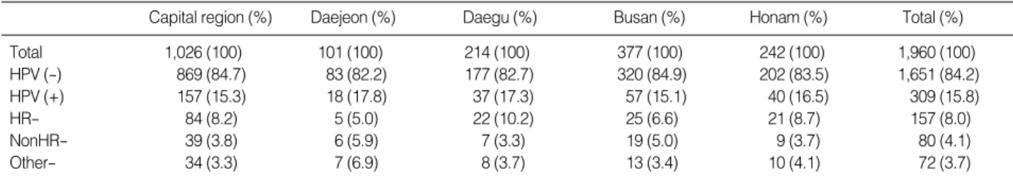
The samples obtained from the 1,960 women with normal cytology were composed of 1,026 (52.3%) cases from the capi-tal region, 377 (19.2%) cases from Busan, 242 (12.3%) cases from Honam, 214 (10.9%) cases from Daegu, and 101 (5.2%) cases from Daejeon. Comparing overall HPV positivity by region, positivity rates were similar for all five areas, ranging from 15.1 % in Busan to 17.8% in Daejeon (Table 2). HPV infection rates were varied widely by hospital (9.7-22.8%; data not shown). Additionally, HR-HPV frequencies in women with normal cytol-ogy differed by region. The average prevalence of HR-HPV infec-tion was 8.0%, with 8.2-10.2% infecinfec-tion rates in the high-fre-quency areas (Daegu, Honam and the capital region), and 5.0-6.6% infection rates in the low-frequency areas (Daejeon and Busan).
HPV prevalence in women with abnormal cytology
Overall HPV prevalence (78.4%) in women with abnormal cytology was correlated with cytological interpretation (p=0.00) (Table 3). The HPV-positivity rate was higher in women with HSIL+/AIS+ (91.7%, 133 of 145) and LSIL (90.7%, 78 of 86) compared to women with ASC-H/AGC (67.9%, 19 of 28) and ASC-US (60.4%, 90 of 149) (p=0.00). In terms of HPV-risk frequency, the HR-HPV infection rate was higher than the LR-HPV rate for all cytological diagnoses. Even among women with normal cytology, HR-HPV infections accounted for approximate-ly 51% (157 of 309) of infections in the HPV-positive women. The proportion of HR-HPV infection was significantly higher in women with HSIL+/AIS+ (73.8%, 107 of 145) compared to LSIL (53.5%, 46 of 86) (p=0.00), while relatively higher in wo-men with ASC-H/AGC (46.4%, 13 of 28) compared to ASC-US (34.2%, 51 of 149) (p>0.05). Region-specific HR-HPV preva-lence was variable and ranged from 39.2-71.3% in women with abnormal cytology: 25.0-60.0% in ASC-US, 35.7-80.0% in ASC-H/AGC, 40.4-87.5% in LSIL, and 65.6-80.0% in HSIL+/ AIS+ (data not shown).
HPV prevalence according to age
The participants were stratified into three age categories: young
Cytology HPV+
(%)
HPV infection pattern (%) Single Multiple Other Total (n=2,368) 629 (26.6) 399 (63.4) 92 (14.6) 138 (21.9) Normal (n=1,960) 309 (15.8) 201 (65.0) 36 (11.7) 72 (23.3) Abnormal* (n=408) 320 (78.4) 198 (61.9) 56 (17.5) 66 (20.6) *atypical squamous or glandular cells and more.
Table 1. HPV infection in women with normal and abnormal cy-tology
HR-, high risk HPV; NonHR-, non-high risk HPV.
Capital region (%) Daejeon (%) Daegu (%) Busan (%) Honam (%) Total (%)
Total 1,026 (100) 101 (100) 214 (100) 377 (100) 242 (100) 1,960 (100) HPV (-) 869 (84.7) 83 (82.2) 177 (82.7) 320 (84.9) 202 (83.5) 1,651 (84.2) HPV (+) 157 (15.3) 18 (17.8) 37 (17.3) 57 (15.1) 40 (16.5) 309 (15.8) HR- 84 (8.2) 5 (5.0) 22 (10.2) 25 (6.6) 21 (8.7) 157 (8.0) NonHR- 39 (3.8) 6 (5.9) 7 (3.3) 19 (5.0) 9 (3.7) 80 (4.1) Other- 34 (3.3) 7 (6.9) 8 (3.7) 13 (3.4) 10 (4.1) 72 (3.7)
Table 2. HPV prevalence according to region for normal cytology
HR-, high risk HPV; NonHR-, non-high risk HPV; ASC-US, atypical squamous cells, undetermined significance; ASC-H, atypical squamous cells, can-not exclude grade squamous intraepithelial lesion; AGC, atypical glandular cells; LSIL, low-grade squamous intraepithelial lesion; HSIL+, high-grade squamous intraepithelial lesion and more; AIS+, adenocarcinoma in situ and more.
Abnormal cytology (%)
Abnormal ASC-US ASC-H/AGC LSIL HSIL+/AIS+
Normal (%) Total 1,960 (100) 408 (100) 149 (100) 28 (100) 86 (100) 145 (100) HPV (-) 1,651 (84.2) 88 (21.6) 59 (39.6) 9 (32.1) 8 (9.3) 12 (8.3) HPV (+) 309 (15.8) 320 (78.4) 90 (60.4) 19 (67.9) 78 (90.7) 133 (91.7) HR- 157 (8.0) 217 (53.2) 51 (34.2) 13 (46.4) 46 (53.5) 107 (73.8) NonHR- 80 (4.1) 37 (9.1) 16 (10.7) 1 (3.6) 13 (15.1) 7 (4.8) Other- 72 (3.7) 66 (16.2) 23 (15.4) 5 (17.9) 19 (22.1) 19 (13.1)
group (women <30 year-of-age, n=143), women between 30-59-years-of-age (n=1,945); and elderly group (≥60 years-of-age, n=280). Table 4 shows the frequency of HPV infection accord-ing to these age categories. The women aged <30 years displayed the highest frequency of HPV infection among women with both normal (21.8%) and abnormal (93.3%) cytology. The second highest frequency was found in womens ≥60-years-of-age for both women with normal (19.3%) and abnormal (82.2%) cytol-ogy. The frequency of HR-HPV infection stratified by age cat-egories also had the same pattern.
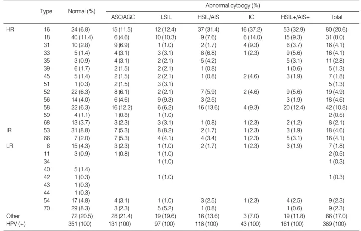
HPV type-specific prevalence by cytological interpretation
Women infected with multiple types of HPV were counted more than once, because each type in a multiple-type infection was considered individually to be a constituent type. Among the 491 HPV-positive women with single or multiple types (except-ing other-type infections), 237 women with normal cytology had a total of 279 HPV infections, and 254 women with abnormal cytology had a total of 323 HPV infections. Table 5 shows type-specific HPV prevalence according to cytological interpretation. The categories ASC-US and ASC-H/AGC were combined into
HR-HPV, high-risk HPV.
Normal cytology (%) Abnormal cytology (%)
No. HPV (+) HR-HPV No. HPV (+) HR-HPV
Age (years)
<30 (n=143) 110 24 (21.8) 16 (14.5) 33 31 (93.9) 21 (63.6)
30-59 (n=1,945) 1,643 245 (14.9) 121 (7.7) 302 229 (75.8) 151 (50.0)
≥60 (n=280) 207 40 (19.3) 20 (9.7) 73 60 (82.2) 45 (61.6)
Table 4. HPV prevalence according to age
HR, high risk; IR, intermediate risk; LR, low risk; ASC, atypical squamous cells; AGC, atypical glandular cells; LSIL, low-grade squamous intraepithelial lesion; HSIL, high-grade squamous intraepithelial lesion; AIS, adenocarcinoma in situ; IC, invasive carcinoma.
Abnormal cytology (%)
ASC/AGC LSIL HSIL/AIS IC HSIL+/AIS+ Total
Type Normal (%) HR 16 24 (6.8) 15 (11.5) 12 (12.4) 37 (31.4) 16 (37.2) 53 (32.9) 80 (20.6) 18 40 (11.4) 6 (4.6) 10 (10.3) 9 (7.6) 6 (14.0) 15 (9.3) 31 (8.0) 31 10 (2.8) 9 (6.9) 1 (1.0) 2 (1.7) 4 (9.3) 6 (3.7) 16 (4.1) 33 5 (1.4) 4 (3.1) 3 (3.1) 8 (6.8) 1 (2.3) 9 (5.6) 16 (4.1) 35 3 (0.9) 4 (3.1) 2 (2.1) 5 (4.2) 5 (3.1) 11 (2.8) 39 6 (1.7) 2 (1.5) 2 (2.1) 1 (0.8) 1 (0.6) 5 (1.3) 45 5 (1.4) 2 (1.5) 2 (2.1) 1 (0.8) 2 (4.6) 3 (1.9) 7 (1.8) 51 1 (0.3) 2 (1.5) 3 (3.1) 5 (1.3) 52 22 (6.3) 8 (6.1) 2 (2.1) 7 (5.9) 2 (4.6) 9 (5.6) 19 (4.9) 56 14 (4.0) 6 (4.6) 9 (9.3) 3 (2.5) 3 (1.9) 18 (4.6) 58 22 (6.3) 16 (12.2) 6 (6.2) 16 (13.6) 4 (9.3) 20 (12.4) 42 (10.8) 59 4 (1.1) 1 (0.8) 1 (1.0) 2 (0.5) 68 13 (3.7) 3 (2.3) 3 (3.1) 1 (0.8) 1 (2.3) 2 (1.2) 8 (2.1) IR 53 31 (8.8) 7 (5.3) 8 (8.2) 2 (1.7) 1 (2.3) 3 (1.9) 18 (4.6) 66 7 (2.0) 7 (5.3) 4 (4.1) 4 (3.4) 1 (2.3) 5 (3.1) 16 (4.1) LR 6 15 (4.3) 3 (2.3) 1 (1.0) 2 (1.7) 1 (2.3) 3 (1.9) 7 (1.8) 11 3 (0.9) 1 (0.8) 1 (1.0) 2 (0.5) 34 1 (1.0) 1 (0.3) 40 5 (1.4) 42 1 (0.3) 1 (1.0) 1 (0.3) 43 1 (0.3) 44 1 (0.3) 54 17 (4.8) 4 (3.1) 1 (1.0) 3 (2.5) 1 (2.3) 4 (2.5) 9 (2.3) 70 29 (8.3) 3 (2.3) 5 (5.2) 1 (0.8) 1 (0.6) 9 (2.3) Other 72 (20.5) 28 (21.4) 19 (19.6) 16 (13.6) 3 (7.0) 19 (11.8) 66 (17.0) HPV (+) 351 (100) 131 (100) 97 (100) 118 (100) 43 (100) 161 (100) 389 (100)
one category, ASC/AGC, because of the very small numbers of ASC-H/AGC study cases. HPV type-specific prevalence was significantly different between women with normal and abnor-mal cytology (p=0.00). In women with norabnor-mal cytology, the five most common types were HPV18, 53, 70, 16, and 52 or 58, in descending order of frequency. In women with abnormal cytol-ogy, HPV16, 58, 18, 52, and 56 or 53 were the five most com-mon types in descending order. Acom-mong women with HSIL+/ AIS+, the most common type was HPV16, followed by 58, 18, 33, and 52. The HPV16-positivity rate was correlated with the degree of cytological atypia (p=0.00). The five most common HPV types were found in variable frequencies according to cyto-logical interpretation, from 42% in women with normal cytol-ogy to 70% in women with IC.
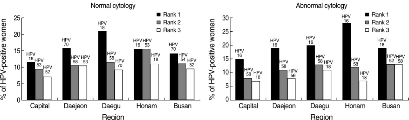
HPV type-specific prevalence by region
Fig. 1 displays the three most common HPV types by region in women with normal and abnormal cytology. Women with normal cytology had a more heterogeneous distribution of HPV types than those with abnormal cytology, but this difference was statistically insignificant (p>0.05). In women with normal cytol-ogy, six different types of HPV (18, 70, 53, 58, 54, and 52) were identified as among the three most common types in each of the five regions. The most common HPV type was HPV 18 in two areas (capital area and Daegu), HPV70 in two areas (Daejeon and Busan), and HPV16 or 53 together in one area (Honam). In wo-men with abnormal cytology, five types of HPV (16, 58, 18, 52, and 56) were among the three most common types in each of the five regions. HPV16 was identified as the most common type in all areas with the exception of Busan, in where the most common type was HPV18. Women with HSIL+/AIS+ were also infected most commonly with HPV16 in each region, again with the exception of Busan.
DISCUSSION
HPV prevalence in women with normal cytology
In this study, the overall prevalence of HPV DNA was 15.8% in women with normal cytology. This finding is similar to that of a large worldwide meta-analysis of women with normal cer-vical cytology.10The meta-analysis showed geographic
variabil-ity in HPV prevalence. Among Asian counties, China and South Korea showed the highest HPV prevalence (13.6%) compared to Taiwan and Japan (7.0%); the Philippines, Thailand and Viet-nam (7.2%); and India (7.5%). The authors inferred that this high HPV prevalence in eastern Asia might be related to liber-al sexuliber-al behavior patterns.
Prior reports conducted in different regions of South Korea have shown variable HPV infection rates in women with nor-mal cytology: 8.5% (75/751) in Busan, in a study using PCR-EIA (enzyme immunoassay) to detect 36 HPV types,1310%
(13/130) in Inchon, in a study using PCR with consensus primers to detect seven HR-HPV types,14and 19.4% (84/432) in
Buc-heon, Gyeonggi-do15or 35.1% (401/1143) in Bundang,
Gyeong-gi-do,16in studies using HPV DNA chip testing (Biomedlab,
Seoul, Korea) to detect 22 HPV types. Among these studies, two were performed in similar areas, but in different locations-Bucheon, Gyeonggi-do and Bundang, Gyeonggi-do- using the same HPV DNA chip kit (Biomedlab). These studies revealed different HPV-infection rates (19.4% vs 35.1%).15,16In our
study, the overall HPV-positivity rates were similar (15.1-17.8 %) across the five regions. However, widely varying HPV infec-tion rates was evident in the hospital studied (9.7- 22.8%), even though all of the participating institutions were university or referral hospitals. HR-HPV prevalence was also variable across the regions. The variability of HPV-positivity rates by hospital and region may be partly related to differences in the nature of
Fig. 1. Regional variation in the three most common HPV types among women with normal and abnormal cytology.
% of HPV-positive women 25 20 15 10 5 0
Capital Daejeon Daegu Honam Busan
Normal cytology Rank 1 HPV 18 HPV 53HPV 52 HPV 70 HPV 58 HPV53 HPV 18 HPV 58 HPV 70 HPV 16HPV53 HPV 18 HPV 70 HPV 54 HPV 52 Rank 2 Rank 3 % of HPV-positive women 30 25 20 15 10 5 0
Capital Daejeon Daegu Honam Busan
Region Region Abnormal cytology Rank 1 HPV 16 HPV 58 HPV 18 HPV 16 HPV 58 HPV 56 HPV 16 HPV 58 HPV 18 HPV 16 HPV 58 HPV 18 HPV 18 HPV 52HPV58 Rank 2 Rank 3
the patients as well as patient age and sexual activity between hospitals and regions.
HPV prevalence in women with abnormal cytology
This study demonstrated that overall HPV prevalence is much higher in women with abnormal cytology (78.4%) than in wo-men with normal cytology (15.8%) (p=0.00). The HPV preva-lence in SIL+/AIS+ women (LSIL and HSIL+/AIS+: 91.3%, 211 of 231) was significantly higher than that in women with ASC/AGC (ASC-US and ASC-H/AGC: 61.6%, 109 of 177) (p= 0.00). There was no significant difference, however, between women with ASC-US (60.4%) and ASC-H/AGC (67.3%), and between women with LSIL (90.7%) and HSIL+/AIS+ (91.7%) (p>0.05). In contrast, the prevalence of HR-HPV was different by cytological interpretation, showing a higher correlation with cytological atypia. As might be expected, the HR-HPV frequen-cy in SIL+/AIS+ women (LSIL and HSIL+/AIS+: 66.2%, 153 of 231) was significantly higher than that detected in women with ASC/AGC (ASC-US and ASC-H/AGC: 36.2%, 64 of 177) (p=0.00). Also, the HR-HPV infection rate was significantly higher in women with HSIL+/AIS+ (73.8%) compared to women with LSIL (53.5%) (p=0.00). Although the difference in preva-lence between women with ASC-US (34.2%) and ASC-H/AGC (46.4%) was not statistically significant (p>0.05), HR-HPV prevalence in women with ASC-H/AGC (46.4%) was interme-diate between that of women with ASC-US (34.2%) and HSIL+/ AIS+ (73.8%). These results are similar to those of other stud-ies.17-19ASC-H cytology, defined as cytological changes
sugges-tive of HSIL, would likely be associated with a higher posisugges-tive predictive value for the detection of underlying CIN2 or 3 than ASC-US, but would be less predictive than HSIL. In women with ASC-H, frequencies of HR-HPV DNA detection and under-lying high-grade CIN have been reported to be intermediate between those of ASC-US and HSIL.17On the other hand, CIN2
or 3 has been identified significantly more frequently in HR-HPV-positive H (32.7%) than in HR-HPV-negative ASC-H (1.2%).18HR-HPV can be associated with both LSIL and
HSIL, but it should be observed with significantly higher fre-quency in women with HSIL.3,19These data suggest that
HR-HPV types are closely related to the development of high-grade CIN and IC of the uterine cervix.
In our study, the overall prevalence of HR-HPV infection in women with LSIL was 53.5%, with a wide range of HR-HPV-positivity rates across regions (40.4-87.5%). Our HR-HPV infec-tion rate result was lower than the ALTS result (83%).20
More-over, the ALTS group described a much narrower range of HR-HPV-DNA positivity among women with LSIL (79.1-86.1%) than the range reported in our study; this may be related to the ages of the participants. The ALTS study population was young (mean age, 24.9-years-of-age) and only 17.6% of women with LSIL in their study were ≥30-years-of-age. In our study, how-ever, the population of women with LSIL (mean age, 42-years-of-age) had a wide age range, with only 16.3% (14 of 86) <30 years old and 58.1% (50 of 86), the majority, ≥40-years-of-age. Compared with younger women, older women are less likely to be HR-HPV positive and more likely to have false-positive LSIL cytology.21,22 While a cytological interpretation of LSIL could
be a very accurate marker for HPV infection in young women, many cases of LSIL in postmenopausal women could represent epithelial alterations of atrophic changes. The peri- or post-meno-pausal uterine cervix exhibits pseudokoilocytosis, including pro-minent perinuclear halos, nuclear hyperchromasia, variation in nuclear size and multinucleation, while showing low positivity for HPV DNA.21In older women with LSIL, the low HR-HPV
detection rate is thought to be associated with age-related epithe-lial alterations, termed postmenopausal squamous atypia.22
There-fore, our data demonstrating a relatively low prevalence and wide range of HR-HPV positivity in women with LSIL cytology could be explained by age-related phenomena. Based on this evidence, well-standardized HPV DNA testing on age-adjusted LSIL or HSIL samples could potentially be utilized as an alternative me-thod for quality-control purposes in cytology laboratories.
In regard to regional variability in women with ASC/AGC positive for HPV, our data also showed a wide range of HR-HPV positivity in women with US (25.0-60.0%) and ASC-H/AGC (35.7-80.0%). Similarly, the ALTS group showed a wide range of HR-HPV positivity among women with ASC-US (31-60%).23Regional variability in the ASC category is largely caused
by the cytological characteristics of ASC classification, because ASC encompasses heterogeneous biologic entities, from normal cytology unrelated to HR-HPV infection to, rarely, IC that is strongly related to HR-HPV infection.
HPV type-specific prevalence
In this study, HPV type-specific prevalence between women with normal and abnormal cytology was significantly different (p=0.00). The five most common HPV types in women with abnormal cytology were HPV16, 58, 18, 52, and 56 or 53. Among those, the prevalence of HPV16 (20.6%) was much hig-her than that of HPV58 (10.8%), HPV18 (8.0%) and HPV52
(4.9%). HPV16 was the most common viral type found in most abnormal cytological categories, including LSIL, HSIL/AIS, and IC. Increased HPV16 prevalence was correlated with an increased severity of cytological atypia: women with normal cytology, 6.8%; ASC/AGC, 11.5%; LSIL, 12.4%; HSIL/AIS, 31.4%; and IC, 37.2% (p=0.00). These observations suggest that HPV16 is the most common type of the HR-HPV and also the single most commonly identified HPV type in high-grade intraepithe-lial lesions and cancers.
Overall, the five most common types of HPV presently detect-ed were 16, 58, 18, 33, and 52 in women with HSIL/AIS, com-pared to HPV16, 18, 31 or 58, and 45 or 52 in IC. Among these HPV types, HPV16, 18, 31, and 45 were more prevalent in IC than HSIL/AIS. These results are consistent with a previous meta-analysis, in which HPV16, 18, and 45 were each more prevalent in women with invasive SCC than in women with HSIL, suggest-ing that an HSIL infected with HPV16, 18, or 45 preferentially progresses to SCC.24Our data also suggest that HPV16-, 18-, 31-,
or 45-infected HSIL preferentially progresses to SCC, although our number of study cases was too small for proper evaluation.
A worldwide meta-analysis reported geographic differences in HPV type-specific prevalence.11In SCC, HPV16, 18, 45, 31,
and 33 were the five most common HPV types in all regions except Asia. In Asia, HPV types 58 and 52 were more frequent-ly identified, especialfrequent-ly in eastern and southeastern areas. Other studies in Asia and Korea have presented the same data.25,26Our
study data confirms these previous reports. HPV types 58 and 52 were frequently identified as one of the five most common HPV types in women with normal and abnormal cytology. HPV types 16, 18, 52, and 58 occupied 58.5% of all HPV infections in women with HSIL/AIS, and 65.1% of all HPV infections in women with IC. Based on this data, HPV types 58 and 52 should also be considered in the development of HPV vaccines for Kore-an women.
Type-specific prevalence in women with normal cytology also has significant geographic regional variations.10,27In normal
cytol-ogy specimen the proportion of the most common HPV16 was variable across regions (12.3-25.5%), and heterogeneity between areas of Asia was significant.27Similarly, our results showed
het-erogeneity of HPV types across regions in women with normal cytology (p>0.05). The five most common types were HPV18, 53, 70, 16, and 52 or 58 in normal cytology, but the rank of HPV prevalence by type was variable across the regions. In contrast, abnormal cytological specimens showed homogeneity in HPV-type distribution across the regions. HPV16 was significantly the most common type in all regions except one (Busan) (p=
0.00). Our results support the findings of another study, which demonstrated that regional heterogeneity decreases with increas-ing lesion severity as HPV16 becomes increasincreas-ingly dominant.28
In summary, a total of 2,368 participants were gathered from five regions. The mean participant age was 45.7 years. The HPV chip test provided useful data regarding HPV positivity and viral type. Overall HPV and HR-HPV positivity were 15.8% and 8.0% in women with normal cytology, 78.4% and 53.2% in women with abnormal cytology, and 91.7% and 73.8% in women with HSIL+/AIS+. HR-HPV positivity showed better correlation with high-grade cytological atypia than did overall HPV positivity, which included non-HR-HPV types. The preva-lence of overall HPV in cases of LSIL (90.7%) was similar to that found in cases of HSIL+/AIS+ (91.7%), but HR-HPV preva-lence was much higher in women with HSIL+/AIS+ (73.8%) than in women with LSIL (53.5%) (p=0.00). This data suggest that HR-HPV types are closely related to the development of the cervical cancer. In descending order of frequency, the five most common HPV types in women with normal cytology were 18, 53, 70, 16, and 52 or 58, and HPV16, 58, 18, 52, and 56 or 53 in women with abnormal cytology. HPV type-specific distribution was correlated with cytological diagnosis. HPV16 was the most common type in abnormal cytological categories, including LSIL, HSIL/AIS, and IC across most regions. Howev-er, women with normal cytology showed increased heterogene-ity of HPV type distribution than those with abnormal cytology (p>0.05). Moreover, contrary to the findings in western countries, HPV58 and 52 were frequently identified as one of the five most common HPV types in women with both normal and abnormal cytology. HPV16, 18, 31, and 45 were more prevalent in IC than in HSIL/AIS, which supports findings that HSIL/AIS infected with HPV16, 18, 31, or 45 preferentially progresses to SCC. Although our study number of abnormal cytology specimens across the regions was not large enough for proper evaluation, our overall findings are consistent with other studies previously cited. Our results may provide important information in the development of cervical screening and vaccination programs.
REFERENCES
1. Wallin KL, Wiklund F, Angstro_m T, et al. Type-specific persistence of human papillomavirus DNA before the development of invasive cervical cancer. N Engl J Med 1999; 341: 1633-8.
2. Munoz N, Bosch FX, de Sanjose S, et al. International Agency for Research on Cancer Multicenter Cervical Cancer Study Group.
Epi-demiologic classification of human papillomavirus types associat-ed with cervical cancer. N Engl J Massociat-ed 2003; 348: 518-27.
3. Sherman ME, Lorincz AT, Scott DR, et al. Baseline cytology, human papillomavirus testing, and risk for cervical neoplasia: a 10-year co-hort analysis. J Natl Cancer Inst 2003; 95: 46-52.
4. Wright TC Jr, Lorincz A, Ferris DG, et al. Reflex human papilloma-virus deoxyribonucleic acid testing in women with abnormal Papan-icolaou smears. Am J Obstet Gynecol 1998; 178: 962-6.
5. Symmans F, Mechanic L, MacConnell P, DaSilva K, Stricker B, Nu-ovo GJ. Correlation of cervical cytology and human papillomavirus DNA detection in postmenopausal women. Int J Gynecol Pathol 1992; 11: 204-9.
6. Saslow D, Runowicz CD, Solomon D, et al. American Cancer Soci-ety guideline for the early detection of cervical neoplasia and cancer. CA Cancer J Clin 2002; 52: 342-62.
7. Guido R, Schiffman M, Solomon D, Burke L. Postcolposcopy man-agement strategies for women referred with low-grade squamous intraepithelial lesions or human papillomavirus DNA-positive atyp-ical squamous cells of undetermined significance: a two-year pros-pective study. Am J Obstet Gynecol 2003; 188: 1401-5.
8. Wright TC Jr, Cox JT, Massad LS, Carlson J, Twiggs LB. Wilkinson EJ. 2001 consensus guidelines for the management of women with cervical intraepithelial neoplasia. Am J Obstet Gynecol 2003; 189: 295-304.
9. Saslow D, Castle PE, Cox JT, et al. American Cancer Society Guide-line for human papillomavirus (HPV) vaccine use to prevent cervi-cal cancer and its precursors. CA Cancer J Clin 2007; 57: 7-28. 10. de Sanjose@ S, Diaz M, Castellsague@ X, et al. Worldwide prevalence
and genotype distribution of cervical human papillomavirus DNA in women with normal cytology: a meta-analysis. Lancet Infect Dis 2007; 7: 453-9.
11. Clifford GM, Smith JS, Plummer M, Munoz N, Franceschi S. Human papillomavirus types in invasive cervical cancer worldwide: a meta-analysis. Br J Cancer 2003; 88: 63-73.
12. Jung WW, Chun T, Sul D, et al. Strategies against human papillo-mavirus infection and cervical cancer. J Microbiol 2004; 42: 255-66. 13. Hong SH, Lee DH, Shin HR. Prevalence of human papillomavirus
infection in women in South Korea: incidence of positive HPV DNA and anti-VLPs in residents of Busan city. Korean J Cytopathol 2004; 15: 17-27.
14. Hwang T. Detection and typing of human papillomavirus DNA by PCR using consensus primers in various cervical lesions of Korean women. J Korean Med Sci 1999; 14: 593-9.
15. Kahng J, Lee HJ. Clinical efficacy of HPV DNA Chip test in the era of HPV vaccination: 1,211 cases, a single institution study. Korean J
Lab Med 2008; 28: 70-8.
16. An HJ, Cho NH, Lee SY, et al. Correlation of cervical carcinoma and precancerous lesions with human papillomavirus (HPV) genotypes detected with the HPV DNA chip microarray method. Cancer 2003; 97: 1672-80.
17. Sherman ME, Solomon D, Schiffman M. Qualification of ASCUS. A comparison of equivocal LSIL and equivocal HSIL cervical cytology in the ASCUS LSIL Triage Study. Am J Clin Pathol 2001; 116: 386-94. 18. Bandyopadhyay S, Austin RM, Dabbs D, Zhao C. Adjunctive human
papillomavirus DNA testing is a useful option in some clinical set-tings for disease risk assessment and triage of females with ASC-H Papanicolaou test results. Arch Pathol Lab Med 2008; 132: 1874-81. 19. The Atypical Squamous Cells of Undetermined
Significance/Low-Grade Squamous Intraepithelial Lesions Triage Study (ALTS) Group. Human papillomavirus testing for triage of women with cytologic evidence of low-grade squamous intraepithelial lesions: baseline data from a randomized trial. J Natl Cancer Inst 2000; 92: 397-402. 20. Jovanovic AS, McLachlin CM, Shen L, Welch WR, Crum CP.
Post-menopausal squamous atypia: a spectrum including “pseudo- koilo-cytosis”. Mod Pathol 1995; 8: 408-12.
21. Symmans F, Mechanic L, MacConnell P, DaSilva K, Stricker B, Nuovo GJ. Correlation of cervical cytology and human papillomavirus DNA detection in postmenopausal women. Int J Gynecol Pathol 1992; 11: 204-9.
22. Solomon D, Schiffman M, Tarone R; ALTS Study group. Compari-son of three management strategies for patients with atypical squa-mous cells of undetermined significance: baseline results from a randomized trial. J Natl Cancer Inst 2001; 93: 293-9.
23. Bae JH, Lee SJ, Kim CJ, et al. Human papillomavirus (HPV) type distribution in Korean women: a meta-analysis. J Microbiol Biotech-nol 2008; 18: 788-94.
24. Clifford GM, Smith JS, Aguado T, Franceschi S. Comparison of HPV type distribution in high-grade cervical lesions and cervical cancer: a meta-analysis. Br J Cancer 2003; 89: 101-5.
25. Bao YP, Li N, Smith JS, Qiao YL, ACCPAB Members. Human papil-lomavirus type distribution in women from Asia: a meta-analysis. Int J Gynecol Cancer 2008; 18: 71-9.
26. Clifford GM, Gallus S, Herrero R, et al. IARC HPV Prevalence Sur-veys Study Group. Worldwide distribution of human papillomavi-rus types in cytologically normal women in the International Agen-cy for Research on Cancer HPV prevalence surveys: a pooled anal-ysis. Lancet 2005; 366: 991-8.
27. Herrero R, Castle PE, Schiffman M, et al. Epidemiologic profile of type-specific human papillomavirus infection and cervical neopla-sia in Guanacaste, Costa Rica. J Infect Dis 2005; 191: 1796-807.