저작자표시-비영리-변경금지 2.0 대한민국 이용자는 아래의 조건을 따르는 경우에 한하여 자유롭게 l 이 저작물을 복제, 배포, 전송, 전시, 공연 및 방송할 수 있습니다. 다음과 같은 조건을 따라야 합니다: l 귀하는, 이 저작물의 재이용이나 배포의 경우, 이 저작물에 적용된 이용허락조건 을 명확하게 나타내어야 합니다. l 저작권자로부터 별도의 허가를 받으면 이러한 조건들은 적용되지 않습니다. 저작권법에 따른 이용자의 권리는 위의 내용에 의하여 영향을 받지 않습니다. 이것은 이용허락규약(Legal Code)을 이해하기 쉽게 요약한 것입니다. Disclaimer 저작자표시. 귀하는 원저작자를 표시하여야 합니다. 비영리. 귀하는 이 저작물을 영리 목적으로 이용할 수 없습니다. 변경금지. 귀하는 이 저작물을 개작, 변형 또는 가공할 수 없습니다.
Diagnostic and clinicopathological
significance of Ki67 mRNA expression
in cervical cancer tissue
Dawn Chung
Department of Medicine
Diagnostic and clinicopathological
significance of Ki67 mRNA expression
in cervical cancer tissue
Directed by Professor Eun Ji Nam
The Master's Thesis
submitted to the Department of Medicine,
the Graduate School of Yonsei University
in partial fulfillment of the requirements for the degree
of Master of Medical Science
Dawn Chung
This certifies that the Master's Thesis of
Dawn Chung is approved.
---
Thesis Supervisor : Eun Ji Nam
---
Thesis Committee Member#1 : Jae Myun Lee
---
Thesis Committee Member#2 : Kwang Hwa Park
The Graduate School
Yonsei University
ACKNOWLEDGEMENTS
First and foremost, I would like to thank God for guiding me
through this merciful journey and for making this dissertation
possible.
I would like to express the deepest appreciation to Professor Eun
Ji Nam, who has the attitude and the substance of genius. She
continually and convincingly conveyed a spirit of adventure in
regard to research and excitement in regard to teaching. Her keen
scientific intuition has inspired and enriched myself as a student, a
researcher and a medical scientist. And I also thank Professor
Kwang Hwa Park and Professor Jae Myun Lee for taking time out
of their busy schedules to provide me with invaluable advices.
I would like to extend my deepest gratitude to Professor Young
Tae Kim who have always supported and encouraged me
academically and spiritually. I truly thank him for always guiding
me in the right direction to be a well-trained gynecologist and a
rational researcher. I greatly appreciate to Professor Hyeyoung
Lee for her crucial opinions and taking her precious time to
review this thesis during the whole period. And special thanks to
Sunyoung Park for her technical assistance provided during
excellent experiments that have been extremely helpful to
complete this thesis.
I have to thank Professor Yong-Won Park, Professor Byung
Seok Lee, Professor Sang Wook Bai, Professor Jae-Hoon Kim,
Professor Sang Wun Kim, Professor Sunghoon Kim, Professor
Young Sik Choi, and Professor Ja-Young Kwon for their grateful
guidance through my whole OBGYN training. I have learned
greatly under their considerable instruction.
I take pride in being a part of the department of Obstetrics and
Gynecology at Severance Hospital of Yonsei University. I wish
the best of luck to all the members of the department.
I owe a deep sense of gratitude to Professor Seong Jin Choi,
Professor Young Jin Lee, and Professor Dong Soo Cha for giving
me unstinting support and encouragement to follow my academic
dreams.
I would like to end by express my deep love to my dearest mother
and father for their boundless devotion. I especially announce that,
I respect my father, Dr. Kun Sub Chung, who has intellectual
endeavor and faithfulness as a senior scientist. He was the first
person who led me to this path of research by stimulating my
curiosity to have a scientific inquiry from the very beginning of my
childhood.
I dearly love you all and thank you.
Dawn Chung
June 1st, 2017
<TABLE OF CONTENTS>
ABSTRACT ···1
I. INTRODUCTION ···3
II. MATERIALS AND METHODS···5
III. RESULTS ···10
IV. DISCUSSION ···19
V. CONCLUSION ···21
REFERENCES ···23
LIST OF TABLES
Table 1. Clinical characteristics of patients ··· 9
Table 2. Ki67 mRNA expression in matched non-cancerous and
cancerous lesions ··· 11
Table 3. Sensitivity, specificity, NPV, and PPV of Ki67 mRNA
levels in cervical cancer and normal tissues ··· 13
Table 4. Association between Ki67 mRNA expression levels and
histologically diagnosed cervical grades ··· 15
Table 5. Ki67 mRNA expression levels correlated with clinico-
pathological parameters in 79 cervical cancer patients ··· 18
LIST OF FIGURES
Figure 1. Receiver operating characteristics (ROC) curve
analysis ··· 14
Figure 2. Dot plots of Ki67 mRNA levels in histologically
ABSTRACT
Diagnostic and clinicopathological significance of Ki67 mRNA
expression in cervical cancer tissue
Dawn Chung
Department of Medicine
The Graduate School, Yonsei University
(Directed by Professor Eun Ji Nam)
Ki67 is a key biomarker associated with cancer cell proliferation and
poor prognosis. We previously evaluated the diagnostic potential of
quantitatively measured Ki67 mRNA levels in formalin-fixed
paraffin-embedded (FFPE) cervical cancer tissue samples. In the present
study, we continued this avenue of research using quantitative reverse
transcription PCR (RT-qPCR) to measure Ki67 mRNA levels in FFPE
cervical tissues and performed an assessment with each clinical
prognostic factor of patients. A total 190 FFPE cervical tissue samples
comprised of 80 squamous cell carcinoma (SCC), 10 adenocarcinoma
(ADC), 30 HSIL, 30 LSIL, and 40 normal cervical tissue samples were
obtained. Using this RT-qPCR assay, the predictive value of Ki67 in
cases with low-grade squamous intraepithelial lesions (LSIL) and those
with high-grade squamous intraepithelial lesions (HSIL) were measured.
As a result, Ki67 mRNA levels were increased in SCC and ADC
cervical cancer tissues (n = 90) compared to those in normal cervical
tissues (n = 40) (P < 0.001). The diagnostic validity of the Ki67 mRNA
assay was evaluated and demonstrated a sensitivity of 93.3% (95%
confidence interval (CI) = 86.1 to 97.5) and a specificity of 97.5% (95%
2
CI = 86.8 to 99.9). Ki67 mRNA expression level was 93.3% for cervical
cancer, 40.0% for HSIL, 13.3% for LSIL, and 2.5% for normal tissue
samples. Furthermore, high levels of Ki67 mRNA expression in cervical
cancer were associated with lymph node status (P = 0.02). In conclusion,
Ki67 mRNA assay can provide an additional accurate approach for
molecular diagnosing cervical cancer, and also predict prognosis of
cervical cancer depending on LSIL and/or HSIL status.
---
Key words: Cervical cancer, Ki67 mRNA, RT-qPCR, molecular diagnosing
3
Diagnostic and clinicopathological significance of Ki67 mRNA
expression in cervical cancer tissue
Dawn Chung
Department of Medicine
The Graduate School, Yonsei University
(Directed by Professor Eun Ji Nam)
I. INTRODUCTION
Cervical cancer is the third most common malignancy in women globally and one of the leading causes of morbidity and mortality in women worldwide. The World Health Organization estimates that approximately 527,600 women are
newly diagnosed and there are 265,700 deaths from cervical cancer every year.1
Human papillomavirus (HPV) is a major cause of cervical cancer and is the most
common sexually transmitted pathogen among women and men.2 So the detection
of HPV is routinely performed in exfoliated infected cervical cells or tissues of patients with cervical cancer or patients with precancerous lesion.3-7 But most high-risk HPV infections resolve spontaneously within 1 to 2 years.
Several studies have tried to investigate the molecular mechanisms underlying cervical cancer carcinogenesis to identify potential diagnostic or prognostic
biomarkers for cervical cancer.8-11 For example, putative molecular markers such
as p16, p53, and Ki67 have been identified in cervical carcinogenesis. Their respective coding genes and proteins have been characterized, and their roles in the process have been studied with the aim of improving diagnosis and treatment of cervical cancer. Some studies found that among these different markers, immunohistochemical (IHC) staining of Ki67 was an effective method for the
4
prognosis of different tumor types.12-15 Ki67 is associated with cell cycle activity and is expressed at varying levels during G1, S, G2, and M phases, but is not expressed in G0.10-16 In the previous research, the potential diagnostic value of
Ki67 in cervical cancer was demonstrated by quantitative measuring of Ki67
mRNA levels in formalin-fixed paraffin-embedded (FFPE) samples.17
The concept of precancerous disease of the cervix was first presented in 1947, when it was noticed that epithelial changes would be identified that had
appearances of invasive cancer but were confined to the epithelium.18 Comparing
to aggressive treatments of carcinoma in situ (CIS), the dysplasias were believed to be less significant and were not treated or were treated only by colposcopic biopsy and cryosurgery. In 1968, the concept of cervical intraepithelial neoplasia (CIN) was presented by Richart, and he suggested these dysplasias have potential for progression.19 Most untreated low-grade CIN lesions regress spontaneously, whereas untreated high‐grade CIN refers to a lesion that may progress to invasive carcinoma.20 Without mitotic activity, proliferating metaplasia could not be called dysplasia or CIN because of very rare progression to invasive cancer. The criteria for diagnosing intraepithelial lesion may be various according to pathologist. However, the key features are cellular immaturity, disorganization, and nuclear abnormality including increased mitotic activity. The extent of immature cellular proliferation, nuclear atypia and mitotic activity identifies the degree of intraepithelial neoplasia.21 The lesion is specified as CIN grade 1, if the extent of mitosis and immature cells is limited to the lower third of the epithelium. Involvement of the middle and upper thirds is designated to CIN grade 2 and 3, respectively. Two-tier system including low-grade and high-grade squamous intraepithelial lesion was introduced as an alternative cytopathologic classification in the late 1980s, and has been translated into histopathological purpose, especially in North America.22-24
5
tumor of female reproductive organs, two-tier system is preferred again. This SIL system was conceptually founded on the presence of two forms of HPV infection, with productive infection causing low-grade squamous intraepithelial lesion (LSIL) and transforming infection leading to high-grade squamous intraepithelial lesion (HSIL), whereas the CIN system focused on identification of CIN lesions
and determination of their grades.25 These LSILs and HSILs are important
precancerous courses in the development of cervical cancer, but their exact
carcinogenesis has had divergent opinions.26, 27 Cox et al performed a
meta-analysis and concluded that the likelihood of progression from LSIL to
HSIL was approximately 10% within 2 years.28 Nevertheless, there is no accurate
approach to identify which LSILs will progress to HSILs. Therefore, a better understanding of cervical cancer development is necessary to improve the management of either LSILs or HSILs.
A purpose of the current study is to investigate the relationship between Ki67 mRNA level and clinicopathological measures of patients with cervical cancer. In this study, the discriminatory power of Ki67 mRNA assay in cervical lesions using quantitative reverse transcription PCR (RT-qPCR) was tested. Then clinical predictive relevance of Ki67 mRNA in each histologic grade of cervix were evaluated by selecting a diagnostic cutoff value using one hundred and ninety FFPE cervical tissue samples.
II. MATERIALS AND METHODS
Patients and samples
One hundred and ninety FFPE cervical tissue samples were collected which were finished pathological diagnosis at the Department of Pathology, Yonsei University Wonju Severance Christian Hospital between January 2010 and
6
December 2014. The study was approved by the Institutional Ethics Committee of Yonsei University Wonju College of Medicine (approval no. CR315052), and all subjects were provided written informed consent. Of the 190 FFPE tissue samples collected, 40 (21.1%) were normal, 30 (15.8%) were LSIL, 30 (15.8%) were HSIL, 10 (5.3%) were adenocarcinoma (ADC), and 80 (42.1%) were squamous cell carcinoma (SCC) (Table 1). FFPE normal tissue samples included 40 chronic cervicitis specimens obtained from patients who underwent a hysterectomy for other benign gynecological diseases such as leiomyoma and adenomyosis. To analyze clinicopathological prognostic parameters of 90 FFPE samples of cervical cancer, electrical medical records of the patients were retrospectively reviewed and 11 FFPE cervical cancer samples had missing medical information because of referral to other institutes.
Deparaffinization of FFPE tissue and total RNA extraction
Three 10-μm sections from each paraffin block of cervical tissue were used for total RNA extraction. Extractions were performed using the Qiagen RNeasy FFPE mini kit (Qiagen, Hilden, Germany) according to the manufacturer’s protocol. RNA purity and concentration were determined by measuring absorbance at 260 nm and 280 nm using a spectrophotometer (Infinite 200, Tecan, Salzburg, Austria). All RNA preparation and handling was performed in a laminar flow hood under RNase-free conditions. Isolated RNA was stored at -70°C.
cDNA synthesis
Complementary DNA (cDNA) was synthesized using the M-MLV Reverse Transcriptase kit (Invitrogen, Carlsbad, CA, USA) and random hexamers (Invitrogen) according to the manufacturer’s recommendations. Briefly, 10 μL of total RNA was added to a master mix containing 10 mM dNTPs at neutral pH, 0.25 μg random hexamers, and 5-μL DEPC-treated water. Reactions were
7
incubated at 65°C for 5 min and chilled on ice. A mixture of 4 μL 5× First-Strand Buffer, 2 μL 0.1 M dithiothreitol, and 1 μL M-MLV reverse transcriptase (RT) was added, and cDNA synthesis was synthesized at 25°C for 10 min, followed by 37°C for 50 min, and 70°C for 15 min.
Ki67 mRNA RT-qPCR assay
Quantitative real-time PCR amplification of the OPTIMYGENE Ki67 mRNA assay (Optipharm, Osong, Korea) was performed in 10 μL 2× Thunderbird probe qPCR mix (Toyobo, Osaka, Japan), 3 μL primer and TaqMan probe mixture, 2 μL template cDNA, and distilled water (DW) to a final volume of 20 μL per sample. Positive and negative controls were included. No-template controls were included in each run and consisted of sterile DW instead of template DNA. PCR cycling was 95°C for 3 min, followed by 40 cycles of 95°C for 3 seconds, and 55°C for 30 seconds. mRNA levels were quantified by determining the cycle threshold (CT), which is defined as the number of PCR cycles required for fluorescence to exceed a value significantly higher than that of the background fluorescence. To avoid false negatives because of mRNA degradation, glyceraldehyde-3-phosphate dehydrogenase (GAPDH) was used as an internal control. The amount of Ki67
mRNA was determined using the comparative CT method (ΔΔCT method), 29
measuring mRNA relative to a reference gene using CFX Manager Software v1.6 (Bio-Rad, Hercules, CA, USA). The amount of Ki67 mRNA was normalized to the internal housekeeping gene GAPDH using the following equation:
ΔΔCT = (ΔCT [target sample] - ΔCT [normal sample])
Statistical analysis
8
Jolla, CA, USA) and SPSS (Statistical Package for the Social Sciences) v23.0 (SPSS Inc., Chicago, IL, USA). The Wilcoxon matched-pairs test was used to compare nonparametric-matched samples, and the Student’s t-test and 95% confidence interval (CI) were used to determine statistical significance. Receiver operating characteristic (ROC) curves were used to predict cutoff values of the marker. Sensitivity and specificity were calculated using MedCalc v12.5 (MedCalc software, Ostend, Belgium). The Pearson’s chi-square test was used to analyze associations between the positivity of Ki67 mRNA expression and histologically diagnosed samples. For all tests, a P value < 0.05 was considered statistically significant.
9
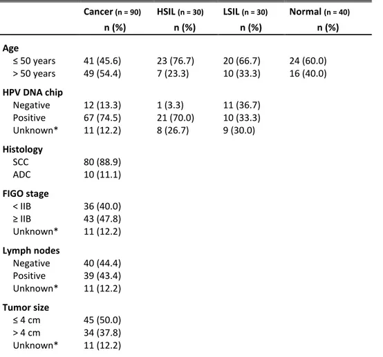
Table 1. Clinical characteristics of patients
Cancer (n = 90) HSIL (n = 30) LSIL (n = 30) Normal (n = 40)
n (%) n (%) n (%) n (%) Age ≤ 50 years 41 (45.6) 23 (76.7) 20 (66.7) 24 (60.0) > 50 years 49 (54.4) 7 (23.3) 10 (33.3) 16 (40.0) HPV DNA chip Negative 12 (13.3) 1 (3.3) 11 (36.7) Positive 67 (74.5) 21 (70.0) 10 (33.3) Unknown* 11 (12.2) 8 (26.7) 9 (30.0) Histology SCC 80 (88.9) ADC 10 (11.1) FIGO stage < IIB 36 (40.0) ≥ IIB 43 (47.8) Unknown* 11 (12.2) Lymph nodes Negative 40 (44.4) Positive 39 (43.4) Unknown* 11 (12.2) Tumor size ≤ 4 cm 45 (50.0) > 4 cm 34 (37.8) Unknown* 11 (12.2)
ADC, adenocarcinoma; LSIL, low-grade squamous intraepithelial lesion; HSIL, high-grade squamous intraepithelial lesion; HPV, human papillomavirus; SCC, squamous cell carcinoma.
10
III. RESULTS
Patient characteristics
Patient characteristics are summarized in Table 1. One hundred and ninety FFPE tissue samples were used in this study of which 90 (47.3%) were cancer, 30 (15.8%) were HSIL, 30 (15.8%) were LSIL, and 40 (21.1%) were normal samples. For cervical cancer cases, data on histology, FIGO stage, tumor size, and lymph node metastasis were retrospectively reviewed from patient electrical medical records (EMR) (Table 1). Among 90 cases of cervical cancer, 80 (88.9%) cases were histologically diagnosed as SCC and 79 cases were analyzed their clinicopathological information archived from EMR. 43 (47.8%) cases showed FIGO stage IIB and more, 39 (43.4%) cases showed positive in lymph node metastasis, and 34 (37.8%) cases had more than 4 cm of tumor size.
Ki67 mRNA levels for matched FFPE non-cancerous and cancerous tissues
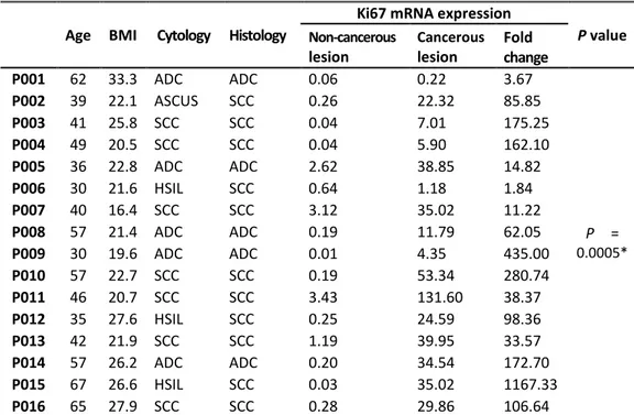
To evaluate the validity of the Ki67 mRNA assay, 16 matched-pairs of non-cancerous and cancerous lesion FFPE samples of the same cervical cancer patient from 16 cervical cancer patients were tested. Using ∆∆CT to determine mRNA levels in matched tissue samples, we found that levels of Ki67 mRNA in cancer tissues were higher than those in matched normal tissues (P = 0.0005). The Ki67 mRNA levels in normal tissues ranged from 0.01 to 3.43 compared to those in cancer tissues ranged from 0.22 to 131.60. The Ki67 mRNA levels in cervical cancer tissues were 1.84 to 1167.33-fold higher compared to levels found in normal tissues (Table 2).
Diagnostic value of Ki67 for cervical cancer
ROC curve analysis was performed to determine the optimal diagnostic cutoff value for the assay to discriminate normal tissues from those with cervical cancer.
11
Table 2. Ki67 mRNA expression in matched non-cancerous and cancerous lesions
Age BMI Cytology Histology
Ki67 mRNA expression
P value Non-cancerous lesion Cancerous lesion Fold change P001 62 33.3 ADC ADC 0.06 0.22 3.67 P = 0.0005* P002 39 22.1 ASCUS SCC 0.26 22.32 85.85 P003 41 25.8 SCC SCC 0.04 7.01 175.25 P004 49 20.5 SCC SCC 0.04 5.90 162.10 P005 36 22.8 ADC ADC 2.62 38.85 14.82 P006 30 21.6 HSIL SCC 0.64 1.18 1.84 P007 40 16.4 SCC SCC 3.12 35.02 11.22 P008 57 21.4 ADC ADC 0.19 11.79 62.05 P009 30 19.6 ADC ADC 0.01 4.35 435.00 P010 57 22.7 SCC SCC 0.19 53.34 280.74 P011 46 20.7 SCC SCC 3.43 131.60 38.37 P012 35 27.6 HSIL SCC 0.25 24.59 98.36 P013 42 21.9 SCC SCC 1.19 39.95 33.57 P014 57 26.2 ADC ADC 0.20 34.54 172.70 P015 67 26.6 HSIL SCC 0.03 35.02 1167.33 P016 65 27.9 SCC SCC 0.28 29.86 106.64
*Ki67 mRNA levels were higher in cancerous tissue lesion than that found in matched Non-cancerous tissue lesion (Wilcoxon matched-pairs test). ADC, adenocarcinoma; ASCUS, Atypical cell of undetermined significance; BMI, body mass index; HSIL, high-grade squamous intraepithelial lesion; SCC, squamous cell carcinoma.
12
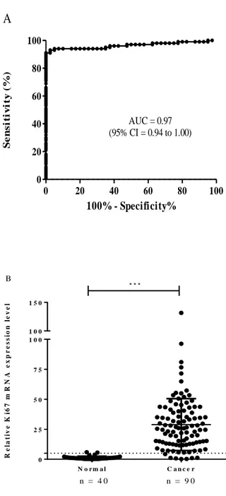
Ki67 mRNA levels were analyzed in 90 FFPE cancer tissues and 40 FFPE normal tissues, and found that the area under the ROC curve (AUC) was 0.97 (95% CI = 0.94 to 1.00, P < 0.001, Figure 1A). Besides, Ki67 mRNA levels in cervical cancer tissues were significantly increased compared to those in normal cervical tissues. Based on these findings, a diagnostic cutoff (threshold) was set of 5 (P < 0.001, Figure 1B). Using a threshold of 5, the assay had a sensitivity of 93.3% (95% CI = 86.1 to 97.5), a specificity of 97.5% (95% CI = 86.8 to 99.9), a positive predictive value of 98.8% (95% CI = 93.6 to 100.0), and a negative predictive value of 86.7% (95% CI = 73.2 to 95.0) (Table 3).
Ki67 mRNA levels in histologically diagnosed FFPE cervical tissues
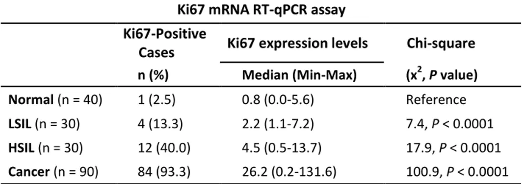
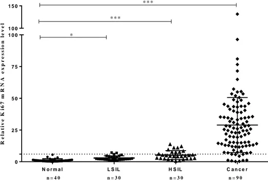
Ki67 mRNA levels were evaluated with histologically diagnosed cervical tissues to determine whether Ki67 mRNA levels could discriminate between normal to precancerous tissues and cancer tissues. Ki67 mRNA levels were ranged from 0.0 to 5.6 (median 0.8) in 40 normal tissues, from 1.1 to 7.2 (2.2) in 30 LSIL tissues, from 0.5 to 13.7 (4.5) in 30 HSIL tissues, and from 0.2 to 131.6 (26.2) in 90 cancer tissues. The highest Ki67 mRNA levels were expressed in cancer tissues with lowering in histological sequence but still elevated levels were checked in HSIL and LSIL tissues compared to the levels in normal tissues (P < 0.0001) (Figure 2 and Table 4). Using a cutoff value of 5, positivity for Ki67 was 2.5% (1/40 cases) for normal, 13.3% (4/30 cases) for LSIL, 40.0% (12/30 cases) for HSIL, and 93.3% (84/90 cases) for cancer. We found that this Ki67 mRNA assay using a diagnostic threshold of 5 discriminated between normal and abnormal cervical lesions (P < 0.001) (Table 4).
13
Table 3. Sensitivity, specificity, NPV, and PPV of Ki67 mRNA levels in
cervical cancer and normal tissues
by RT-qPCR assay Ki67 mRNA
(n = 130) 95% CI Sensitivity 93.30% 86.1-97.5 Specificity 97.50% 86.8-99.9 PPV 98.80% 93.6-100.0 NPV 86.70% 73.2-95.0
14 0 20 40 60 80 100 0 20 40 60 80 100 AUC = 0.97 (95% CI = 0.94 to 1.00)
A
100% - Specificity% S e n s it iv it y ( % ) N o r m a l C a n c e r 0 2 5 5 0 7 5 1 0 0 1 0 0 1 5 0 n = 4 0 n = 9 0 B * * * R e la ti v e K i6 7 m R N A e x p r e s s io n l e v e lFigure 1. Receiver operating characteristics (ROC) curve analysis. A. The area
under the ROC curve (AUC) was 0.97 (95% CI = 0.94 to 1.00, P < 0.001).
B. Ki67 mRNA levels were significantly higher in FFPE cancer tissues
compared to that found in FFPE normal tissues (t-test, P < 0.001) using a diagnostic threshold of 5 (shown as a horizontal dotted line). ***P < 0.001.
15
Table 4. Association between Ki67 mRNA expression levels and histologically
diagnosed cervical grades
Ki67 mRNA RT-qPCR assay
Ki67-Positive
Cases Ki67 expression levels Chi-square
n (%) Median (Min-Max) (x2, P value)
Normal (n = 40) 1 (2.5) 0.8 (0.0-5.6) Reference
LSIL (n = 30) 4 (13.3) 2.2 (1.1-7.2) 7.4, P < 0.0001
HSIL (n = 30) 12 (40.0) 4.5 (0.5-13.7) 17.9, P < 0.0001
Cancer (n = 90) 84 (93.3) 26.2 (0.2-131.6) 100.9, P < 0.0001
LSIL, low-grade squamous intraepithelial lesion; HSIL, high-grade squamous intraepithelial lesion; SD: Standard deviation.
16 N o r m a l L S IL H S IL C a n c e r 0 2 5 5 0 7 5 1 0 0 1 0 0 1 5 0 n = 4 0 n = 3 0 n = 3 0 n = 9 0 * * * * * * * R e la t iv e K i6 7 m R N A e x p r e s s io n l e v e l
Figure 2. Dot plots of Ki67 mRNA levels in histologically diagnosed FFPE
cervical tissues. Ki67 mRNA levels in FFPE normal tissues were significantly lower than that found in low-grade squamous intraepithelial lesion (LSIL), high-grade squamous intraepithelial lesion (HSIL), and cervical cancer tissues (t-test, P <0.0001).
17
Ki67 mRNA levels and clinicopathological parameters in cervical cancer
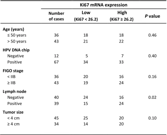
The median value of Ki67 mRNA levels in cervical cancer tissues was 26.2. To inquire into whether there was an association between Ki67 mRNA expression levels in cervical cancer tissues and their clinicopathological parameters, such as age, HPV infection status, FIGO stage, lymph node metastasis, and tumor size, the Ki67 mRNA levels were divided into two groups: a low Ki67 (below the median Ki67 mRNA level) group and a high Ki67 (above the median Ki67 mRNA level) group. Among the parameters, lymph node metastasis showed statistically significant relation with a high Ki67 group (P = 0.02) (Table 5).
18
Table 5. Ki67 mRNA expression levels correlated with clinicopathological
parameters in 79 cervical cancer patients
Ki67 mRNA expression
Number of cases Low High P value (Ki67 < 26.2) (Ki67 ≥ 26.2) Age (years) ≤ 50 years 36 18 18 0.46 > 50 years 43 21 22 HPV DNA chip Negative 12 5 7 0.40 Positive 67 34 33 FIGO stage < IIB 36 20 16 0.16 ≥ IIB 43 19 24 Lymph node Negative 40 24 16 0.02 Positive 39 15 24 Tumor size < 4 cm 45 25 20 0.10 ≥ 4 cm 34 14 20
19
IV. DISCUSSION
Cervical cancer is a worldwide leading cause of cancer mortality in 35-55 year old women. To avoid unnecessary or invasive treatment such as ablation, conization and/or hysterectomy in benign precancerous lesions with transient HPV infection, the optimal screening strategy for cervical cancer should efficiently and accurately identify precursor lesions that will progress to invasive cancers.30 Ki67, a nuclear protein, is expressed in proliferating vertebrate cells so that nuclear staining of this nuclear antigen is widely used as a surrogate marker, measuring proportions of dividing cells to grade tumors. But estimating of Ki67 expression level is not inconsistent and has some limitations in conventional histopathological fields.31
Therefore, the purpose of this article was to evaluate Ki67 mRNA expression levels with histological grades to identify and understand the relationship between the Ki67 mRNA assay and clinicopathological parameters of cervical cancer, and assess the performance of this assay as a diagnostic tool for cervical cancer.
First of all, Ki67 mRNA expression levels of matched non-cancerous and cancerous portion FFPE samples obtained from 16 cervical cancer patients were compared to validate the assay. There were statistically significant differences in Ki67 mRNA expression levels between matched non-cancerous and cancerous samples (P = 0.0005) (Figure 1). Comparing to Ki67 mRNA expression levels in normal tissues, those of cervical cancer tissues enabled to differentiate regardless of their cell types. There have been few other studies comparing Ki67 mRNA expression in normal and cervical cancer samples so far. Similar but earlier studies have widely proved the validation of Ki67 mRNA assay in breast cancer
samples as a distinctive prognostic biomarker. Yamamoto et al reported that
20
to estimate prognosis, as well as conventional Ki67 labeling index of breast cancer.8
Using 90 cervical cancer and 40 normal FFPE tissue samples, this study revealed that the Ki67 mRNA assay was able to accurately discriminate between cervical cancer and normal tissues with a high sensitivity of 93.3% (95% CI 86.1 to 97.5) and a high specificity of 97.5% (95% CI 86.8 to 99.9) (Table 3). Several studies have demonstrated that Ki67 expression using immunoquantification can provide greater discrimination power not only between normal and cancer tissues but also between LSIL and HSIL. And the reported average positivity rates found in normal, LSIL, and HSIL were 7.9%, 49%, and 90%, respectively.11, 32 In this research, the proportion of Ki67 mRNA expression cases were 2.5%, 13.3%, and 40.0% in normal, LSIL, and HSIL respectively. In cervical cancer the proportion was 93.3%, and these data were all statistically significant (P < 0.001) (Table 4).
Moreover, increased Ki67 mRNA expression in cervical cancer samples was significantly higher than those of normal, LSIL, and HSIL samples (Figure 2). Since less than 10% of LSIL cases progress to HSIL, cytological and/or histological follow-up are more frequently needed only in LSIL cases with tendency of progression. Chen et al and Zhou et al attempted to predict the progression using Ki67 immunocytochemistry and immunohistochemistry tests and showed similar results distinguishing LSIL and HSIL.33, 34 In the aspect of burdening the national healthcare system, such molecular diagnosis accompanied with pathological diagnosis may reduce overtreatment for follow-up in LSIL patients, the majority of who do not progress to HSIL.
According to clinicopathological prognostic parameter analysis, separating patients into high Ki67 expression (median ≥ 26.2) and low Ki67 expression (median < 26.2) groups, lymph node metastasis was associated with Ki67 mRNA levels (Table 5). Shokouh studied that Ki67 IHC was correlated to lymph
21
node status in breast cancer.9 Yang et al described that Ki67
immunohistochemical marker was correlated for predicting lymph node metastasis in endometrial cancer.35 Cervical cancer in this study as well as breast cancer in others also showed the interrelation of high Ki67 and lymph node metastasis, which reflected a poor prognosis of cancer.
Through many former studies, IHC staining of Ki67 can be applied to complementary test of HPV testing.36, 37 Due to cervical cancer screening guidelines of PAP smear and HPV co-testing, HPV test is now widely utilized. But there are few additional predictors able to determine the risk amount of carcinogenesis by high-risk HPV infection. Even though the further study about direct comparison Ki67 mRNA assay with Ki67 IHC staining as a treatment responder is needed, it is meaningful for this research to demonstrate that Ki67 mRNA assay can provide additional quantitative information as a molecular diagnostic marker of cervical cancer, and also predict prognosis of each grade of precancerous lesions.
V. CONCLUSION
In summary, to identify the diagnostic and clinicopathological predictive value of Ki67 mRNA assay in uterine cervical cancer and precancerous lesions, RT-qPCR was utilized for 190 FFPE cervical specimens in the research. The validation which referred to discriminating power of Ki67 mRNA for cervical cancer diagnosis was verified and the assay showed high sensitivity, specificity and positive predict value by using a diagnostic cutoff value of 5. Among normal cervical tissue, LSIL, HSIL and cervical cancer tissue samples, Ki67 mRNA expression level of cervical cancer tissue samples was remarkably highest and Ki67 mRNA expression was lowered as tissue samples’ grade went down.
22
Lymph node metastasis from among clinicopathological parameters of cervical cancer had statistically significant relation with high Ki67 mRNA expression
As a result, Ki67 mRNA expression analysis of FFPE cervical specimens using RT-qPCR was proved its clinicopathological performance as a discriminative biomarker in uterine cervical disease. Ki67 mRNA is also able to be a promising prognostic predictor which in cervical cancer and precancerous lesions as an important part of gene expression profiling. Based on collaboration with this Ki67 mRNA assay, further research for predicting a recurrence of HSIL and/or cervical cancer might be expected in the near future.
23
REFERENCES
1. Torre LA, Bray F, Siegel RL, Ferlay J, Lortet-Tieulent J, Jemal A. Global
cancer statistics, 2012. CA Cancer J Clin 2015; 65: 87-108.
2. Schiffman M, Castle PE, Jeronimo J, Rodriguez AC, Wacholder S.
Human papillomavirus and cervical cancer. Lancet 2007; 370: 890-907.
3. Salimovic-Besic I, Tomic-Cica A, Smailji A, Hukic M. Comparison of the
detection of HPV-16, 18, 31, 33, and 45 by type-specific DNA- and E6/E7 mRNA-based assays of HPV DNA positive women with abnormal Pap smears. J Virol Methods 2013; 194: 222-8.
4. Munkhdelger J, Choi Y, Lee D, Kim S, Kim G, Park S, et al. Comparison
of the performance of the NucliSENS EasyQ HPV E6/E7 mRNA assay and HPV DNA chip for testing squamous cell lesions of the uterine cervix. Diagn Microbiol Infect Dis 2014; 79: 422-7.
5. Castro FA, Koshiol J, Quint W, Wheeler CM, Gillison ML, Vaughan LM, et al. Detection of HPV DNA in paraffin-embedded cervical samples: a comparison of four genotyping methods. BMC Infect Dis 2015; 15: 544.
6. Brink AA, Snijders PJ, Meijer CJ. HPV detection methods. Dis Markers
2007; 23: 273-81.
7. Eide ML, Debaque H. HPV detection methods and genotyping techniques
in screening for cervical cancer. Ann Pathol 2012; 32: e15-23, 401-9.
8. Yamamoto S, Ibusuki M, Yamamoto Y, Fu P, Fujiwara S, Murakami K, et
al. Clinical relevance of Ki67 gene expression analysis using formalin-fixed paraffin-embedded breast cancer specimens. Breast Cancer 2013; 20: 262-70. 9. Shokouh TZ, Ezatollah A, Barand P. Interrelationships between Ki67,
HER2/neu, p53, ER, and PR status and their associations with tumor grade and lymph node involvement in breast carcinoma subtypes retrospective observational analytical study. Medicine (Baltimore) 2015; 94: e1359.
10. Li LT, Jiang G, Chen Q, Zheng JN. Ki67 is a promising molecular target in the diagnosis of cancer. Mol Med Rep 2015; 11: 1566-72.
24
and Ki67 expression in normal, dysplastic and neoplastic uterine cervical epithelium and HPV infection. Pathol Res Pract 2014; 210: 482-7.
12. Luttmer R, Dijkstra MG, Snijders PJ, Berkhof J, van Kemenade FJ,
Rozendaal L, et al. p16 / Ki67 dual-stained cytology for detecting cervical (pre)cancer in a HPV-positive gynecologic outpatient population. Mod Pathol 2016; 29: 870-8.
13. Pan D, Wei K, Ling Y, Su S, Zhu M, Chen G. The prognostic role of
Ki-67/MIB-1 in cervical cancer: a systematic review with meta-analysis. Med Sci Monit 2015; 21: 882-9.
14. Piri R, Ghaffari A, Azami-Aghdash S, Ali-Akbar YP, Saleh P,
Naghavi-Behzad M. Ki-67/MIB-1 as a prognostic marker in cervical cancer-a systematic review with meta-analysis. Asian Pac J Cancer Prev 2015; 16: 6997-7002.
15. Ancuta E, Ancuta C, Cozma LG, Iordache C, Anghelache-Lupascu I,
Anton E, et al. Tumor biomarkers in cervical cancer: focus on Ki-67 proliferation factor and E-cadherin expression. Rom J Morphol Embryol 2009; 50: 413-8.
16. Scholzen T, Gerdes J. The Ki-67 protein: from the known and the unknown.
J Cell Physiol 2000; 182: 311-22.
17. Wang HY, Kim G, Cho H, Kim S, Lee D, Park S, et al. Diagnostic
performance of HPV E6/E7, hTERT, and Ki67 mRNA RT-qPCR assays on formalin-fixed paraffin-embedded cervical tissue specimens from women with cervical cancer. Exp Mol Pathol 2015; 98: 510-6.
18. Pund ER, Nieburgs H, Nettles JB, et al. Preinvasive carcinoma of the cervix uteri: seven cases in which it was detected by examination of routine endocervical smears. Arch Pathol Lab Med 1947; 44: 571–7.
19. Richart RM. Natural history of cervical intraepithelial neoplasia. Clin
Obstet Gynecol 1968; 10: 748.
20. Nasiell K, Roger V, Nasiell M. Behavior of mild cervical dysplasia during
long‐term follow‐up. Obstet Gynecol 1986; 67: 665–9.
25
PA: Wolters Kluwer Health/Lippincott Williams & Wilkins; 2012. p.575-618.
22. Solomon D. The 1988 Bethesda System for reporting cervical/vaginal
cytologic diagnoses. Acta Cytol 1989; 33: 567–74.
23. Tabbara S, Saleh AD, Andersen WA, Barber SR, Taylor PT, Crum CP. The
Bethesda classification for squamous intraepithelial lesions: histologic, cytologic, and viral correlates. Obstet Gynecol 1992; 79: 338–46.
24. Stoler MH, Schiffman M. Interobserver reproducibility of cervical
cytologic and histologic interpretations: realistic estimates from the ASCUS-LSIL Triage Study. JAMA 2001; 285: 1500.
25. Herrington CS. The terminology of pre-invasive cervical lesions in the UK
cervical screening programme. Cytopathology 2015 Dec; 26(6): 346-50.
26. Weinstein LC, Buchanan EM, Hillson C, Chambers CV. Screening and
prevention: cervical cancer. Prim Care 2009; 36: 559-74.
27. Safaeian M, Solomon D, Castle PE. Cervical cancer prevention-cervical
screening: science in evolution. Obstet Gynecol Clin North Am 2007; 34: 739-60.
28. Cox JT. The development of cervical cancer and its precursors: what is the
role of human papillomavirus infection? Curr Opin Obstet Gynecol 2006; 18 Suppl 1: s5-13.
29. Livak KJ, Schmittgen TD. Analysis of relative gene expression data using
real-time quantitative PCR and the 2(T)(-Delta Delta C) method. Methods 2001; 25: 402-8.
30. ACS-ASCCP-ASCP Cervical Cancer Guideline Committee. American Cancer Society, American Society for Colposcopy and Cervical Pathology, and American Society for Clinical Pathology screening guidelines for the prevention and early detection of cervical cancer. CA Cancer J Clin 2012; 62: 147-72. 31. Sobecki M, Mrouj K, Colinge J, Gerbe F, Jay P, Kransiska L, et al.
Cell-cycle regulation accounts for variability in Ki-67 expression levels. Cancer Res. 2017 May 15; 77(10):2722-34
32. Pacchiarotti A, Ferrari F, Bellardini P, Chini F, Collina G, Dalla P, et al. Prognostic value of p16-INK4A protein in women with negative or CIN1
26
histology result: a follow-up study. Int J Cancer 2014; 134: 897-904.
33. Chen CC, Huang LW, Bai CH, Lee CC. Predictive value of p16/Ki-67
immunocytochemistry for triage of women with abnormal Papanicolaou test in cervical cancer screening: a systematic review and meta-analysis. Ann Saudi Med 2016; 36: 245-51.
34. Zhou WQ, Sheng QY, Sheng YH, Hou WJ, Xu GX, Wu YM, et al. Expressions of survivin, P16(INK4a), COX-2, and Ki-67 in cervical cancer progression reveal the potential clinical application. Eur J Gynaecol Oncol 2015; 36(1): 62-8.
35. Yang B, Shan B, Xue X, Wang H, Shan W, Ning C, et al. Predicting lymph node metastasis in endometrial cancer using serum CA125 combined with immunohistochemical markers PR and Ki67, and a comparison with other prediction models. PLoS One 2016; 11: e0155145.
36. Agoff SN, Lin P, Morihara J, Mao C, Kiviat NB, Koutsky LA. p16 (INK4a) expression correlates with degree of cervical neoplasia: A comparison detection of high-risk with Ki-67 expression and HPV types. Mod Pathol 2003; 16: 665-73.
37. Zappacosta R, Colasante A, Viola P, D’Antuono T, Lattanzio G, Capanna S,
et al. Chromogenic in situ hybridization and p16/Ki67 dual staining on formalin-fixed paraffin-embedded cervical specimens: correlation with HPV-DNA test, E6/E7 mRNA test, and potential clinical applications. Biomed Res Int 2013; 2013: 453606.
27
ABSTRACT (IN KOREAN)
자궁경부암 조직에서 발현되는 Ki67 mRNA의
진단적, 임상적 의의
<지도교수 남 은 지>
연세대학교 대학원 의학과
정 다 은
Ki67은 암의 증식과 예후와 관련된 중요한 바이오마커로
널리 알려져 있다. 이전의 연구들을 통하여 포르말린고정
파라핀 포매된 자궁경부암 조직샘플로부터 Ki67 mRNA 발현을
정량적으로 측정할 수 있는 가능성을 평가하였다.
선행연구를 토대로, 본 연구에서는 실시간 중합효소연쇄반응
정량검사를 이용하여 포르말린고정 파라핀 포매된 자궁경부
조직샘플의 Ki67 mRNA 발현을 정량적으로 측정, 비교하였다.
정상 자궁경부 및 자궁경부 병변별 Ki67 mRNA 발현량을
비교하여 Ki67 mRNA의 예측적 가치를 확인하고자 하였다.
또한, 자궁경부암 샘플에서의 Ki67 mRNA 발현량과 해당
암환자들의 임상적 예후인자들 간의 관계를 분석하였다.
전체 190건의 자궁경부 검체가 연구에 사용되었으며 이
중에는 80건의 편평상피세포암, 10건의 선암, 30건의 고등급
상피내병변, 30건의 저등급 상피내병변, 40건의 정상 자궁경부
조직 (대조군)이 포함되었다. 실험 결과, 정상 자궁경부 조직 (N
28