저작자표시-비영리-변경금지 2.0 대한민국 이용자는 아래의 조건을 따르는 경우에 한하여 자유롭게 l 이 저작물을 복제, 배포, 전송, 전시, 공연 및 방송할 수 있습니다. 다음과 같은 조건을 따라야 합니다: l 귀하는, 이 저작물의 재이용이나 배포의 경우, 이 저작물에 적용된 이용허락조건 을 명확하게 나타내어야 합니다. l 저작권자로부터 별도의 허가를 받으면 이러한 조건들은 적용되지 않습니다. 저작권법에 따른 이용자의 권리는 위의 내용에 의하여 영향을 받지 않습니다. 이것은 이용허락규약(Legal Code)을 이해하기 쉽게 요약한 것입니다. Disclaimer 저작자표시. 귀하는 원저작자를 표시하여야 합니다. 비영리. 귀하는 이 저작물을 영리 목적으로 이용할 수 없습니다. 변경금지. 귀하는 이 저작물을 개작, 변형 또는 가공할 수 없습니다.
Dissertation for the degree of Doctor of Philosophy
Feasibility of cartilage tissue engineering
using cellulose membrane in vivo bioreactor
By
Xue Guang Li
Major in Medicine
Department of Medical Sciences
The Graduate School, Ajou University
Feasibility of cartilage tissue engineering
using cellulose membrane in vivo bioreactor
By
Xue Guang Li
A Dissertation of Submitted to the Graduate School of Ajou
University in Partial Fulfillment of the Requirements for the
Degree of Doctor of Philosophy
Supervised by
Byoung-Hyun Min M.D., Ph.D.
Major in Medicine
Department of Medical Sciences
The Graduate School, Ajou University
I
ABSTRACT
Articular cartilage is the connective tissue of diarthrodial joints that function is to provide a smooth, lubricated surface for articulation and to distributes loads. Articular cartilage degenerates due to multiple factors, such as trauma, bone malalignment, overweight, osteoarthritis and inflammatory arthritis. Articular cartilage is devoid blood vessels, lymphatics and nerves, and once damaged, it is difficult to heal itself. Current treatments include marrow tapping techniques, osteochondral auto/allo-grafting and cell-based techniques. But the result is generally a fibrocartilage, and the treatment is not satisfactory. Although cartilage tissue engineering as a promising new treatment method is being widely studied, there are still many hurdles to be solved. For example, degradation of the cartilage matrix and immune problems. This study aims to devise a new in vivo bioreactor for better culture of cartilage tissue. First, we evaluated whether the diffusion chamber made of cellulose membrane and silicone tube under the skin of nude mice was similar to the joint cavity environment, and whether it supported chondrogenesis. Secondly, we cultured cartilage tissue from xenogeneic cells in rabbit subcutaneous diffusion chamber and implanted it into the cartilage defect. Focusing on chondrogenesis, immunogenicity and cartilage healing, thereby establishing a in vivo bioreactor.
II
CHAPTER I :
Nude mouse subcutaneous model, known as heterotopic chondrogenesis, is popularly used for cartilage tissue engineering. Yet this model frequently has not been interpreted real bioreactor for chondrogenesis by different environment to joint, such as adhesion of implanted tissue to surrounding tissue allowing vascular invasion, with resulting in osteogenic differentiation. The purpose of this study was to create an insulated subcutaneous cavity to reduce such limitations. Study design included two groups; the free cavity (subcutaneous) and insulated cavity (in vivo bioreactor) on subcutaneous transplantation. Rabbit chondrocytes were pellet-cultured at an initial cell count of 3 × 10⁵ for 2 weeks. For the subcutaneous group, the cell pellets were surgically inserted subcutaneously. For the in vivo (IV) bioreactor, pellets were inserted in the Cellulose membrane chamber was composed of a 8 mm × 8 mm silicone tube and cellulose membrane. First evaluated the IV bioreactor fluid appearance, component and liquidity, and then evaluate chondrogenesis of the pellets using gross observation, cell viability, histology, biochemical analysis and mechanical test. The fluid color and transparency of IV bioreactor were similar to synovial fluid (SF) and the component was also close to SF compared to the serum. The IV bioreactor group showed more hyaline like tissue with less osteogenic differentiation. The IV bioreactor group offers a novel way to achieve heterotopic chondrogenesis, similar to the synovial joint environment.
III
To regenerate tissue engineered cartilage as a source for the restoration of cartilage defects, we used a human fetal cartilage progenitor cell (hFCPC) pellet for improve the chondrogenesis and modulation the immune response with a in vivo (IV) bioreactor system, that was buried subcutaneously in the host and then implanted into a cartilage defect. In vivo bioreactor (IVB) was composed of silicone tube and cellulose nanopore-size membrane. FCPC pellets were first cultured in vitro for 3 days, and then cultured in
vitro, subcutaneous and IV bioreactor for 3 weeks. First evaluated the IV bioreactor
fluid appearance, component and liquidity, and then evaluate chondrogenesis and immunogenicity of the pellets using gross observation, cell viability, histology, biochemical analysis, RT-PCR, and Western Blot, finally evaluates the cartilage repair and synovial inflammation using histology. The fluid color and transparency of IV bioreactor were similar to synovial fluid (SF) and the component was also close to SF compared to the serum. IV bioreactor system not only promotes the synthesis of cartilage matrix and maintains cartilage phenotype, but also delays the occurrence of calcification compared with subcutaneous. A IV bioreactor, which has been predominantly adopted to study cell differentiation, was effective in preventing host immune rejection.
Keywords: Cartilage tissue engineering; In vivo bioreactor; Cellulose membrane; Fetal
IV
TABLE OF CONTENTS
ABSTRACT---I TABLE OF CONTENTS---IV LIST OF FIGURES---VII LIST OF TABLES---VⅢ Background---11.1 Cartilage tissue engineering---1
1.2 In vivo bioreactor---1
1.3 Cellulose membrane---2
1.4 Aims of study---3
Chapter I.---5
2.1 Introduction---5
2.2 Materials and Methods---7
2.2.1 Cell isolation and culture---7
2.2.2 Pellet culture---7
2.2.3 Preparation of cellulose membrane chamber---7
2.2.4 Ectopic chondrogenesis in the subcutaneous and IV bioreactor environment---8
2.2.5 Permeability assay---9
2.2.6 In vivo bioreactor fluid component analysis---9
2.2.7 Gross observation and size measurement of the pellets---10
2.2.8 Cell viability assay---10
2.2.9 Histology and Immunohistochemistry---10
2.2.10 Biochemical assay---11
2.2.11 Mechanical test---12
2.2.12 Statistical analysis---13
V
2.3.1 Set up the in vivo bioreactor---14
2.3.2 IV bioreactor fluid characteristics and Confirm the fluid liquidity---16
2.3.3 Gross observation, size measurement and cell viability of the pellets---20
2.3.4 Histological observation of the pellets---22
2.3.5 Immunohistochemical observation of the pellets---23
2.3.6 Calcification of the pellets---25
2.3.7 Biochemical analysis for the content of DNA, GAGs and collagen---27
2.3.8 Compressive strength---29
2.4 Discussion---31
Chapter II.---35
3.1 Introduction---35
3.2 Materials and Methods---38
3.2.1 Cell isolation and culture---38
3.2.2 Pellet culture---38
3.2.3 Preparation of cellulose membrane chamber---39
3.2.4 Ectopic chondrogenesis in the subcutaneous and IV bioreactor environment--39
3.2.5 Cartilage defect repair---40
3.2.6 Measurement of transmittance---40
3.2.7 Permeability assay---41
3.2.8 In vivo bioreactor fluid component analysis---41
3.2.9 Gross observation and size measurement of the pellets---42
3.2.10 Cell viability assay---42
3.2.11 Histology and Immunohistochemistry---42
3.2.12 Biochemical assay---47
3.2.13 Reverse Transcription Polymerase Chain Reaction (RT-PCR)---47
3.2.14 Western Blot analysis---48
3.2.15 Statistical analysis---49
VI
3.3.1 Set up the in vivo bioreactor---51
3.3.2 IV bioreactor fluid characteristics and Confirm the fluid liquidity---54
3.3.3 Gross observation, size measurement and cell viability of the pellets---57
3.3.4 Histological observation of the pellets---60
3.3.5 Immunohistochemical observation of the pellets---61
3.3.6 Calcification of the pellets---62
3.3.7 Biochemical analysis for the content of DNA, GAGs and collagen---67
3.3.8 Molecular analysis of immunogenicity---69
3.3.9 Macroscopic and histological observation of the ectopic engineered cartilage for cartilage repair in vivo---73
3.3.10 Immunohistochemical observation of the ectopic engineered cartilage for cartilage repair in vivo---75
3.3.11 Histological observation of the synovium---76
3.4 Discussion---80
Conclusion---84
VII
LIST OF FIGURES
CHAPPTER I.
Fig 1.1. SEM images of the cellulose membrane---8
Fig 1.2. Set up the in vivo bioreactor---15
Fig 1.3. IV bioreactor fluid characteristics and Confirm the fluid liquidity---17
Fig 1.4. Gross observation, size measurement and cell viability of the pellets---21
Fig 1.5. Histological and immunohistochemical observation of the pellets---24
Fig 1.6. Calcification of the pellets---26
Fig 1.7. Biochemical analysis---28
Fig 1.8. Compressive strength---30
CHAPTER II.
Fig 2.1. Schematic illustration of the overall design of this study---52Fig 2.2. IV bioreactor fluid characteristics and Confirm the fluid liquidity---55
Fig 2.3. Gross observation, size measurement and cell viability of the pellets---58
Fig 2.4. Histological and immunohistochemical observation of the pellets---65
Fig 2.5. Biochemical analysis---68
Fig 2.6. Molecular analysis of immunogenicity---71
Fig 2.7. Macroscopic, histological and immunohistochemical observation of neocartilage in the cartilage defect---78
VIII
LIST OF TABLES
CHAPPTER I.
Table 1.1. IV bioreactor fluid component analysis---18
Table 1.2. Confirm the IV bioreactor fluid liquidity---19
CHAPTER II.
Table 2.1 Scheme for the histopathological assessment of the three features of chronic synovitis---44Table 2.2 The Bern score---45
Table 2.3 Cartilage repair score---46
Table 2.4 Sequences of primers used for RT-PCR analysis---48
Table 2.5 List of primary antibodies used in this study---49
1
Background
1.1 Cartilage tissue engineering
Cartilage is difficult to self-repair, current treatment methods include microfracture, mosaicplasty and autologous chondrocyte implantation (ACI) [1, 2]. However, these methods have disadvantages such as difficulty in obtaining adequate amounts of autologous cells or tissues, damage to the supply sites, and fibrocartilage formation [3, 4]. Therefore, the hope is to use the allogeneic or xenogeneic cells to create tissue-engineered cartilage. However, these cells have the problem of causing immune rejection to be solved [5]. Furthermore, Chondrocytes dedifferentiate when expanding
in vitro, resulting in loss of phenotypic and extracellular matrix (ECM) components [6].
Previously, we have reported that human fetal cartilage progenitor cells (hFCPCs) having high yield, proliferation, multipotent differentiation and maintains the chondrogenic phenotype abilities, in cartilage tissue formation [7]. In addition, it has been reported that cells from the fetus do not cause immune rejection [8]. Therefore, FCPC can as a novel cell source for cartilage regeneration.
1.2 In vivo bioreactor
In order to solve the dedifferentiation of chondrocytes in monolayer culture, pellet culture was widely used in chondrocytes culture [9]. However, chondrocyte pellet culture method can easily cause cartilage mineralization in vitro [10]. Pellet method can
2
be used to cultivate cartilage tissue in two ways, that is in vitro or in vivo tissue engineering. In traditional tissue engineering, autologous stem cells seed on a bio active scaffold and cultivate in a bioreactor [11]. Subcutaneous environment was often used to animal model of regenerate ectopic tissue-engineered cartilage, but due to the cell ingrowth and vascular invasion of the host, lead to matrix is des troyed and calcified [12]. Therefore, a new trend to avoid the above problems is in
vivo bioreactor (IVB), which can effectively prevent host immune rejection, host cell
invasion and vascular invasion [12]. This using the body as a bioreactor, the traditional three elements (scaffold, cell and growth factor) were cultured, and the regeneration environment of body provided help for its growth [13]. Until now, the IVB concept has
been applied to a series of laboratory and clinical studies to induce regeneration of
various tissues. Such as the trachea [14], full-thickness skin [15], bladder [16], esophagus [17], skeletal muscle [18], bone [19] and cartilage [20]. The basic principle
of the IVB strategy is toselect the appropriate anatomical site to provide the necessary
microenvironment for regeneration [13]. However, so far there has been no IVB that mimicking the articular cavity environment of native cartilage growth, and the analysis of the IVB microenvironment, and the study on the regeneration of ectopic tissue-engineered cartilage in the IVB by using xenogeneic chondrocytes.
3
Cellulose is the most abundant biopolymer on earth, due to its biocompatible, biodegradable and tuneable tensile strength, it has been applied in medical tissue engineering in recent years [21]. The cellulose membrane used in this study is composed of fairly long cellulose fibrils connected with each other and has a highly porous three-dimensional network [22]. The silicone is a kind of bio-inert material with good biocompatibility and low immune response, which is often used in medical implants. Therefore, it is suitable for the fabrication of IVB chamber in this study.
1.4 Aims of this study
This study aims to devise a new in vivo bioreactor for better culture of tissue engineered cartilage.
• Chapter I.
In this study, component and liquidity were analyzed of the cellulose membrane IV bioreactor fluid and evaluated the chondrogenesis of young rabbit chondrocytes pellets cultured in the bioreactor and compared with subcutaneous.
• Chapter II.
In this study, component and liquidity were analyzed of the cellulose membrane IV bioreactor fluid and evaluated the chondrogenesis, immunogenicity and the ability to healing cartilage defects of FCPCs pellets cultured in the bioreactor and compared with
4
5
CHAPTER I
2.1 Introduction
Because articular cartilage is devoid the nutritional support of the vascular system, once the lesions is often unable to heal itself [1, 2]. The treatment of articular cartilage lesions has always been a challenge for orthopedists. Current treatments include marrow tapping techniques, osteochondral auto/allo-grafting and cell-based techniques. But the result is generally a fibrocartilage, and the treatment is not satisfactory [23].
Cartilage tissue engineering is being widely studied as a promising new treatment option [24]. The research stage of tissue engineering is divided into several stages from
in vitro research to human clinical trials [25]. As in vivo culture system, heterotopic
chondrogenesis is the first stage for confirming chondrogenic potential [26]. As the Heterotopic Chondrogenesis model, immunodeficient nude mice are widely used in the
study of cartilage reconstruction[27]. Tissue-engineered cartilage is usually implanted
under the skin of nude mice for study. However, the implanted cartilage tissue is prone to early calcification due to the invasion of subcutaneous blood vessels [28]. Preventing cartilage calcification is a challenge for tissue engineered cartilage. Current methods include the use of antiangiogenic factors and physical blocking of vascular invasion [12, 20].
It has been reported that IV bioreactor can effectively prevent invasion of host cells and blood vessels and maintain cartilage characteristics [12]. This using the body as a bioreactor, the traditional three elements (scaffold, cell and growth factor) were cultured,
6
and the regeneration environment of body provided help for its growth [13]. Until now,
the IV bioreactor concept has been applied to a series of laboratory and clinical studies
to induce regeneration of various tissues. However, so far there has been no IV bioreactor that mimicking the articular cavity environment of native cartilage growth, and the analysis of the IV bioreactor microenvironment. Cellulose is the most abundant biopolymer on earth, due to its biocompatible, biodegradable and tuneable tensile strength, it has been applied in medical tissue engineering in recent years [21]. The cellulose membrane used in this study is composed of fairly long cellulose fibrils connected with each other and has a highly porous three-dimensional network [22].
In this study, component and liquidity were analyzed of the cellulose membrane IV bioreactor fluid and evaluated the chondrogenesis of young rabbit chondrocytes pellets cultured in the bioreactor and compared with subcutaneous.
7
2.2 Materials and Methods 2.2.1 Cell isolation and culture
Rabbit articular cartilages were obtained from knee joints of 2-week-old males, New Zealand white rabbits (KOATECH, Pyeongtaek, Korea). Cartilage tissues were minced into small pieces and treated with 0.1% collagenase type II (Worthington Biochemical Corp, Freehold, NJ, USA) for 12 h and suspended in Dulbecco’s modified Eagle Medium (DMEM; HyClone, Logan, UT, USA) containing 10% fetal bovine serum
(FBS; Hyclone) and 1% antibiotics. Chondrocytes were seeded at 1 x 10⁶ cells/plate
(150mm) and cultured at 37 ℃ in a 5% CO2 incubator. Cells were passaged at 80%
confluence, and pellets were made in P2-P3.
2.2.2 Pellet culture
Aliquots of 3 × 10⁵ cells/0.5 ml were centrifuged at 500 g for 10 min in 15 ml
polypropylene tubes. After 1 day, cell pellets were cultured in chondrogenic defined medium (DMEM supplemented with ITS mixture, 50 μg/ml ascorbate-2 phosphate, 100 nM dexamethasone, 40 mg/ml proline, and 1.25 mg/ml BSA) without TGF-β, a typical chondrogenic inducer. Pellets were cultured for 2 weeks.
2.2.3 Preparation of cellulose membrane chamber
For this study, our own cellulose membrane chamber was manufactured. Cellulose membrane chamber was composed of silicone tube (8 mm × 6 mm × 8 mm, outer
8
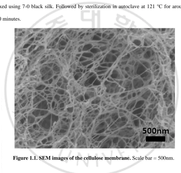
diameter × inner diameter × height) (Korea Ace Scientific, Seoul, Korea) and cellulose membrane. The pore size of cellulose membrane is about 50-400 nm (Figure 1), provided by the bioenergy research center, college of life sciences, Kyung Hee University. Both ends of the silicone tube were sealed with cellulose membrane and fixed using 7-0 black silk. Followed by sterilization in autoclave at 121 ℃ for around 20 minutes.
Figure 1.1. SEM images of the cellulose membrane. Scale bar = 500nm.
2.2.4 Ectopic chondrogenesis in the subcutaneous and IV bioreactor environment
This study was approved by the ethics committee for animal research of the Laboratory Animal Research Center of Ajou University Medical Center (approval No.
9
2013-0067). Six-week-old male nude mouse (n=30) were anesthetized with a mixture of
Zoletil and Rumpun. manufactured cellulose membrane chambers (two
chambers/mouse) were implanted under back skins of nude mice. In this study total of 120 pellets were divided into two environment: nude mouse subcutaneous and in vivo bioreactor (IVB). After 3 days, a total of 120 pellets were put into the subcutaneous and cellulose membrane chamber (1 pellets/chambers). Mice were sacrificed at each time point of 1, 3, and 6 weeks post-implantation. Retrieved pellets were used for analyses, gross observation, pellets size measurement, cell viability assay, histological analysis, immunohistochemical analysis, biochemical assay and mechanical property assay, respectively.
2.2.5 Permeability assay
In order to test diffusion across the cellulose membrane, we confirmed the diffusion patterns of glucose through the cellulose membrane using a two-chamber system developed previously in our laboratory [29]. Briefly, the cellulose membrane was fixed between two specific chambers. We loaded 4500 mg/L glucose DMEM on one side of the chamber and loaded distilled water (DW) on the other side of the chamber. After 2, 4, 8, 12, and 24 hours measured the glucose concentration in the DW in the Ajou University Hospital.
10
Total protein and glucose in IV bioreactor fluid were measured at the Ajou University Hospital. Hyaluronan (HA) content in IV bioreactor fluid was measured using ELISA kit according to the manufacture’s instructions (R&D Systems, Minneapolis, USA). Lactate content in IV Bioreactor fluids was measured using EnzyChromTM L-Lactate Assay Kit (ECLE-100) according to the manufacturer’s instructions (BioAssay Systems, Hayward, CA, USA). Experiments were conducted in triplicate, and optical densities were used to normalize the lactate production results.
2.2.7 Gross observation and size measurement of the pellets
Retrieved pellets (n = 5/time point/group) were observed in terms of their shape, color and size. The size of pellets was determined using the Image J program.
2.2.8 Cell viability assay
The pellets (n = 5/time point/group) were incubation at 48-wel plate in serum free DMEM medium 200 μl alamarBlue (Invitrogen, California, USA) 20 μl, 37 ℃ in a 5%
CO2 incubator. After 5h of incubation, the absorbance of duplicate samples (100μl) of
each well was measured in a 96well plate, at wavelengths of 570 and 600 nm, using a microplate reader (Infinite M200, Tecan, CH).
2.2.9 Histology and Immunohistochemistry
11
dehydrated, and then embedded in paraffin wax. Sections, each 4 μm in thickness, were prepared and stained with hematoxylin/eosin for visualizing cell distribution and morphology, Safranin-O/fast green for accumulation of sulfated proteoglycans (for the evaluation of Safranin O-Fast green stained cartilaginous pellet using the Bern score [30], and Von Kossa for calcified pellets (for quantitative analysis of the calcification area using the Image J program). For immunohistochemical analyses, sections of
samples prepared as described above were treated with 3% Hydrogen peroxide (H2O2)
for 10 min and reacted with a pepsin solution (Golden Bridge International, Inc, Mukilteo, WA, USA) for 10 min. After blocking the sections with 1% BSA in phosphate-buffered saline (PBS) for 1 h, sections were incubated with primary antibodies (1:200) for 1.5 h at room temperature. Primary antibodies were mouse
anti-rabbit collagen type II monoclonal antibody(Calbiochem, San Diego, CA, USA). With
2 times washing in PBS, sections were incubated with a biotinylated secondary antibody (1:200) for 30 min and peroxidase-conjugated streptavidin solution for 30 min at room temperature (both from Golden Bridge International). Finally, sections were reacted with a 3,3´-diaminobenzidine (DAB) solution (Golden Bridge International) and counterstained with Mayer’s hematoxylin (Sigma, St Louis, MO) and then mounted for microscopic observation (Nikon E600, Japan).
2.2.10 Biochemical assay
12
5/time point/group) were fully digested for 24 h at 60 ℃ in papain digestion solution containing 125 ug/ml of papain with 5 Mm L-cysteine-HCl and 5 Mm EDTA in 100Mm
Na2HPO4 (all from sigma, USA). The DNA content was measured using the Picogreen
assay. Total Sgag content was measured using a 1,9-dimethylmethylene blue (DMB) colorimetric method [31]. The papain-digested samples were reacted with DMB solution for 30 min, and the absorbance was measured at 530 nm using a spectrophotometer. The values were normalized by a standard curve using shark
chondroitin sulfate (~50 ug; Sigma) and expressed as a ratio to dry weight. Total
collagen content was determined by measuring hydroxyproline assay, as described
previously but with modifications [32].The pellets were reacted with 4-N HCL and 4-N
NaOH by autoclaving at 121 ℃ for 20 min. The pellets were then treated with 0.056 M chloramines T reagent for 25 min at room temperature. The Ehrlich’s aldehyde reagent was reacted with samples at 65 ℃ for 20 min in order to generate chromospheres. The amount of hydroxyproline was measured at 550 nm. The values were normalized by a standard curve using Trans-4-hydroxy-L-proline (~50 ug; Sigma) and expressed as a ratio to dry weight.
2.2.11 Mechanical test
Pellets (n = 5/time point/group) were subjected to the unconfined compression test using Universal Testing Machine (Model H5K-T; HTE, England). A pellet was placed
13
automatically stopped after moving for the programmed length in between the top and bottom platen. The peak load was obtained from the load-displacement curve and individual compressive strength was calculated. Samples in each group (n = 5) were tested for different time points (1, 3, and 6 weeks).
2.2.12 Statistical analysis
Data from the size of pellets, bern score, biochemical assays, and mechanical property were analyzed for statistical significance by Tukey-Kramer Multiple Comparisons Test using GraphPad software. The experiments were repeated at least three times (n = 5). Data are expressed as mean ± standard deviation (SD). Statistical significance was assigned as *p < 0.05.
14
2.3 Results
2.3.1 Set up the in vivo bioreactor
In order to confirm own self-regenerative capacity to regenerate new cartilage, we created the in vivo bioreactor using cellulose membrane and silicon tube in nude mouse subcutaneous (Fig. 2A-B). The young rabbit chondrocytes pellet culture within the chamber at 3 days after implantation, a thin fibrous tissue encapsulated with a vascular network surrounded all chambers, and the only minimal fibrotic reaction was seen in the subcutaneous pockets surgically created. The body fluid was full inside the IV Bioreactor chamber. The IV Bioreactor fluid is clear, slightly yellow and free of particulate (Fig. 2C). The pellet culture 6 weeks after, the fluid state was similar to that before implantation and round white pellet was observed. Pellet implanted under the skin can also be visualized (Fig. 2D).
15
Fig 1.2. Set up the in vivo bioreactor. Young rabbit chondrocytes pellets were used to
construct ectopic cartilage in the IV bioreactor system. (A) A gross image of the IV bioreactor chamber (size 21 mm × 10 mm) composed of silicone tube and cellulose membrane. (B) A gross image of the two IV bioreactor chamber implanted under the skin of a nude mouse. (C) 3 days after IV bioreactor chamber implantation, pellets were placed into the IV bioreactor for culture. (D) A gross image of IV bioreactor was obtained after 6 weekS of pellets culture.
16
2.3.2 IV bioreactor fluid characteristics and Confirm the fluid liquidity
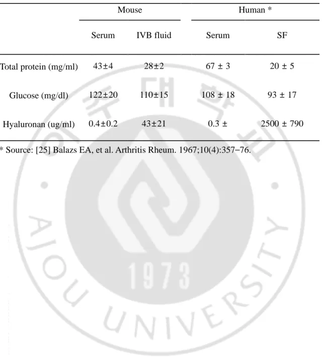
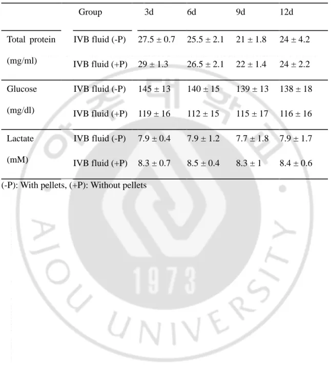
The appearance (Fig. 3A) of the IV bioreactor fluid are shown. IV Bioreactor fluid recovered at after pellets culture 1, 3 and 6 weeks. The IV Bioreactor fluid is clear, slightly yellow and free of particulate, at all time points, similar to no pellet culture cellulose membrane chamber fluid. The contents of total protein, glucose, and hyaluronic acid in IV Bioreactor fluid before pellets culture were measured and compared with those in nude mouse serum, and the contents of human SF and serum were referenced (Table 1). The results showed that these components in the IV bioreactor group were closer to synovial fluid than serum. Before and implantation 3 weeks after the permeability of the cellulose membrane used in the IV bioreactor was compared for 24 hours (Fig. 3B). The color thickens with the time of the right chamber (DW) and the color of both chambers has changed similarly at 24 hours in both groups. The concentration of the glucose in the right chamber was similarly increased with time at both groups (Fig. 3C). Increased quickly from the first 2 hours and relatively slowly since then. Total protein, glucose and lactic acid contents in IV bioreactor fluids with and without pellets were compared at 3, 6, 9 and 12 days (Table 2). The contents of these components were similar between both groups at the same time point, and each group maintained similar levels at all time points. These results indicated that the IV bioreactor fluid content was closer to SF than serum. And after 3 weeks of implantation, the cellulose membrane can be seen that the permeability is good as before. And the IV bioreactor system maintained homeostasis.
17
Fig 1.3. IV bioreactor fluid characteristics and Confirm the fluid liquidity. (A) The
IV bioreactor fluid appearance. (B) The permeability of the cellulose membrane. (C) The concentration of glucose that was transferred from the left chamber to the right through the cellulose membrane.
18
Table 1.1. IVB fluid component analysis
Mouse Human *
Serum IVB fluid Serum SF
Total protein (mg/ml) 43±4 28±2 67 ± 3 20 ± 5
Glucose (mg/dl) 122±20 110±15 108 ± 18 93 ± 17
Hyaluronan (ug/ml) 0.4±0.2 43±21 0.3 ± 2500 ± 790
19
Table 1.2. Confirm the IVB fluid liquidity
Group 3d 6d 9d 12d Total protein (mg/ml) IVB fluid (-P) 27.5 ± 0.7 25.5 ± 2.1 21 ± 1.8 24 ± 4.2 IVB fluid (+P) 29 ± 1.3 26.5 ± 2.1 22 ± 1.4 24 ± 2.2 Glucose (mg/dl) IVB fluid (-P) 145 ± 13 140 ± 15 139 ± 13 138 ± 18 IVB fluid (+P) 119 ± 16 112 ± 15 115 ± 17 116 ± 16 Lactate (mM) IVB fluid (-P) 7.9 ± 0.4 7.9 ± 1.2 7.7 ± 1.8 7.9 ± 1.7 IVB fluid (+P) 8.3 ± 0.7 8.5 ± 0.4 8.3 ± 1 8.4 ± 0.6
20
2.3.3 Gross observation, size measurement and cell viability of the pellets
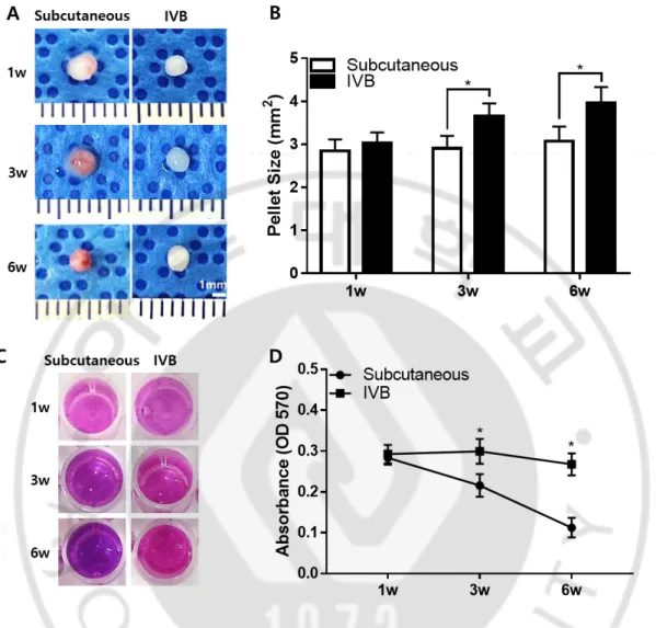
The effect of the IV bioreactor was compared with subcutaneous on the growth of young rabbit chondrocyte pellets. The pellets were first cultured in vitro for 2 weeks, then cultured subcutaneous and IV bioreactor for 1, 3 and 6 weeks. IV bioreactor group pellets showed whitish, hyaline cartilage-like circular morphology at all time points, but subcutaneous group pellets showed a subcutaneous tissue adhesion or turned a blood stained red color and became worse over time (Fig. 4A). In the gross images and size measurement, the size of the IV bioreactor group significantly increased with time, but the subcutaneous group did not change. The estimated size of the IV bioreactor group
(3.654 ± 0.298, 3.960 ± 0.372 mm2) was larger than that of the subcutaneous group
(2.906 ± 0.294, 3.072 ± 0.343 mm2) (p < 0.05) at 3 and 6 weeks (Fig. 4B). The pellet
viability was measurement (Fig. 4C). The reagent changed from purple to pink of a ll groups at all time points and measured the absorbance (Fig. 4D). The absorban ce increased with time at IV bioreactor group, but the subcutaneous group was d ecreased at 3 and 6 weeks. These results indicated the ability of the IV bioreactor to maintain the growth of young rabbit chondrocyte pellets observed above by gross and viability analysis.
21
Fig 1.4. Gross observation, size measurement and cell viability of pellets retrieved from the subcutaneous and IV bioreactor experiment. The pellets were cultured for 2
weeks in vitro before implantation in the subcutaneous and IV bioreactor of nude mouse. The implanted pellets (n = 5/time point/group) were retrieved at 1, 3 and 6 weeks after implantation. (A) The gross images of pellets are presented. (B) The size of each pellet was measured in the subcutaneous (open bar) and IV bioreactor (solid bar) groups using Image J program. (C) The cell viability was examined using Alamar blue assay. (D) Absorbance of Alamar blue at 570 nm. The data are presented as a mean ± standard deviation (SD) from 5 independent experiments (n = 5). *p < 0.05.
22
2.3.4 Histological observation of the pellets
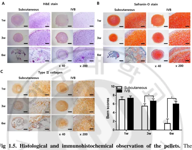
Chondrogenesis of the pellets cultured subcutaneous and IV bioreactor was further examined by histological and immunohistochemical analysis (Fig. 5A-B). Safranin-O/fast green staining showed that the accumulation of sulfated GAGs was no significant difference between the IV bioreactor and subcutaneous groups at 1 week. Over time, the intensity and area of Safranin-O staining decreased slightly in the IV bioreactor group, but significantly decreased in the subcutaneous group. The Bern score of the IV bioreactor group (6.7 ± 0.5, 6.0 ± 0.6) was larger than that of the subcutaneous group (5.5 ± 0.5, 1.5 ± 1.0) (p < 0.05) at 3 and 6 weeks (Fig. 5B). In the high magnification images (x 200), the lacunar structure characteristic of native cartilage can be seen during the entire culture process in the metachromatically stained area of the IV bioreactor group (Fig. 5A-B). This morphology was observed for cells of the subcutaneous group in 1 and 3 weeks of culture, but the cells structures were severely degraded in the 6 weeks of culture.
23
2.3.5 Immunohistochemical observation of the pellets
The immunostaining for type Ⅱ collagen further examined to confirm the chondrogenic phenotypes of the pellets (Fig. 5C). The type Ⅱ collagen retained with time IV bioreactor group, but in a subcutaneous group it significantly decreased, reaching about 20% at 6 weeks in both its intensity and stained area than 1 week. The IV bioreactor group showed strong expression of type Ⅱ collagen in the whole pellets at 6 weeks and stronger than the subcutaneous group. These results indicated that the IV bioreactor not only better supports the chondrogenesis but also maintain the chondrogenic phenotype of young rabbit chondrocyte pellets.
24
Fig 1.5. Histological and immunohistochemical observation of the pellets. The
pellets were cultured for 2 weeks in vitro before implantation in the subcutaneous and IV bioreactor of nude mouse. The implanted pellets (n = 5/time point/group) were retrieved at 1, 3 and 6 weeks after implantation. The pellets were processed to prepare thin sections, each 4 μm in thickness. (A) Hematoxylin/eosin to observe the distribution of cells. (B) Safranin-O/fast green to observe accumulation of sulfated proteoglycans, and the Bern scores were evaluated. (C) Immunostained with an antibody type Ⅱ collagen. The stained images are presented as a whole pellet (left columns, × 40) and at high magnification (right columns, × 200). Scale bar = 500 μm for × 40 and 100 μm for × 200 images. Data are presented as a mean ± SD from 5 independent experiments in the histograms (n = 5). *p < 0.05.
25
2.3.6 Calcification of the pellets
To examine if the loss of chondrogenic phenotypes was correlated with calcification of the matrix, Von Kossa staining was performed, and using the Image J program confirmed that the percentages of calcification area (Fig. 6). In the subcutaneous and IV bioreactor groups, black stains, indicative of calcified mineral deposits, was first observed in the peripheral region at 1 week and spread into the central region at 6 weeks. The stained area was more intense and broader in the subcutaneous group (20.7 ± 6.4%, 84.7 ± 3.7%) than in the IV bioreactor (3.9 ± 2.6%, 16.5 ± %) (p < 0.05) at 3 and 6 weeks. This result also indicated that the IV bioreactor maintains the chondrogenic phenotype and delays calcification of young rabbit chondrocyte pellets.
26
Fig 1.6. Calcification of the pellets. Alizarin Red stain to observe calcium deposits in the pellets. The stained images are presented as a whole pellet (left columns, × 40) and at high magnification (right columns, × 200). Scale bar = 500 μm for × 40 and 100 μm for × 200 images. Data are presented as a mean ± SD from 5 independent experiments in the histograms (n = 5). *p < 0.05.
27
2.3.7 Biochemical analysis for the content of DNA, GAGs and collagen
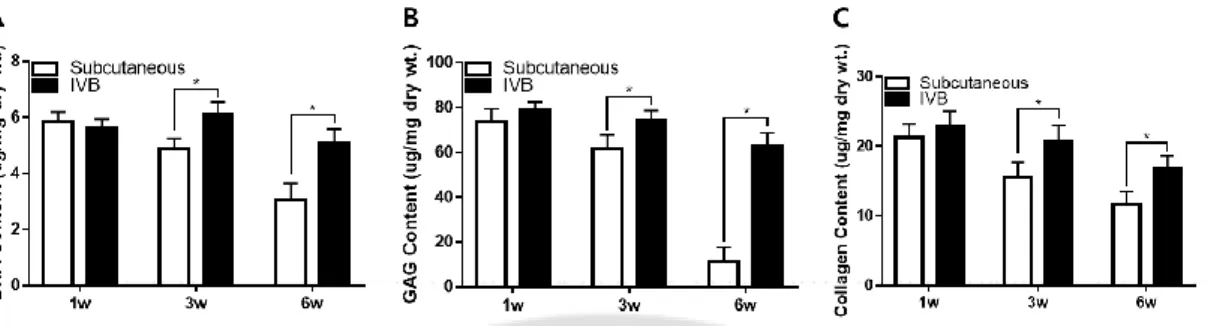
The content of DNA, GAGs, and collagen was measured quantitatively by chemical assays of the retrieved pellets (Fig. 7). The DNA content in the IV bioreactor group maintained at similar high levels until 6 weeks, but subcutaneous group was decreased rapidly from 3 weeks (Fig. 7A). The DNA content of the IV bioreactor group (6.10 ± 0.45 μg/mg, 5.11 ± 0.48 μg/mg) was higher than of the subcutaneous group (4.87 ± 0.38 μg/mg, 3.05 ± 0.60 μg/mg) (p < 0.05) at 3 and 6 weeks. This result suggests that the number of cells maintained with time IV Bioreactor group, but the subcutaneous group was decreased from 3 weeks. The content of GAGs and collagen showed a similar pattern overall to that of DNA content. The GAGs content in the IV bioreactor group maintained at similar high levels until 6 weeks, but subcutaneous group was decreased rapidly from 3 weeks (Fig. 7B). The IV bioreactor group (74.22 ± 4.59 μg/mg, 62.86 ± 5.74 μg/mg) was higher than of the subcutaneous group (61.90 ± 5.79 μg/mg, 11.42 ± 6.13 μg/mg) (p < 0.05) at 3 and 6 weeks. The collagen content in the IV bioreactor group maintained at similar high levels until 6 weeks, but subcutaneous group was decreased rapidly from 3 weeks (Fig. 7C). The IV bioreactor group (20.86 ± 2.18 μg/mg, 16.74 ± 1.93 μg/mg) was higher than of the subcutaneous group (15.54 ± 2.13 μg/mg, 11.66 ± 1.91 μg/mg) (p < 0.05) at 3 and 6 weeks. These results support the ability of the IV bioreactor to maintain chondrogenesis of young rabbit chondrocyte pellets observed above by biochemical analysis.
28
Fig 1.7. Biochemical analysis of subcutaneous and IV bioreactor pellets for total content of DNA (A), GAGs (B) and collagen (C). The pellets were cultured for 2
weeks in vitro before implantation in the subcutaneous and IV bioreactor of nude mouse. The implanted pellets (n = 5/time point/group) were retrieved at 1, 3 and 6 weeks after implantation. The amount of each component was calculated using standard curves for each assay and normalized by the dry weight of each pellet. Data are presented as a mean ± SD from 5 independent experiments in the histograms (n = 5). *p < 0.05.
29
2.3.8 Compressive strength
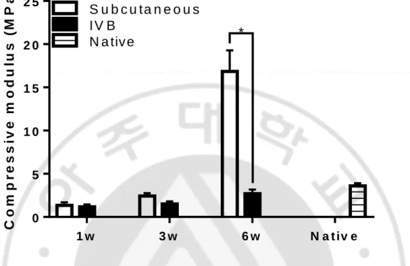
In order to confirm the physical properties of tissue engineered cartilage, compressive strength was measured (Fig. 8). The compressive strength in the IV bioreactor group maintained at similar levels until 6 weeks, but subcutaneous group was increased rapidly at 6 weeks. The subcutaneous group (16.84 ± 2.45 μg/mg) was higher than of the IV bioreactor group (2.70 ± 0.48 μg/mg) (p < 0.05) at 6 weeks. This result support the ability of the IV bioreactor to maintain physical properties of young rabbit chondrocyte pellets observed above by compressive strength.
30 1 w 3 w 6 w N a t iv e 0 5 1 0 1 5 2 0 2 5 C o m p r e s s iv e m o d u lu s ( M P a ) S u b c u ta n e o u s I V B N a tiv e *
Fig 1.8. Compressive strength. The pellets were cultured for 2 weeks in vitro before
implantation in the subcutaneous and IV bioreactor of nude mouse. The implanted pellets (n = 5/time point/group) were retrieved at 1, 3 and 6 weeks after implantation. The unconfined compression test using Universal Testing Machine (Model H5K-T; HTE, England). Data are presented as a mean ± SD from 5 independent experiments in the histograms (n = 5). *p < 0.05.
31
2.4 Discussion
In this study, a kind of IV bioreactor mimicking the environment of articular cavity was prepared under the subcutaneously of nude mouse using silicone tube and cellulose membrane (Fig. 2A-D), and the chondrogenesis was compared with that of traditional artificial cartilage manufacturing environment, namely subcutaneously. The cells used for artificial cartilage were young rabbit chondrocytes, and the culture method was pellet culture. Nude mouse model has been widely used for research on cartilage regeneration due to ease of handling, no immune response and relative cost effectiveness [27].
The macroscopic observation of the IV bioreactor fluid after pellet culture showed that the color and transparency similar to non-pellet culture fluid (Fig. 3A). There was no difference in the permeability of cellulose membrane used in IV bioreactor be fore and after implantation (Fig. 3B-C). The SEM images of the freeze-dried cellulose membrane showed that the membrane presented a three-dimensional network structure, with the width of fibrils of approximately 20-30 nm and the pore size ranging
from 50 to 400 nm [22].This cellulose membrane was first used in IV bioreactor b
y us. We chose it as the IV bioreactor membrane for two reasons. The first is that the size of pores can be passed through most nutrients, but the cells cannot. The second is that cellulose is a hydrophilic material, non-specific adsorption of proteins is low, mammalian cells are not easy to adsorb cellulose surface, so as to avoid the blocking of pores [33-36]. The results of the IV bioreactor environmental stability experiment
32
showed that the total protein, glucose and lactic content in the IV bioreactor wit h and without pellets showed no significant difference at the same time point, a nd remained at a certain level over time (Table 2). This indicates that nutrients r equired for pellet growth are constantly added to the IV bioreactor, and metaboli tes generated are removed, which is a relatively stable environment. The size of pellets in IV bioreactor environment increased with time, while the size of pellet s in subcutaneous environment no significant change. (Fig. 4A-B). Moreover, the r esults of cell activity assay showed that IV bioreactor environment was more sui table for the growth of chondrocytes than subcutaneous environment (Fig. 4C-D). The results of subcutaneous group may be related to the degradation of pellet m atrix, while the results of IV bioreactor group may be related to the fluid compo nent of IV bioreactor. IV bioreactor component analysis showed that protein, glu cose and HA were close to synovial fluid than serum (Table 1). The nutrient supp ly of natural articular cartilage mainly comes from synovial fluid [37]. glucose p lays an important role in chondrogenesis and maintenance as a nutrient [38]. wh en HA was added to the culture medium, increase DNA, sulfated glycosaminogly can and type Ⅱ collagen synthesis [39].
IVB environment promotes the synthesis of cartilage matrix and maintains the cartilage phenotype until 3 weeks, while subcutaneous cultured pellet degenerate and reduce the phenotypes after 6 weeks (Fig 5 and 7). Subcutaneous implantation is widely used in cartilage tissue engineering. Bone marrow-derived mesenchymal progenitor
33
cells can differentiate into chondrocytes after being implanted subcutaneously with appropriate scaffold [40]. However, subcutaneous engineered cartilage has less matrix synthesis and is prone to loss of cartilage phenotype and function [41, 42]. It may be because the IV bioreactor environment is similar to the joint cavity environment and the IV bioreactor fluid contains substances required for chondrogenesis. The key advantage of IV bioreactor is that, depending on the design, the body can offer a constant stream of its own stem cells and growth factors to created regenerative microenvironment for tissue growth [13].
IVB environment not only promotes the synthesis of cartilage matrix and maintains cartilage phenotype, but also delays the occurrence of calcification compared with subcutaneous (Fig. 6 and 8). Calcification is a multifactorial process caused by an imbalance between inhibitors and promineralization factors. Loss of proteoglycans often leads to articular cartilage calcification, because proteoglycans are effective mineralizing inhibitors [43]. The loss of chondrogenic phenotypes appears to be associated with increased matrix calcification (Von Kossa staining). Chondrocyte phenotypic changes include hypertrophy differentiation, apoptosis, altered responses to growth factors, inflammatory cytokines and mediators [44]. In this study, calcification in the subcutaneous group may be associated with host cell ingrowth and vascular invasion [45], while the reasons for delayed calcification in the IV bioreactor group may be related to some serum protein contained in the IV bioreactor, which may play an inhibitory role in the early stage of the calcification process after penetrating the matrix
34 [46].
According to our experimental results, the IV bioreactor environment mimicking the articular cavity is similar to the environment of native cartilage in structure and component (Fig. 2, 3, Table 1 and 2). Compared with the subcutaneous group, the IV bioreactor group promoted cell growth and matrix synthesis during the chondrogenesis of young rabbit chondrocyte pellets, maintained cartilage phenotype (Fig. 4, 5 and 7). IV bioreactor also delayed calcification of young rabbit chondrocyte pellet compared to subcutaneous (Fig. 6 and 8).
35
CHAPTER II
3.1 Introduction
Articular cartilage has a limited ability to heal after being damaged. As it has very little blood supply, cartilage has difficulty self-repairing after damage [1, 2]. Cell-based autologous or allogeneic transplantation has been employed to treat cartilage defects [3, 4]. The method of using autologous cells requires an invasive procedure for harvesting and has a limited amount of harvesting. Moreover, use of allogeneic MSCs has some limitations, an immune response may be elicited, leading to the failure of the cell therapy [5]. Furthermore, Chondrocytes dedifferentiate when expanding in vitro, resulting in loss of phenotypic and extracellular matrix (ECM) components [6].
Three factors play an important role in the engineering of cartilage tissue. Cells are so matic cells or stem cells [13]. Biomaterials are synthetic or natural materials. These ele ments are combined optimally and cultured in a bioreactor to produce cartilage similar t o natural cartilage [12]. Tissue engineered cartilage may be an applicable resource for h ealing cartilage defects, which would be more promising if the cartilage could be forme d in an immunocompetent host [12]. However, inflammatory reactions, which might des troy chondrocytes and subsequently deform the tissue, obstructed reconstruction of tissu e in immunocompetent animals. Moreover, host cell ingrowth would impair the quality of reconstructed tissue. Therefore, a new trend to avoid the above problems is in vivo bi oreactor (IV bioreactor), which can effectively prevent vascular invasion, host cell invas
36
ion and immune rejection [13]. This using the body as a bioreactor, the traditional three elements (scaffold, cell and growth factor) were cultured, and the regeneration environ ment of the body provided help for its growth. This would be applicable for allografts b ecause the immune system cannot recognize allografts once host cell invasion is inhibite d. Cellulose is a kind of bio-inert material with good biocompatibility and low i m m u n e r e s p o n s e , w h i c h i s o f t e n u s e d i n m e d i c a l i m p l a n t s [21]. The cellulose membrane of IV bioreactor used in this study is composed of fairly long c ellulose fibrils connected with each other and has a highly porous three-dimensional network [22]. On the other hand, the selection of favorable stem cells in car tilage tissue engineering is of great importance. In previous stem cell treatments, stem c ells not only have the ability for regeneration in chronic tissue damage but also have a re gulatory effect on the immune environment. Implanted stem cells can regulate the immu ne environment during tissue repair for tissue regeneration [7]. Previously, we have repo rted that human fetal cartilage progenitor cells (hFCPCs) having high yield, proliferatio n, multipotent differentiation and maintains the chondrogenic phenotype abilities, in cart ilage tissue formation [7]. Pellet culture was widely used in chondrocytes culture [9]. H owever, the chondrocyte pellet culture method can easily cause cartilage mineralization
in vitro [10]. The subcutaneous environment was often used to the animal model o
f regenerate ectopic tissue-engineered cartilage, but due to the cell ingrowth and vascular invasion of the host, lead to a matrix is destroyed and calcified [12].
37
oreactor fluid and evaluated the chondrogenesis, immunogenicity and the ability to heali ng cartilage defects of FCPCs pellets cultured in the bioreactor and compared with in vit
ro and subcutaneous.
38
3.2 Materials and Methods 3.2.1 Cell isolation and culture
Human fetal cartilage progenitor cell (hFCPC) were isolated from the human fetal cartilage tissue at 12 weeks of gestation, as previously described [7]. The fetal cartilage tissue was obtained from patients after elective abortion, as approved by the institutional review board (IRB) of the Ajou University Medical Center (AJIRB-CRO-16-139). Cartilage pieces were digested in 0.1% collagenase type II (Worthington Biochemical Corp, Freehold, NJ, USA) in high-glucose Dulbecco’s modified Eagle Medium (DMEM; HyClone, Logan, UT, USA) containing 1% fetal bovine serum (FBS; HyClone) at 37 ℃ under 5% CO2. After 16 h, isolated cells were cultured in DMEM
supplemented with 1% antibiotic-antimycotic and 10% FBS at a density of 8 × 10³
cells/cm2. After 3 days, non-adherent cells were removed and the medium was changed.
Cells were passaged at 80% confluence by 0.05% Trypsin–EDTA (Gibco, Gaithersburg, MD, USA) treatment.
3.2.2 Pellet culture
Aliquots of 3 × 10⁵ cells/0.5 ml were centrifuged at 500 g for 10 min in 15 ml
polypropylene tubes. After 1 day, cell pellets were cultured in chondrogenic defined medium (DMEM supplemented with ITS mixture, 50 μg/ml ascorbate-2 phosphate, 100 nM dexamethasone, 40 μg/ml proline, and 1.25 mg/ml BSA) without TGF-β, a typical chondrogenic inducer. Pellets were cultured for 3 days.
39
3.2.3 Preparation of cellulose membrane chamber
For this study, our own cellulose membrane chamber was manufactured. Cellulose membrane chamber was composed of a silicone tube (21 mm × 15 mm × 10 mm, outer diameter × inner diameter × height) (Korea Ace Scientific, Seoul, Korea) and cellulose membrane. The pore size of the cellulose membrane is about 50-400 nm, provided by the bioenergy research center, college of life sciences, Kyung Hee University [22]. Both ends of the silicone tube were sealed with a cellulose membrane and fixed using 7-0 black silk. Followed by sterilization in an autoclave at 121 ℃ for around 20 minutes.
3.2.4 Ectopic chondrogenesis in the subcutaneous and IV bioreactor environment
This study was approved by the ethics committee for animal research of the Laboratory Animal Research Center of Ajou University Medical Center (approval No. 2014-0068). Authors complied with institutional ethical use protocols (NIH Guide for Care and Use of Laboratory Animals). 3.5 kg female adult New Zealand white rabbits (n = 24 for subcutaneous and IV bioreactor groups; OrientBio, Seongnam, Korea) were anesthetized with a mixture of Zoletil and Rumpun. manufactured cellulose membrane chambers (two chambers/rabbit) were implanted under back skins of rabbits. In this study total of 360 pellets (pellet size 1.14 ± 0.04 mm) were divided into three environments: in vitro, rabbit subcutaneous and IV bioreactor. After 3 days, a total of 240 pellets were put into the subcutaneous and cellulose membrane chamber (5
40
pellets/chambers). Rabbits were sacrificed at each time point of 1 day, 1, and 3 weeks post-implantation. Retrieved pellets were used for analyses, gross observation, pellets size measurement, cell viability assay, histological analysis, immunohistochemical analysis, biochemical, reverse transcription polymerase chain reaction (RT-PCR) analysis and western blot analysis, respectively.
3.2.5 Cartilage defect repair
The same 45 adult New Zealand White rabbits were used for cartilage defect repair (OrientBio). Under general anesthetic, the knee was exposed after lateral skin incision and muscle detachment. A cylindrical cartilage defect (diameter: 2.0 mm, depth: 0.5 mm) was created in the trochlear groove of the femur. Then, 1 week cultured engineered cartilage tissues from in vitro, autologous IV bioreactor and subcutaneous, were implanted into the defects and the wound was stitched layer to layer. The control group was implanted with nothing at the defect. After surgery, all of the rabbits were kept in individual cages at constant temperature and humidity, with unrestricted access to a standard diet and water. Rabbits could walk freely with full weight bearing. Both analgesics and antibiotics were administered for 3 days after surgery. The animals were sacrificed using an intravenous injection of a euthanasia solution at 4, 8 and 12 weeks after surgery, then the repaired cartilage and synovial membrane was harvested for gross observation, H&E staining, Safranin-O staining and Immunohistochemistry (IHC) staining of collagen type II (COL II), collagen type I (COL I) and collagen type Ⅹ (COL
41 Ⅹ).
3.2.6 Measurement of transmittance
Initially, the transparency of each fluid was visually estimated. Subsequently, the transmission spectra of each fluid were observed using a UV-Vis spectrophotometer (Jasco V-650, Japan). The spectral distribution was measured in the visible wavelength range (400-800 nm). The results were normalized with water.
3.2.7 Permeability assay
In order to test diffusion across the cellulose membrane, we confirmed the diffusion patterns of glucose through the cellulose membrane using a two-chamber system developed previously in our laboratory [29]. Briefly, the cellulose membrane was fixed between two specific chambers. We loaded 4500 mg/L glucose DMEM on one side of the chamber and loaded distilled water (DW) on the other side of the chamber. After 2, 4, 8, 12, and 24 hours measured the glucose concentration in the DW in the Ajou University Hospital.
3.2.8 In vivo bioreactor fluid component analysis
Total protein and glucose in IV bioreactor fluid were measured at the Ajou University Hospital. Hyaluronan (HA) content in IV bioreactor fluid was measured using ELISA kit according to the manufacture’s instructions (R&D Systems, Minneapolis, USA).
42
Lactate content in IV bioreactor fluids was measured using EnzyChromTM L-Lactate Assay Kit (ECLE-100) according to the manufacturer’s instructions (BioAssay Systems, Hayward, CA, USA). Experiments were conducted in triplicate, and optical densities were used to normalize the lactate production results.
3.2.9 Gross observation and size measurement of the pellets
Retrieved pellets (n = 5/time point/group) were observed in terms of their shape, color, and size. The size of the pellets was determined using the Image J program.
3.2.10 Cell viability assay
The pellets (n = 5/time point/group) were incubation at 48-well plate in serum-free DMEM medium 200 μl alamarBlue (Invitrogen, California, USA) 20 μl, 37 ℃ in a 5%
CO2 incubator. After 5h of incubation, the absorbance of duplicate samples (100μl) of
each well was measured in a 96well plate, at wavelengths of 570 and 600 nm, using a microplate reader (Infinite M200, Tecan, CH).
3.2.11 Histology and Immunohistochemistry
Samples (n = 5/time point/group) were fixed with 4% formaldehyde for 24 h, dehydrated, and then embedded in paraffin wax. Sections, each 4 μm in thickness, were prepared and stained with hematoxylin/eosin for visualizing cell distribution and morphology (for assessment of synovitis using Krenn’s synovitis score system [47],
43
Table 1), Safranin-O/fast green for accumulation of sulfated proteoglycans (for the evaluation of Safranin O-Fast green stained cartilaginous pellet using the Bern score [30], Table 2 and cartilage repair score using the O’Driscoll score system [48], Table 3), and Alizarin Red for calcified pellets (for quantitative analysis of the calcification area using the Image J program). For immunohistochemical analyses, sections of samples
prepared as described above were treated with 3% Hydrogen peroxide (H2O2) for 10
min and reacted with a pepsin solution (Golden Bridge International, Inc, Mukilteo, WA, USA) for 10 min. After blocking the sections with 1% BSA in phosphate-buffered saline (PBS) for 1 h, sections were incubated with primary antibodies (1:200) for 1.5 h at room temperature. The primary antibodies used were type II, type I and type X collagen (all from Abcam, Cambridge, UK; supporting information Table 4). With 2 times washing in PBS, sections were incubated with a biotinylated secondary antibody (1:200) for 30 min and peroxidase-conjugated streptavidin solution for 30 min at room temperature (both from Golden Bridge International). Finally, sections were reacted with a 3,3´-diaminobenzidine (DAB) solution (Golden Bridge International) and counterstained with Mayer’s hematoxylin (Sigma, St Louis, MO) and then mounted for microscopic observation (Nikon E600, Japan).
44
45
46
47
3.2.12 Biochemical assay
For DNA and sulfated glycosaminoglycan (sGAG) contents assays, pellets (n = 5/time point/group) were fully digested for 24 h at 60 ℃ in papain digestion solution containing 125 μg/ml of papain with 5 mM L-cysteine-HCl and 5 mM EDTA in 100
mM Na2HPO4 (all from Sigma, USA). The DNA content was measured using the
Picogreen assay. Total sGAG content was measured using the Blyscan GAG assay kit (Biocolor, Carrickfergus, UK). The papain-digested pellets were reacted with Blyscan dye reagent for 30 min and centrifuged for 10 min (12,000 rpm). The deposits were dissolved with dissociation reagent and absorbance was read at 656 nm. For collagen content assay, pellets (n = 5/time point/group) were fully digested for 2 days at 4 ℃ in 0.1M of HCl digestion solution containing 1 mg/ml pepsin (Sigma). Collagen content was measured using the Sircol collagen assay kit (Biocolor). The HCL-digested pellets were reacted with Sircol dye reagent for 30 min and centrifuged for 10 min (12,000 rpm). The deposits were washed with ice-cold acid-salt wash reagent and centrifuged for 10 min (12,000 rpm). The deposits were dissolved with Alkali reagent and absorbance was read at 555 nm.
3.2.13 Reverse Transcription Polymerase Chain Reaction (RT-PCR)
The pellets (n = 5/time point/group) were rinsed in PBS and lysed in 500 μl TRIzol (Invitrogen, CA, USA) for 10 min. Total RNA was extracted according to the
48
manufacturer’s instructions. Then, cDNA was synthesized using total RNA and iScript cDNA synthesis kit (Bio-Rad, Hercules, CA, USA). Reverse transcriptase-polymerase
chain reaction (RT-PCR) was performed for human leukocyte antigens-ABC
(HLA-ABC), CD80, CD86 and glyceraldehyde-6-phosphate dehydrogenase (GAPDH) using AccuPower PCR PreMix (Bioneer, Korea) according to the manufacturer’s instructions. The sequences of the primers and the reaction conditions are described with detailed information in Table 4.
Table 2.4.
3.2.14 Western Blot analysis
Western blotting for HLA-ABC, CD80, CD86, and β-Actin. Total proteins were extracted from pellets and using Radioimmunoprecipitation assay (RIPA) lysis buffer (Rockland, Gilbertsville, PA, USA) and the protein concentrations were quantified using the Bradford assay (Bio-Rad). The protein extracts were subjected to electrophoresis
49
through a 4–20% precast polyacrylamide gel (Bio-Rad). Subsequently, the proteins were transferred onto polyvinylidene fluoride (PVDF) membranes (Bio-Rad) and the membranes were blocked in 5% nonfat dry milk in Tris-buffered saline (TBS) containing 0.1% Tween-20 (TBST). Membranes were incubated for 2 h at 37 ℃ with primary antibodies against β-Actin (GeneTex, Irvine, CA, USA; dilution of 1:2000), HLA-ABC, CD80, and CD86 (all from Abcam; dilution of 1:2000; Table 5). The membranes were then incubated with horseradish peroxidase (HRP)-conjugated anti-rabbit or anti-mouse secondary antibodies (both from GeneTex; dilution of 1:2000) at 37 ℃ for 1 h.
50
3.2.15 Statistical analysis
Data were analyzed for statistical significance by one-way and two-way analysis of variance (ANOVA) Tukey’s multiple comparisons test using GraphPad Prism 7.00 software. The experiments were repeated at least three times (n = 5). Data are expressed as mean ± standard deviation (SD). Statistical significance was assigned as *p < 0.05.
51
3.3 Results
3.3.1 Set up the in vivo bioreactor
In order to confirm own self-regenerative capacity to regenerate new cartilage, we created the in vivo bioreactor using cellulose membrane and silicon tube in rabbit subcutaneous (Fig. 1A-D). The FCPC pellet culture within the chamber at 3 days after implantation, a thin fibrous tissue encapsulated with a vascular network surrounded all chambers, and the only minimal fibrotic reaction was seen in the subcutaneous pockets surgically created. The body fluid was full inside the IV Bioreactor chamber. (Fig. 1E). The pellet culture 1 week after, it was transplanted autologous cartilage defect (Fig. 1F). Pellet was opaque and whitish, like native cartilage, and no vascular invasion was observed. The shape of the newly formed tissue changed little compared with that before implantation.
52
Figure 2.1. Schematic illustration of the overall design of this study. FCPC pellets
were used to construct ectopic cartilage in the IV BIOREATOR system for cartilage repair. (A) A gross image of the cellulose membrane (size 21 mm × 0.15 mm). (B) A gross image of the silicone tube (size 21 mm × 10 mm). (C) A gross image of the IV bioreactor chamber suture by 7-0 black silk. (D) A gross image of the two IV bioreactor chamber implanted under the skin of a rabbit. (E) 3 days after IV bioreactor chamber implantation, pellets were placed into the IV bioreactor for culture, and the gross image
53
of IV bioreactor was obtained after 1 week of pellets culture. (F) A gross image of the 1 week culture of pellets transplanted into the cartilage defect (defect size 2 mm × 0.5 mm).
54
3.3.2 IV bioreactor fluid characteristics and Confirm the fluid liquidity
The appearance (Fig. 2A) and transparency (Fig. 2B) of the IV bioreactor fluid and native synovial fluid (SF) are shown. IV bioreactor fluid recovered at after pellets culture 1day, 1 and 3 weeks. The control was a failed cellulose membrane chamber that was yellow-brown, turbid and contained small particles, but the IV bioreactor fluid is clear, slightly yellow and free of particulate, at all time points, similar to SF. The light transmittance results showed that the synovial fluid was the highest, followed by the IV bioreactor fluid at 1 day, 1 and 3 weeks, and the lowest was the control. The transmittance was 97%, 96%, 94%, 89% and 63% respectively at 800 nm. The contents of total protein, glucose, and hyaluronic acid in IV Bioreactor fluid before pellets culture were measured and compared with those in rabbit SF and serum, and the contents of human SF and serum were referenced (Table 6). The results showed that these components in the IV bioreactor group were closer to synovial fluid than serum. Before and implantation 3 weeks after the permeability of the cellulose membrane used in the IV bioreactor was compared for 24 hours (Fig. 2C). The color thickens with the time of the right chamber (DW) and the color of both chambers has changed similarly at 24 hours in both groups. The concentration of the glucose in the right chamber was similarly increased with time at both groups (Fig. 2D). Increased quickly from the first 2 hours and relatively slowly since then. These results indicated that the IV bioreactor fluid content was closer to SF than serum.
55
Figure 2.2. IV bioreactor fluid characteristics and Confirm the fluid liquidity. The
IV bioreactor fluid appearance (A) and transparency (B). (C) The permeability of the cellulose membrane. (D) The concentration of glucose that was transferred from the left chamber to the right through the cellulose membrane.