저작자표시-비영리-변경금지 2.0 대한민국 이용자는 아래의 조건을 따르는 경우에 한하여 자유롭게 l 이 저작물을 복제, 배포, 전송, 전시, 공연 및 방송할 수 있습니다. 다음과 같은 조건을 따라야 합니다: l 귀하는, 이 저작물의 재이용이나 배포의 경우, 이 저작물에 적용된 이용허락조건 을 명확하게 나타내어야 합니다. l 저작권자로부터 별도의 허가를 받으면 이러한 조건들은 적용되지 않습니다. 저작권법에 따른 이용자의 권리는 위의 내용에 의하여 영향을 받지 않습니다. 이것은 이용허락규약(Legal Code)을 이해하기 쉽게 요약한 것입니다. Disclaimer 저작자표시. 귀하는 원저작자를 표시하여야 합니다. 비영리. 귀하는 이 저작물을 영리 목적으로 이용할 수 없습니다. 변경금지. 귀하는 이 저작물을 개작, 변형 또는 가공할 수 없습니다.
Protective effects of regular exercise and Korea
red ginseng against high-fat diet-increased
renal injury in ᴅ-galactose-induced aging rats
by
Chan-Sik Kim
Major in Medicine
Department of Medical Sciences
The Graduate School, Ajou University
Protective effects of regular exercise and Korea
red ginseng against high-fat diet-increased
renal injury in ᴅ-galactose-induced aging rats
by
Chan-Sik Kim
A Dissertation Submitted to The Graduate School of Ajou
University in Partial Fulfillment of The Requirements for The
Degree of Ph. D. in Medicine
Supervised by
Soo Hwan Lee, Ph.D.
Yi-Sook Jung, Ph.D.
Major in Medicine
Department of Medical Sciences
The Graduate School, Ajou University
This certifies that the dissertation
of Chan-Sik Kim is approved.
SUPERVISORY COMMITTEE
The Graduate School, Ajou University
December, 13th, 2014
i
- ABSTRACT-
Protective effects of regular exercise and Korea red ginseng against
high-fat diet-increased renal injury in ᴅ-galactose-induced aging
rats
A decline in renal function is seen commonly in aging. Aging further increase oxidative stress in the kidney and are associated with reduced renal function. Aging is progressive accumulation of oxidative agents. Advanced glycation end products (AGEs) and advanced lipoxidation end products (ALEs) formation has been implicated in the aging process. Obesity induced by a high-fat diet (HFD) may reduce renal function. However, the impact of obese on the age-related renal disease is not well understood. Exercise reduces oxidative stress. Korean red ginseng (KRG) has been reported to ameliorate oxidative tissue injury and has an anti-aging effect. The purpose of this study was to investigate whether HFD would accelerate ᴅ-galactose (GAL)-induced renal injury and to examine the preventive effects of a regular exercise and KRG on GAL/HFD -induced renal injury.
In the first experiment, age-related renal injury was induced by an administration with GAL (100 mg/kg, i.p.) in the absence or presence of high-fat diet (60% kcal as fat) for 9 weeks. The exercise group was trained on a motorized treadmill for 60 min/day, 5 times/week over the same period. In the second experiment, in vitro inhibitory effect of KRG on AGEs-cross-linking was examined by enzyme-linked immunosorbent assay (ELISA), and KRG (200 mg/kg/day) was given to GAL plus HFD-induced aging rats for 9 weeks.
ii
Immunohistochemical staining for 8-OHdG (a specific marker of oxidative DNA damage) and CMLs (a marker of both glycation and lipoxidation reactions) revealed that GAL-treated rats fed a HFD showed aggravated renal injury associated with more pronounced renal AGEs/ALEs formation and oxidative DNA damage. In TUNEL assay, the numbers of TUNEL-positive cell in the GAL/HFD group were significantly higher than the GAL group. The expression of activated caspase-3 protein and Bax/Bcl-2 ratio also were significantly increased in the GAL/HFD group than that in the GAL group. Moreover, imuunohistochemical staining for synaptopodin and WT-1, well-known podocyte markers, revealed that HFD aggravates the loss of podocytes in renal glomeruli. However, the regular exercise restored all these renal changes in HFD plus GAL-treated rats.
KRG inhibited AGEs and collagen cross-link at ten-fold less concentration (IC50=55.65 μg/ml) than aminoguanidine (IC50=563.54 μg/ml), a well-known glycation inhibitor. When
rats were fed with a HFD for 9 weeks in GAL-induced aging rats, renal AGEs accumulation, extracellular high mobility group box 1 protein (HMGB1), a signal of tissue damage) and receptor for AGE (RAGE) were extensively expressed in renal tissues of the GAL/HFD group than that in the GAL group. HMGB1 was clearly translocated from the nucleus to the cytoplasm in renal tubular epithelial cells. However, treatment of HFD plus GAL-induced aging rats with KRG restored all these renal changes.
In summary, when rats were fed with a HFD for 9 weeks in GAL-induced aging rats, oxidative DNA damage, protein glycations, renal cell apoptosis and cytoplasmic translocation of HMGB1 were caused in renal glomerular cells and tubular epithelial cells. However, the regular exercise and KRG treatment restored all these renal changes in
iii
GAL/HFD-treated rats. Therefore, this study suggested that long-term HFD may accelerate the deposition of AGEs/ALEs and oxidative renal injury in GAL-treated rats. This HFD-increased renal injury in GAL-induced aging rats could be suppressed by regular exercise and KRG through the repression of oxidative injury.
Key words: Age-related renal injury; ᴅ-Galactose; Exercise; High-fat diet; Korean red ginseng; Oxidative stress
iv
TABLE OF CONTENTS
ABSTRACT ... i
TABLE OF CONTENTS ... iv
LIST OF FIGURES ... vii
ABBREVIATION ... ix
I. INTRODUCTION ... 1
A. Age-related oxidative renal injury ... 1
B. AGEs and oxidative stress ... 2
C. High mobility group box protein-1 and receptor for AGE ... 3
D. Role of podocyte in glomerular pathobiology ... 3
E. Exercise and renal injury ... 4
F. Korean red ginseng and renal injury ... 5
G. Obesity-related renal injury ... 6
H. Aims of study... 6
II. MATERIALS AND METHODS ... 8
A. KRG preparation ... 8
v
C. Analysis of metabolic data ... 9
D. In vitro assay of the cross-linking of glycated proteins ... 10
E. Immunohistochemical staining ... 10
F. Double staining for TUNEL and Wilms tumor antigen-1 ... 11
G. Apoptosis analysis ... 11
H. Statistical analysis ... 12
III. Results ... 13
A. Body weight and blood lipid profile ... 13
B. CML accumulation in renal tissues ... 16
C. Oxidative DNA damage in renal tissues ... 18
D. Apoptosis assay in renal tissues ... 20
E. Expression of Bax and Bcl-2 in renal tissues ... 22
F. Caspase-3 activation... 25
G. Glomerular podocyte loss ... 27
H. Inhibitory effect of KRG on glycated proteins cross-linking in vitro. ... 31
I. Body weight and blood lipid profile ... 33
J. Oxidative DNA damage in renal tissues ... 36
vi
L. Apoptosis assay in renal tissues ... 40
M. HMGB1 cytoplasmic translocalization in renal tissues ... 42
N. RAGE Expression in renal tissues ... 44
IV. DISCUSSION ... 47
V. CONCLUSION ... 53
VI. REFERENCES ... 54
vii
LIST OF FIGURES
Fig. 1. Glomerular changes after podocyte injury ... 4
Fig. 2. Initial and final body weights of each experimental group ... 14
Fig. 3. Blood lipid levels of each experimental group ... 15
Fig. 4. Renal Nε-carboxymethyllysine (CML) accumulation. ... 17
Fig. 5. Detection of oxidatively modified DNA. ... 19
Fig. 6. In situ detection of fragmented DNA. ... 21
Fig. 7. Renal Bax and Bcl-2 expression. ... 23
Fig. 8. Quantitative analysis of Bax and Bcl-2 immunostaining signal intensity. ... 24
Fig. 9. Activation of caspase-3. ... 26
Fig. 10. Glomerular podocyte loss. ... 28
Fig. 11. Anti-apoptotic effect of exercise in the renal podocyte. ... 29
Fig. 12. Inhibitory effect of KRG on a glycated protein cross-linking in vitro. ... 32
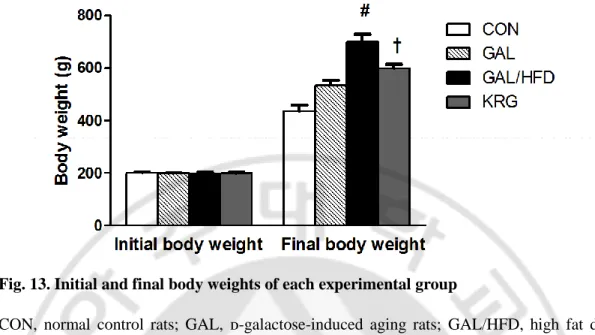
Fig. 13. Initial and final body weights of each experimental group ... 34
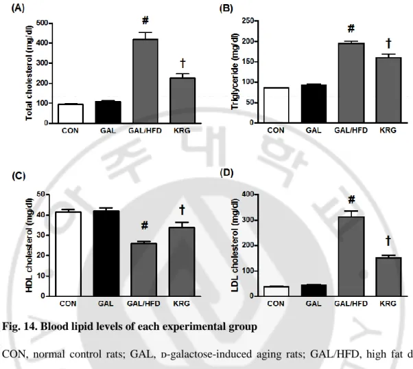
Fig. 14. Blood lipid levels of each experimental group... 35
Fig. 15. Detection of oxidatively modified DNA. ... 37
viii
Fig. 17. In situ detection of fragmented DNA. ... 41
Fig. 18. Immunohistochemical localization of extracellular high mobility group box 1 protein (HMGB1). ... 43
Fig. 19. Immunohistochemical localization of receptor for AGE (RAGE). ... 45
Fig. 20. Schematic diagram for effects of regular exercise and KRG on age-related oxidative renal injury. ... 46
ix
ABBREVIATION
AGEs: Advanced glycation end-products ALEs: advanced lipoxidation end-products CML: Nε-carboxymethyllysine
HFD: High-fat diet
HMGB1: High mobility group box 1 protein HDL: High-density lipoprotein
LDL: Low-density lipoprotein IL-6: interleukin-6
KRG: Korean red ginseng
NF-κB: Transcription factor nuclear factor-κB RAGE: receptor for advanced glycation end-products RCS: Reactive carbonyl species
ROS: Reactive oxygen species TG: Triglyceride
TUNEL: Terminal deoxynucleotidyl transferase dUTP nick end labeling TNF-α: tumor necrosis factor-α
WT-1: Wilms tumor antigen-1 8-OHdG: 8-hydroxyguanine
1
I. INTRODUCTION
A. Age-related oxidative renal injury
Aging as an extremely complex biological phenomenon is demonstrated by accumulation of deleterious changes during the time with an increase in the chance of disease and death. The free radical and oxidative stress theory of aging is recognized as one of the most plausible and promising explanations for the process of aging (Hamid et al., 2012). The pathology of aging and age-related diseases involves oxidative stress at an early stage in its development (Barja, 2004) as confirmed by a decrease in antioxidant defences and an increase in oxidative damage (Moreira et al., 2005). Some recent reports suggests that oxidative stress can cause functional changes in kidneys leading to renal disorders (Fardoun et al., 2006) and cardiovascular disorders (Suh et al., 2001).
Recently, ᴅ-galactose-induced aging rats have been reported to be a reliable animal model for renal aging because rats chronically injected with ᴅ-galactose for a period of 6 weeks showed significant similarities with the naturally aged rats in terms of impaired redox homeostasis (Aydin et al., 2012). ᴅ-Galactose is a physiological nutrient and a reducing sugar that reacts with free amines of amino acids in proteins to form advanced glycation end products (AGEs) through nonenzymatic glycation (Chen et al., 2006). As such, oversupply of galactose could contribute to generation of ROS through oxidative metabolism of ᴅ-galactose as well as through glycation end products (Song et al., 1999; Lu et al., 2006).
2 B. AGEs and oxidative stress
Advanced glycation end products (AGEs) are stable end products of a non-enzymatic glycation reaction (Thorpe and Baynes, 2003). AGEs formation is accelerated under hyperglycemic condition as well as under oxidative stress. The advanced lipoxidation end products (ALEs) are similar adducts but are formed from oxidatevely damaged lipids (Vlassara, 2005). Hyperlipidemia leads to intracellular accumulation of fatty acids and cholesteryl esters (Weinberg, 2006) and chemical modification, particularly oxidation, of both proteins and lipids (Rice-Evans et al., 1996). Reactive carbonyl species (RCS), such as aldehydes acrolein, malondialdehyde and glyoxal, react with proteins to generate these stable adducts (Ahmed, 2005). Glyoxal is a dicarbonyl compound deriving also from autoxidation of Amadori products (glycoxidation) and reacting with lysine residues with formation of Nε-carboxymethyllysine (CML), which is therefore both an ALE and an AGE (Uchida, 2000). AGEs/ALEs accumulation is associated with aging and several age-associated diseases such as Alzheimer’s disease and renal insufficiency (Nass et al., 2007; Thornalley, 2008). The receptor for AGEs (RAGE) was shown to be predominantly involved in mediating AGE/ALE-induced tissue injury (Yan et al., 2003). The interaction between AGE/ALE and RAGE trigger signaling pathways leading to a chronic inflammation and cell damage (Moore and Freeman, 2006). Moreover, an increased lipid accumulation in renal tissues causes mesangial cell activation resulting in glomerular hypertrophy and matrix deposition, podocyte injury and proteinuria (Joles et al., 2000).
3
C. High mobility group box protein-1 and receptor for AGE
High mobility group box protein-1 (HMGB1), also known as amphoterin, is best known as an architectural transcription factor because of its ability to regulate gene activity through DNA binding (Reeves and Wolffe, 1996). The receptor for advanced glycation end products (RAGE) is a member of the immunoglobin superfamily of cell surface receptors that is activated by several ligands including HMGB1 but also by AGEs, S100 proteins, and amyloid β-peptide (Aβ) (Bierhaus et al., 2005). At the time, the consequences of HMGB1 interaction with RAGE were unknown, but it was discovered later that HMGB1 signaling through RAGE promotes chemotaxis and the production of cytokines in a process that involves the activation of the transcription factor nuclear factor-κB (NF-κB) (Palumbo et al., 2007).
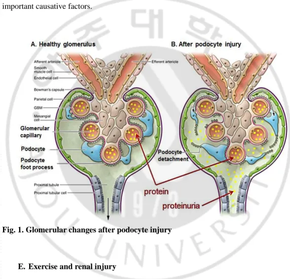
D. Role of podocyte in glomerular pathobiology
Glomerular visceral epithelial cells, which are also called podocytes, function as critical size and charge barriers of protein excretion from glomerulus, therefore, injuries in podocyte can cause marked proteinuria (Laurens et al., 1995; Pavenstadt, 1998). There is a growing body of evidence suggesting that podocyte injury plays an important role in not only proteinuria but also the progression to end-stage renal disease in diabetic nephropathy (Hoshi et al., 2002) and hypertensive glomerulosclerosis (Kretzler et al., 1994). Floege et al have provided data to support the concept that the glomerulosclerosis of aging is “a podocyte disease” although the mechanism by which this might occur was not elucidated (Floege et al., 1997). Since podocytes lack the ability of proliferation, they do not recover from their
4
disappearance induced by podocyte injury, resulting in the decrease of podocytes from the glomerulus, podocytopenia, which is one of the important processes of glomerulosclerosis. Apoptosis is known to be the major course of podocytopenia (Kretzler, 2005). Although there are several factors influencing apoptosis in podocytes, oxidative stress is one of the important causative factors.
Fig. 1. Glomerular changes after podocyte injury
E. Exercise and renal injury
Exercise reduces morbidity and mortality from various cardiovascular and kidney diseases in the elderly population (Larson and Bruce, 1987; Odden, 2010). It has beneficial metabolic actions including reduction in plasma triglycerides, increases in the high-density lipoprotein to low-density lipoprotein ratio and insulin sensitivity, and improves cardiac and
5
renal function (Heath et al., 1983; Sun et al., 2008; Barcellos et al., 2012). Exercise is reported to reduce oxidative stress in rats and mice (Navarro et al., 2004; Asghar et al., 2007). Moderate exercise by inducing the expression of antioxidant enzymes reduces oxidative stress (Gomez-Cabrera et al., 2008). In addition, regular moderate exercise reduces renal CML contents in obese Zucker rats (Boor et al., 2009). However, the mechanism as to how exercise reduces oxidative stress and AGE/ALE formation is not known.
F. Korean red ginseng and renal injury
Panax ginseng, one of the best health foods for vitality and combating fatigue, increases energy and eliminates chronic fatigue while improving health. Ginseng has been used as a tonic for more than 2000 years. It is well known as an energy booster and dietary supplement in Asian countries. Among the several kinds of Panax ginseng products, Korean red ginseng (KRG) has been found to have the most potent pharmacological effects against immune deficiency, metabolic syndrome, and cancer (Park et al., 2012; Paul et al., 2012; Kim et al., 2013). KRG is produced by steaming and drying fresh ginseng. During this process, ginsenosides undergo chemical changes that have the potential to bring about special physiologic activities in vivo (Kim, 2001). Currently, more than 30 different ginsenosides from KRG have been isolated and characterized, and these ginsenosides are known to have different pharmacologic effects (Sun et al., 2009). In addition, KRG extract is reported to reduce AGEs formation in hyperglycemic rats (Quan et al., 2013). Although some clinical and experimental studies have reported that Panax ginseng has a preventive effect on the age-related organ dysfunction and anti-glycation activity (Hwang et al., 2010; Heo et al.,
6
2011; Ramesh et al., 2012; Quan et al., 2013), the effects on age-related renal injury and cross-link formation of glycated proteins unknown.
G. Obesity-related renal injury
Genetic and environmental factors play a role in the development of obesity, and diet is one of the main environmental factors that contribute to this disease. Human studies have shown that increased fat intake is associated with body weight gain which can lead to obesity and other related metabolic diseases. Animal rodent models are therefore useful tools for studying obesity as they will readily gain weight when fed high-fat diets (HFD) (Van Heek et al., 1997; Buettner et al., 2007). HFD also causes renal lipid accumulation and renal injury (Jiang et al., 2005). Renal lipid accumulation may enhance oxidative stress and inflammation in the kidney and contribute to the development of renal injury (Trovati and Cavalot, 2004; Saiki et al., 2005). Reactive oxygen species (ROS) play critical roles in the development and progression of kidney damage (Forbes et al., 2008; Nistala et al., 2008). Obesity-related nephropathy is associated with the increase of proinflammatory cytokines, such as tumor necrosis factor-α (TNF-α) and interleukin-6 (IL-6), due to increased ROS production (Chow et al., 2007). Although the links between hypertension, diabetes and age-related renal disease are established, the relationship between HFD and oxidative stress in age-related renal injury still remain unknown.
H. Aims of study
7
renal injury induced by GAL. In addition, we evaluated the renoprotective effects of exercise and KRG in the GAL/HFD-induced aging rats.
8
II. MATERIALS AND METHODS
A. KRG preparation
KRG extracts were provided by Korea Ginseng Corporation (Daejon, Korea). The KRG extracts contained the following seven glycosides, known as ginsenosides (mg/g): Rg1 (0.71), Rb1 (4.62), Rg3(s) (2.14), Re (0.93), Rc (2.41), Rb2 (1.83), and Rd (0.89).
B. Animals and experimental design
Male SD rats (8-weeks old) were used in this study. They were all individually housed in a temperature-controlled room with a 12-h light/dark cycle and had free access to drinking water. This animal experiment is divided into two experiments. The first experiment is to investigate whether HFD would accelerate oxidative renal injury in ᴅ-galactose (GAL)-induced aging rats and to examine the renopreventive effects of a regular exercise. After acclimation for 2 weeks, the rats were divided into four groups: (1) normal control rats (CON, n = 8), (2) galactose-induced oxidative stressed rats (GAL, n = 8), (3) HFD + galactose-induced oxidative stressed and obese rats (GAL/HFD, n=8) and (4) HFD + ᴅ-galactose-induced oxidative stressed and obese rats exercised regularly on a treadmill (EXE, n=8). The rats in the CON and GAL groups were fed a standard laboratory chow (3.34 kcal/g, PMI Nutrition International, MO, USA). To induce obesity, the rats in the GAL/HFD and EXE groups were fed a high-fat diet containing 60% kcal fat (5.24 kcal/g, D12492, Research Diets, NJ, USA) for 9 weeks. To induce oxidative stress, the rats in the GAL, GAL/HFD,
9
EXE groups were injected intraperitoneally with ᴅ-galactose (100 mg/kg/day), while the rats in the CON group were injected with an equal volume of the vehicle (0.9% saline). The rats in the EXE groups were subjected to a treadmill exercise for 8 weeks (12 meter·/min-1 ·60 min-1, 15 degree grade, 5 days/week) according to a published protocol (Asghar et al., 2002), and the rats in other sedentary groups were not exercised. The exercise training was begun 1 week after the onset of HFD feeding.
The second experiment is to to investigate whether HFD would accelerate AGE/ALE-related renal injury in GAL-induced aging rats and to examine the renopreventive effects of a long-term treatment of KRG. The rats in the GAL group, the GAL/HFD group and the KRG group were injected intraperitoneally with GAL (100 mg/kg/day), while the rats in the CON group were injected with an equal volume of the vehicle (0.9% saline). The treatment of KRG was begun 1 week after the onset of HFD feeding, and KRG was administered orally to the rats for 8 weeks. The body weight was monitored consecutively. All experimental procedures were performed under the supervision of our Institutional Animal Care and Use Committee.
C. Analysis of metabolic data
The body weight was monitored consecutively. One day before autopsy, the animals were fasted for 15 h at least and immediately anesthetized and killed. Blood samples were collected from the tail vein, and total cholesterol, triglyceride (TG), High-density lipoprotein cholesterol (HDL) and Low-density lipoprotein cholesterol (LDL) were measured using an automated analyzer (Wako, Tokyo, Japan).
10
D. In vitro assay of the cross-linking of glycated proteins
For the AGEs cross-linking inhibition assay, AGE-BSA (TransGenic Inc, Kobe, Japan) was incubated in either the presence or absence of KRG or aminoguanidine, a well-known AGE inhibitor, in collagen-coated 96-well plates. Collagen-AGE-BSA cross-linking was detected using a mouse anti-AGEs antibody (6D12, Wako, Osaka, Japan), a horseradish peroxidase-linked goat anti-mouse IgG antibody and a H2O2 substrate containing the ABTS chromogen. The inhibition of collagen-AGE-BSA cross-linking was expressed as the percent decrease in optical density (OD = 410 nm). We calculated the IC50 concentration (µg/ml) as the concentration at which collagen-AGE-BSA cross-linking was inhibited by 50%.
E. Immunohistochemical staining
Immunohistochemistry was performed as previously described (Sohn et al., 2007). Antibodies were a mouse anti-CML (TransGenic, Kobe, Japan), a mouse anti-AGEs (6D12, Wako, Osaka, Japan), a mouse anti-8-hydroxygluanine (8-OHdG) antibody (Santa Cruz Biotechnology, CA, USA), a rabbit anti- high mobility group box 1 protein (HMGB1, 1:200, Epitomics, CA, USA), a rabbit anti-RAGE and a rabbit anti-synaptopodin (Santa Cruz Biotechnology, CA, USA). For detection of CML, AGEs, 8-OHdG and synaptopodin, the sections were incubated with the Envision kit (DAKO, CA, USA) and visualized by 3,3’-diaminobenzidine tetrahydrochloride. Negative controls for immunohistochemistry were run by incubating the sections with nonimmune serum instead of the primary antibody. The intensity of immunohistochemical staining of CML and 8-OHdG was analyzed in eight
11
randomly selected mm2 areas of renal cortex, and the positive signal areas of synaptopodin was determined per one glomerulus in a total of 40 glomeruli using Image J software (NIH, MD, USA).
F. Double staining for TUNEL and Wilms tumor antigen-1
In order to confirm the apoptotic cell death in renal podocytes, a sequential immunostaining was performed. The first staining of TUNEL was performed with a kit (DeadEnd apoptosis detection system, Promega, WI, USA) according to the manufacturer’s instructions. Apoptotic cells were detected with FITC-conjugated streptavidin (Santa Cruz Biotechnology, CA, USA). The second sequence of staining using rabbit anti-Wilms tumor antigen-1 (WT-1, Santa Cruz Biotechnology, CA, USA) was performed on the same section with tetramethyl rhodamine isothiocyanate (TRITC)-conjugated goat anti-rabbit IgG antibody (Santa Cruz Biotechnology, CA, USA). To prevent cross-reactivity between two staining sequences the slides were incubated with normal mouse serum (DAKO, CA, USA) after the first staining. For morphometric analysis, the positive cell numbers of WT-1 per one glomerulus in a total of 40 glomeruli was determined using Image J software.
G. Apoptosis analysis
To evaluate apoptosis in renal tissues, the TUNEL assay was performed with a kit according to the manufacturer’s instructions. Apoptotic cells were detected with peroxidase conjugated streptavidin. The number of TUNEL-positive cells per unit area (mm2) was then determined in counted in a total of 5 fields.
12 H. Statistical analysis
Comparisons between groups were performed using one-way analysis of variance (ANOVA) followed by Tukey's multiple comparison test using GraphPad Prism 4.0 software (Graph pad, CA, USA).
13
III. Results
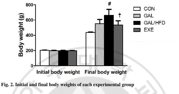
A. Body weight and blood lipid profile
Body weights at the beginning and at the end of the experiment are shown in Fig. 2. The body weight in the GAL group was slightly increased compared to the control group. In rats received both the GAL and HFD, body weight was significantly increased compared to the control rats and reduced by exercise training. Total cholesterol, TG and LDL levels were significantly increased in the GAL/HFD group (P<0.01 vs. CON group). EXE group showed significantly reduced in total cholesterol, TG and LDL levels as compared to the GAL/HFD group. However, no differences in lipid levels were noted between the CON group and the GAL group (Fig. 3).
14
Fig. 2. Initial and final body weights of each experimental group
CON, normal control rats; GAL, ᴅ-galactose-induced oxidative stressed rats; GAL/HFD, high fat diet plus ᴅ-galactose-induced oxidative stressed and obese rats, EXE, high-fat diet plus ᴅ-galctose-induced oxidative stressed and obese rats exercised regularly. Values in the bar graphs represent means ± SE, n = 8. #p<0.01 compared with GAL group; †p<0.01 compared with GAL/HFD group.
15 Fig. 3. Blood lipid levels of each experimental group
CON, normal control rats; GAL, ᴅ-galactose-induced oxidative stressed rats; GAL/HFD, high fat diet plus ᴅ-galactose-induced oxidative stressed and obese rats, EXE, high-fat diet plus ᴅ-galctose-induced oxidative stressed and obese rats exercised regularly. Values in the bar graphs represent means ± SE, n = 8. #p<0.01 compared with GAL group; †p<0.01 compared with GAL/HFD group.
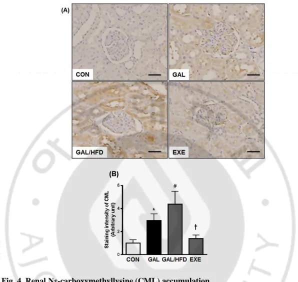
16 B. CML accumulation in renal tissues
To determine whether HFD accelerate renal injury, we performed the immunohistochemical staining for CML as a marker for AGE/ALE, in renal tissues. As shown in Fig. 4, the immunoreactivities of CML in the GAL and GAL/HFD groups were significantly higher compared to the CON group. The expressions of CML were mainly distributed in the regions of the glomerulus and renal tubules. Especially, high expression of CML was observed in the proximal tubular epithelial cells. Moreover, the staining intensity of the GAL/HFD group was significantly higher than the GAL group, which indicated that the HFD had an accelerating effect on the renal accumulation of AGE/ALE and oxidative renal injury induced by GAL. Next, we evaluated whether the regular exercise were found preventive effect on the accumulation of CML in renal tissues. The regular exercise suppressed the accumulation of CML compared to the GAL/HFD group (Fig. 4B).
17
Fig. 4. Renal Nε-carboxymethyllysine (CML) accumulation.
(A) Representative CML immunohistochemistry in renal tissues. Original magnifications: X200 (Scale bar; 50 μm). (B) Quantitative analysis of CML immunostaining signal intensity. The intensity of immunohistochemical staining of CML was analyzed in eight randomly selected mm2 areas of renal cortex. CON, normal control rats; GAL, ᴅ-galactose-induced oxidative stressed rats; GAL/HFD, high fat diet plus ᴅ-galactose-induced oxidative stressed and obese rats, EXE, high-fat diet plus ᴅ-galctose-induced oxidative stressed and obese rats exercised regularly. Values in the bar graphs represent means ± SE, n = 8. *p<0.01 compared with CON group; #p<0.01 compared with GAL group; †p<0.01 compared with GAL/HFD group.
18 C. Oxidative DNA damage in renal tissues
To determine whether HFD would interact with GAL to accelerate the age-related oxidative renal injury, we examine the oxidative DNA damage in renal tissues by immunostaining of 8-OHdG. The oxidation of guanine to form 8-OHdG acts as a marker of oxidative DNA damage (Beckman and Ames, 1997). As shown in Fig. 5, the 8-OHdG marker exhibited nuclear and/or perinuclear localization in the renal tubular epithelial cells. The immunoreactivity of 8-OHdG in the GAL and GAL/HFD groups were significantly higher compared to the CON group. Moreover, the staining intensity of the GAL/HFD group was significantly higher than the GAL group, which indicated that the HFD had an accelerating effect on the age-related oxidative renal injury induced by GAL. The regular exercise suppressed the expressions of 8-OHdG compared to the GAL/HFD group (Fig. 5B).
19 Fig. 5. Detection of oxidatively modified DNA.
(A) Representative 8-OHdG immunohistochemitry in renal tissues. Original magnifications: X200 (Scale bar; 50 μm). (B) Quantitative analysis of 8-OHdG immunostaining signal intensity. The intensity of immunohistochemical staining of 8-OHdG was analyzed in eight randomly selected mm2 areas of renal cortex. CON, normal control rats; GAL, ᴅ-galactose-induced oxidative stressed rats; GAL/HFD, high fat diet plus ᴅ-galactose-ᴅ-galactose-induced oxidative stressed and obese rats, EXE, high-fat diet plus ᴅ-galctose-induced oxidative stressed and obese rats exercised regularly. Values in the bar graphs represent means ± SE, n = 8. *p<0.01 compared with CON group; #p<0.01 compared with GAL group; †p<0.01 compared with GAL/HFD group.
20 D. Apoptosis assay in renal tissues
TUNEL staining used widely as a marker for apoptosis (Charriaut-Marlangue and Ben-Ari, 1995). In the CON group, a TUNEL-positive nucleus was barely detected. In the GAL and GAL/HFD groups, many TUNEL-positive cells were observed in renal glomeruli and tubular epithelial cells. The numbers of TUNEL-positive cells in the GAL/HFD group were significantly higher than the GAL group. However, the regular exercise prevented the increase in the positive cells that was seen in the CON group (Fig. 6).
21 Fig. 6. In situ detection of fragmented DNA.
(A) Representative TUNEL staining in renal tissues. Original magnifications: X200 (Scale bar; 50 μm). (B) Quantitative analysis of TUNEL-positive cells. CON, normal control rats; GAL, induced oxidative stressed rats; GAL/HFD, high fat diet plus ᴅ-galactose-induced oxidative stressed and obese rats, EXE, high-fat diet plus ᴅ-galctose-ᴅ-galactose-induced oxidative stressed and obese rats exercised regularly. Values in the bar graphs represent means ± SE, n = 8. *p<0.01 compared with CON group; #p<0.01 compared with GAL group; †p<0.01 compared with GAL/HFD group.
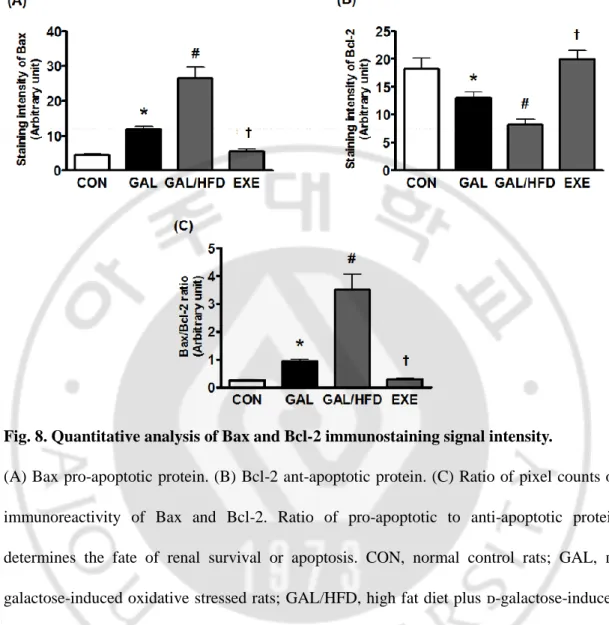
22 E. Expression of Bax and Bcl-2 in renal tissues
To determine the mechanism underlying the anti-apoptotic effects of regular exercise, we measured the expression of pro-apoptotic Bax protein and anti-apoptotic Bcl-2 protein in renal glomeruli and tubular epithelial cells. The results revealed that Bax and Bcl-2 were mainly expressed in the cytoplasm of renal glomeruli and tubular epithelial cells, and the intensity of Bax expression in the GAL and GAL/HFD groups was higher than that in the control group (Fig. 7). The intensity of Bcl-2 expression in the GAL and GAL/HFD groups was lower than that of the control group. However, the regular exercise were restored the expression of these proteins compared to the GAL/HFD group (Fig. 8).
23 Fig. 7. Renal Bax and Bcl-2 expression.
Representative Bax and Bcl-2 immunohistochemistry in renal tissues. Bax pro-apoptotic protein in the GAL and GAL/HFD groups was higher than that in the control group. Bcl-2 anti-apoptotic protein in the GAL and GAL/HFD groups was lower than that of the control group. Original magnifications: X200 (Scale bar; 50 μm). CON, normal control rats; GAL, induced oxidative stressed rats; GAL/HFD, high fat diet plus ᴅ-galactose-induced oxidative stressed and obese rats, EXE, high-fat diet plus ᴅ-galctose-ᴅ-galactose-induced oxidative stressed and obese rats exercised regularly.
24
Fig. 8. Quantitative analysis of Bax and Bcl-2 immunostaining signal intensity.
(A) Bax pro-apoptotic protein. (B) Bcl-2 ant-apoptotic protein. (C) Ratio of pixel counts of immunoreactivity of Bax and Bcl-2. Ratio of pro-apoptotic to anti-apoptotic protein determines the fate of renal survival or apoptosis. CON, normal control rats; GAL, ᴅ-galactose-induced oxidative stressed rats; GAL/HFD, high fat diet plus ᴅ-ᴅ-galactose-induced oxidative stressed and obese rats, EXE, high-fat diet plus ᴅ-galctose-induced oxidative stressed and obese rats exercised regularly. Values in the bar graphs represent means ± SE, n = 8. *p<0.01 compared with CON group; #p<0.01 compared with GAL group; †p<0.01 compared with GAL/HFD group.
25 F. Caspase-3 activation
Caspases are a family of at least 10 proteases that are important effectors in apoptotic signaling and play a key role in the apoptotic pathway. Caspase-3 activation is needed for a highly specific proteolytic cleavage of a functionally important nuclear repair enzyme, PARP, known as a substrate of activated caspase. As shown in Fig. 9, the activated caspase-3 in the GAL and GAL/HFD groups were significantly higher compared to the CON group. However, the regular exercise suppressed the activation of caspase-3 compared to the GAL/HFD group (Fig. 9).
26 Fig. 9. Activation of caspase-3.
(A) Representative activated caspase-3 immunohistochemitry in renal tissues. Original magnifications: X200 (Scale bar; 50 μm).μm. (B) Quantitative analysis of activated caspase-3 immunostaining signal intensity. CON, normal control rats; GAL, ᴅ-galactose-induced oxidative stressed rats; GAL/HFD, high fat diet plus ᴅ-galactose-induced oxidative stressed and obese rats, EXE, high-fat diet plus ᴅ-galctose-induced oxidative stressed and obese rats exercised regularly. Values in the bar graphs represent means ± SE, n = 8. *p<0.01 compared with CON group; #p<0.01 compared with GAL group; †p<0.01 compared with GAL/HFD group.
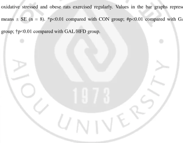
27 G. Glomerular podocyte loss
The loss of glomerular podocytes precedes and predicts the onset of clinical nephropathy (Meyer et al., 1999). To evaluate the loss of podocytes in renal tissues, we performed imuunohistochemical staining for synaptopodin and WT-1, well-known podocyte markers (Mundel et al., 1997; Schiffer et al., 2004). Average numbers of podocytes per glomerular section were determined by counting cells and measuring areas that were positively labeled with two podocyte markers. In the GAL group, synaptopodin and WT-1 positive cell counts tended to decrease compared with the CON group. The staining areas of synaptopodin and the number of WT-1-positive cells in the GAL/HFD group were significantly lower than the GAL group (Fig. 10A and 11A). In double staining, some TUNEL-positive cells were localized within the region where WT-1 was positively stained. This mixed color staining (TUNEL; green, WT-1; red) was obtained in podocytes, confirming the apoptosis of podocytes (Fig. 11A). The regular exercise visibly increased the positive cells and areas in the kidney glomeruli (Fig. 10B and 10B).
28 Fig. 10. Glomerular podocyte loss.
(A) Representative synaptopodin immunohistochemitry in renal tissues. Original magnifications: X200 (Scale bar; 50 μm). (B) Quantitative analysis of synaptopodin immunostaining signal intensity. CON, normal control rats; GAL, ᴅ-galactose-induced oxidative stressed rats; GAL/HFD, high fat diet plus ᴅ-galactose-induced oxidative stressed and obese rats, EXE, high-fat diet plus ᴅ-galctose-induced oxidative stressed and obese rats exercised regularly. Values in the bar graphs represent means ± SE (n = 8). *p<0.01 compared with CON group; #p<0.01 compared with GAL group; †p<0.01 compared with GAL/HFD group.
29
Fig. 11. Anti-apoptotic effect of exercise in the renal podocyte.
(A) Representative photomicrographs of double-staining for TUNEL (green) and WT-1 (red). Mixed yellow-colored cells in the merged images (arrows) indicate that apoptotic cell death may have occurred in podocyte. Original magnifications: X200 (Scale bar; 50 μm). (B) Quantitative analysis of WT-1 immunostaining signal intensity. CON, normal control rats; GAL, induced oxidative stressed rats; GAL/HFD, high fat diet plus ᴅ-galactose-induced oxidative stressed and obese rats, EXE, high-fat diet plus ᴅ-galctose-ᴅ-galactose-induced oxidative stressed and obese rats exercised regularly. Values in the bar graphs represent means ± SE (n = 8). *p<0.01 compared with CON group; #p<0.01 compared with GAL group; †p<0.01 compared with GAL/HFD group.
30
31
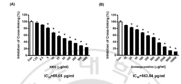
H. Inhibitory effect of KRG on glycated proteins cross-linking in vitro.
To investigate whether KRG could inhibit the AGEs cross-link, AGE-BSA was incubated with KRG in collagen-coated plates. KRG (IC50=55.65 μg/ml) exhibited much stronger inhibitory activity on AGE-BSA binding with collagen than aminoguanidine (IC50=563.54 μg/ml), a well-known glycation inhibitor (Fig. 12A and B).
32
Fig. 12. Inhibitory effect of KRG on a glycated protein cross-linking in vitro.
Inhibition of KRG (A) and aminoguanidine (B) on AGE-BSA and collagen cross-linking. *P<0.01 vs. untreated group.
33 I. Body weight and blood lipid profile
Body weights at the beginning and at the end of the experiment are shown in Fig. 13. The body weight in the GAL group was slightly increased compared to the control group. In rats received both the GAL and HFD, body weight was significantly increased compared to the control rats and reduced by KRG treatment. Total cholesterol, TG and LDL levels were significantly increased in the GAL/HFD group (P<0.01 vs. CON group). KRG group showed significantly reduced in total cholesterol, TG and LDL levels as compared to the GAL/HFD group. However, no differences in lipid levels were noted between the CON group and the GAL group (Fig. 14).
34
Fig. 13. Initial and final body weights of each experimental group
CON, normal control rats; GAL, ᴅ-galactose-induced aging rats; GAL/HFD, high fat diet plus ᴅ-galactose-induced aging rats, KRG, high-fat diet plus ᴅ-galctose-induced aging rats treated with KRG (200 mg/kg/day). Values in the bar graphs represent means ± SE, n = 8. #p<0.01 compared with CON group; †p<0.01 compared with GAL/HFD group.
35
Fig. 14. Blood lipid levels of each experimental group
CON, normal control rats; GAL, ᴅ-galactose-induced aging rats; GAL/HFD, high fat diet plus ᴅ-galactose-induced aging rats, KRG, high-fat diet plus ᴅ-galctose-induced aging rats treated with KRG (200 mg/kg/day). Values in the bar graphs represent means ± SE, n = 8. #p<0.01 compared with CON group; †p<0.01 compared with GAL/HFD group.
36 J. Oxidative DNA damage in renal tissues
To determine whether HFD would interact with GAL to accelerate the age-related oxidative renal injury, we examine the oxidative DNA damage in renal tissues by immunostaining of 8-OHdG. The oxidation of guanine to form 8-OHdG acts as a marker of oxidative DNA damage (Beckman and Ames, 1997). As shown in Fig. 15, the 8-OHdG marker exhibited nuclear and/or perinuclear localization in the renal tubular epithelial cells. The immunoreactivity of 8-OHdG in the GAL and GAL/HFD groups were significantly higher compared to the CON group. Moreover, the staining intensity of the GAL/HFD group was significantly higher than the GAL group, which indicated that the HFD had an accelerating effect on the age-related oxidative renal injury induced by GAL. The treatment with KRG suppressed the expression of 8-OHdG compared to the GAL/HFD group (Fig. 15B).
37 Fig. 15. Detection of oxidatively modified DNA.
(A) Representative 8-OHdG immunohistochemistry in renal tissues. Original magnifications: X200 (Scale bar; 50 μm). (B) Quantitative analysis of 8-OHdG immunostaining signal intensity. CON, normal control rats; GAL, ᴅ-galactose-induced aging rats; GAL/HFD, high fat diet plus ᴅ-galactose-induced aging rats, KRG, high-fat diet plus ᴅ-galctose-induced aging rats treated with KRG (200 mg/kg/day). Values in the bar graphs represent means ± SE, n = 8. *p<0.01 compared with CON group; #p<0.01 compared with GAL group; †p<0.01 compared with GAL/HFD group.
38 K. Protein glycations in renal tissues
To determine whether HFD would interact with GAL to accelerate cross-link formation of glycated proteins in vivo, we performed the immunohistochemical staining for AGEs in renal tissues. As shown in Fig. 16, the immunoreactivities of AGEs in the GAL and GAL/HFD group were significantly higher compared to the CON group. The expressions of AGEs were mainly distributed in the regions of the glomerulus and renal tubules. Especially, high expression of AGEs was observed in the proximal tubular epithelial cells. Moreover, the staining intensity of the GAL/HFD group were significantly higher than the GAL group, which indicated that the HFD had an accelerating effect on the renal AGEs cross-linking induced by GAL. Next, we evaluated whether KRG were found preventive effect on the AGEs cross-linking in renal tissues. KRG exhibited inhibitory activity on AGEs cross-link in vitro (Fig.12). The treatment with KRG suppressed the expression of AGEs compared to the GAL/HFD group (Fig. 16B).
39
Fig. 16. Renal advanced glycation end products (AGEs) cross-linking and deposition. (A) Representative AGEs immunohistochemistry in renal tissues. Original magnifications: X200 (Scale bar; 50 μm). (B) Quantitative analysis of AGEs immunostaining signal intensity. CON, normal control rats; GAL, ᴅ-galactose-induced aging rats; GAL/HFD, high fat diet plus ᴅ-galactose-induced aging rats, KRG, high-fat diet plus ᴅ-galctose-induced aging rats treated with KRG (200 mg/kg/day). Values in the bar graphs represent means ± SE, n = 8. *p<0.01 compared with CON group; #p<0.01 compared with GAL group; †p<0.01 compared with GAL/HFD group.
40 L. Apoptosis assay in renal tissues
To characterize the injury of renal cells, we applied the staining for TUNEL. TUNEL analysis detects cells in which DNA is fragmenting and used widely as a marker for apoptosis (Charriaut-Marlangue and Ben-Ari, 1995). In the CON group, a TUNEL-positive nucleus was barely detected. In the GAL and GAL/HFD groups, many TUNEL-positive cells were observed in renal glomeruli and tubular epithelial cells. The numbers of TUNEL-positive cells in the GAL/HFD group were significantly higher than the GAL group. However, treatment with KRG prevented the increase in the positive cells that was seen in control renal tissues (Fig. 17).
41 Fig. 17. In situ detection of fragmented DNA.
(A) Representative TUNEL staining in renal tissues. Original magnifications: X200 (Scale bar; 50 μm). (B) Quantitative analysis of TUNEL-positive cells. CON, normal control rats; GAL, ᴅ-galactose-induced aging rats; GAL/HFD, high fat diet plus ᴅ-galactose-induced aging rats, KRG, high-fat diet plus ᴅ-galctose-induced aging rats treated with KRG (200 mg/kg/day). Values in the bar graphs represent means ± SE, n = 8. *p<0.01 compared with CON group; #p<0.01 compared with GAL group; †p<0.01 compared with GAL/HFD group.
42
M. HMGB1 cytoplasmic translocalization in renal tissues
HMGB1, a nuclear DNA-binding protein, can be released extracellularly and acts as a pro-inflammatory cytokine or alarm signal for tissue damage (Ulloa and Messmer, 2006). In renal tissue of rat with diabetic nephropathy, HMGB1 is extensively released from renal glomerular cells and renal tubular epithelial cells (Kim et al., 2011). To investigate the pathogenic status of HMGB1 in age-related renal injury, we applied the immunohistochemical staining in renal tissues. In the CON group, HMGB1 was expressed only in nuclei, but HMGB1 was detected in the nuclei and diffusely in the cytoplasm in renal tissues of the GAL and GAL/HFD groups. This cytoplasmic translocation of HMGB1 was markedly inhibited by KRG administration (Fig.18).
43
Fig. 18. Immunohistochemical localization of extracellular high mobility group box 1 protein (HMGB1).
Representative HMGB1 immunohistochemistry in renal tissues. In the GAL and GAL/HFD groups, HMGB1 was expressed in both the cytoplasmic and nuclear patterns in renal glomerular cells and tubular epithelial cells, although in the CON group, HMGB1 was expressed only in the cell nuclei. CON, normal control rats; GAL, ᴅ-galactose-induced aging rats; GAL/HFD, high fat diet plus galactose-induced aging rats, KRG, high-fat diet plus ᴅ-galctose-induced aging rats treated with KRG (200 mg/kg/day). Original magnifications: X200 (Scale bar; 50 μm).
44 N. RAGE Expression in renal tissues
RAGE is a potential receptor for HMGB1. The interaction between RAGE and HMGB1 is key players in cellular response to stress conditions, such as inflammation (Schiraldi et al., 2012). Thus, we measured the expression levels of RAGE in age-related renal injury. As shown in Fig. 19, RAGE was expressed in cell surface of renal tubular epithelial cells. The immunoreactivity of RAGE in the GAL and GAL/HFD groups were significantly higher compared to the CON group. Moreover, the staining intensity of the GAL/HFD group was significantly higher than the GAL group, which indicated that the HFD had an accelerating effect on the age-related renal injury induced by GAL. The treatment with KRG suppressed the expression of RAGE compared to the GAL/HFD group (Fig. 19).
45
Fig. 19. Immunohistochemical localization of receptor for AGE (RAGE).
(A) Representative RAGE immunohistochemistry in renal tissues. Original magnifications: X200 (Scale bar; 50 μm). (B) Quantitative analysis of RAGE immunostaining signal intensity. CON, normal control rats; GAL, ᴅ-galactose-induced aging rats; GAL/HFD, high fat diet plus ᴅ-galactose-induced aging rats, KRG, high-fat diet plus ᴅ-galctose-induced aging rats treated with KRG (200 mg/kg/day). Values in the bar graphs represent means ± SE, n = 8. *p<0.01 compared with CON group; #p<0.01 compared with GAL group; †p<0.01 compared with GAL/HFD group.
46
Fig. 20. Schematic diagram for effects of regular exercise and KRG on age-related oxidative renal injury.
47
IV. DISCUSSION
The present study provides novel evidence that a HFD accelerated the renal accumulation of AGE/ALE and podocyte loss in renal tissues of GAL-induced aging rats. We showed that rats, when fed with a HFD for 9 weeks, become obese and have an enhanced oxidative stress and cross-link formation of glycated proteins in the renal tissues of the GAL-induced aging rats. Our results described the link between an HFD and oxidative stress in age-related renal injury. In addition, we showed that the regular exercise and KRG has the renopreventive effect in this animal model.
ᴅ-Galactose, a reducing sugar, metabolized at normal concentration, but the excessive levels of ᴅ-galactose can be converted into aldose and hydroperoxide under the catalysis of galactose oxidase, resulting in the generation of a superoxide anion and oxygen-derived free radicals (Wu et al., 2008). When ᴅ-galactose is injected to rats for 6 ~ 9 weeks, free radical production is increased in renal tissues (Lei et al., 2008). Mice and rats injected with ᴅ-galactose have been used as an animal model of oxidative stress (Hua et al., 2007; Lu et al., 2007; Fan et al., 2009; Zhang et al., 2009).
Extensive renal accumulation of lipids associated with increased renal lipid synthesis via a SREBP-1c-dependent pathway has been demonstrated in animals fed HFD (40–60% kcal fat) (Wei et al., 2004; Kume et al., 2007). In our study, we demonstrated that the elevated levels of circulating lipids and the enhanced AGE/ALE accumulation. Moreover, GAL-injected rats fed a HFD showed accelerated renal injury associated with more pronounced
48
renal ALE/AGE content and oxidative DNA damage. Our data indicate that a GAL/HFD induced an extensive formation and renal deposition of AGE/ALE, such as CML. This product is known to produce carbonyl and oxidative stress which may cause injury both directly and via receptor-mediated mechanisms (Iacobini et al., 2009). The accelerated renal injury observed in rats in the GAL/HFD group compared with the GAL group indicates that a HFD plays a pivotal role in GAL-induced oxidative renal injury. The hyperlipidemia induced by a HFD was associated with the enhanced deposition of AGE/ALE in the aging process.
AGE/ALE has been reported to induce apoptosis in various cell lines, such as mesangial cells and endothelial cells (Yamagishi et al., 2002; Kaji et al., 2003). Renal cell apoptosis or necrosis can inevitably affect glomerular filtration rate and endothelial function, resulting in renal failure (Bonegio and Lieberthal, 2002; de Vries et al., 2003; Favreau et al., 2010). In this study, the HFD can amplify the GAL-induced apoptotic renal cell death. Oxidative stress or pro-apoptotic cytokine through interaction between AGE/ALE and RAGE were involved in podocyte apoptosis (Yamamoto and Yamamoto, 2012). Among the three intrinsic cells in glomerulus, podocyte is one of the important ingredients of filtration barrier which has special cytobiological trait and physiological function. The injury of podocyte can inevitably lead to the occurrence of proteinuria (Zhang et al., 2007). Podocyte apoptosis has been demonstrated to correlate with worsening renal function (Susztak et al., 2006). Consistent with this interpretation, the result of the present study demonstrates that the increased renal levels of CML and 8-OHdG in GAL/HFD-treated rat are involved, at least in part, in the injury of podocytes. Although there was disappearance of podocyte
49
markers from glomeruli, apoptosis was observed by TUNEL staining predominantly in tubular epithelial cells. Liu et al. also showed that ᴅ-galactose treatment in mice induced extensive tubular damage by presence of necrotic tubular epithelial cells (Liu et al., 2010). These results suggest that GAL-induced renal injury was occurred predominantly in both proximal and distal tubular epithelial cells.
Activated caspase-3 is the final mediator in the extrinsic and intrinsic pathways of apoptosis. Increased oxidative stress can improve the level of cleaved caspase-3 and promote apoptosis in aged kidney (Kiray et al., 2009). Bax and Bcl-2 are considered to be the principal factors in determining whether the execution of apoptosis proceeds (Carambula et al., 2002). Bcl-2 is an important anti-apoptotic proto-oncogene and Bax is a pro-apoptotic one. The ratio of pro-apoptotic Bax to anti-apoptotic Bcl-2-like protein is believed to be important in determining cell survival versus cell death (Oltvai et al., 1993). The ratio of Bax to Bcl-2 is altered in age-related chronic kidney disease in a way that favors apoptosis (Percy et al., 2009). The ratio of increased Bax/Bcl-2 damages the integrity of mitochondria, causing release of cytochrome c from mitochondria, thereby leading to the activation of caspase-3 and caspase-9 (Desagher and Martinou, 2000). In this study, pro-apoptotic Bax and caspase-3 were significantly increased, otherwise anti-apoptotic Bcl-2 were deceased in renal tissues of GAL-treated rats. Also all these renal changes were highly aggravated in the GAL/HFD group than that in the GAL group.
In our first animal experiment, we examined the effect of intervention by the regular exercise on the increases in AGE/ALE accumulation, oxidative DNA damage and podocyte loss in kidney tissues. The intervention by the regular exercise inhibited increases in
50
AGE/ALE deposition, renal 8-OHdG levels and podocyte loss in HFD/GAL-treated rats. A substantial body of evidence showed the renoprotective effects of the physical exercise in diabetic nephropathy (Kanauchi et al., 1994; Ward et al., 1994; Chiasera et al., 2000; Koh et al., 2011; Rodrigues et al., 2011)and chronic kidney disease (George et al., 2009; Coelho et al., 2010; de Souza et al., 2012). Physical exercise has a positive influence on physical capacity, hypertension, left ventricular function, lipid and glucose metabolism and oxidative status (Pechter et al., 2003; Moinuddin and Leehey, 2008). Coelho et al. showed that physical training performed before the onset of chronic kidney disease is capable of improving oxidative stress parameters, possibly by reducing oxidant production (Coelho et al., 2010). In addition, the moderate regular exercise reduced the burden of AGEs in diabetes- and obesity-associated nephropathy rats. Exercise-induced higher energy demands might decrease the pool of reactive intermediates for glycation or lipoxidation (Boor et al., 2009). Moreover, the regular exercise ameliorated the activated caspase-3, enhanced pro-apoptotic Bax and the decreased Bcl-2 protein in the GAL and GAL/HFD groups. Thus, it suggests that regular exercise has a potential anti-apoptotic effect by its antioxidant activity. Our results indicated that the reduction of AGE/ALE accumulation by the regular exercise might have been a major mechanism of exercise-associated renoprotection in GAL/HFD-induced oxidative stressed and obese rats.
In addition, our second experiments showed the effect of intervention by KRG on the increases in protein glycation. It was previously reported that KRG has anti-AGEs activity under hyperglycemic conditions (Quan et al., 2013). Similarly, we showed that the intervention by KRG inhibited AGEs cross-link and renal AGEs deposition in
GAL/HFD-51
treated rats. Our results indicated that the reduction of AGEs cross-linking by KRG might be a major mechanism of renoprotection in GAL/HFD-induced aging rats.
A ubiquitous nuclear protein, HMGB1, can be released by various immune cells in response to infection or injury (Lotze and Tracey, 2005) and is a danger signal to alert the immune system (Scaffidi et al., 2002). HMGB1 is downstream of apoptosis on the final common pathway to organ damage. In primary apoptosis, there is almost no release of HMGB1; however, in conditions with inappropriate clearance of apoptotic material, apoptotic cells may undergo secondary necrosis leading to HMGB1 release (Bell et al., 2006). As an extracellular signaling molecule, HMGB1 elicits its effects through the RAGE which has been shown to interact with HMGB1 with high affinity (Hori et al., 1995). In an animal model of adenine-induced nephropathy, granuloma formation was found to be associated with high HMGB1 expression and up-regulation of RAGE in the kidney (Oyama et al., 2010). Our result is the first evidence that HMGB1 is extensively expressed in renal tissues of the GAL/HFD-treated rats, and HMGB1 was released from renal glomerular cells and renal tubular epithelial cells. The distribution of HMGB1 in these cells was clearly translocated from the nucleus to the cytoplasm. The level of RAGE expression also was significantly increased in renal tissues of GAL-treated rats and highly increased in the GAL/HFD group than that in the GAL group. This observation indicated that renal glomerular cells and renal tubular epithelial cells might secrete HMGB1 and represent the major source of HMGB1 in age-related renal injury, suggesting that obesity may aggravate the age-related renal injury induced by GAL. The treatment of KRG also inhibited the cytoplasmic translocation of HMGB1 and expression of RAGE in the GAL/HFD-treated rats.
52
These results also showed that KRG may exert a renoprotective effect on the age-related renal injury by its antioxidant activity.
53
V. CONCLUSION
In conclusion, this study indicates that HFD may enhance GAL-induced renal injury. We also demonstrated that the regular exercise and KRG protects against aged-related renal injury. These effects could be attributed, at least in a part, to reduction of AGE/ALE deposition and oxidative stress. We suggest here that the regular exercise and KRG might be an easy and effective strategy to attenuate renal injury related with aging and obesity.
54
VI. REFERENCES
1. Ahmed N: Advanced glycation endproducts--role in pathology of diabetic complications. Diabetes Res Clin Pract 67: 3-21, 2005
2. Asghar M, George L, Lokhandwala MF: Exercise decreases oxidative stress and inflammation and restores renal dopamine D1 receptor function in old rats. Am J Physiol Renal Physiol 293: F914-919, 2007
3. Asghar M, Hussain T, Lokhandwala MF: Higher basal serine phosphorylation of D1A receptors in proximal tubules of old Fischer 344 rats. Am J Physiol Renal Physiol 283: F350-355, 2002
4. Aydin S, Yanar K, Atukeren P, Dalo E, Sitar ME, Uslu E, Caf N, Cakatay U: Comparison of oxidative stress biomarkers in renal tissues of D-galactose induced, naturally aged and young rats. Biogerontology 13: 251-260, 2012
5. Barcellos FC, Santos IS, Mielke GI, del Vecchio FB, Hallal PC: Effects of exercise on kidney function among non-diabetic patients with hypertension and renal disease: randomized controlled trial. BMC Nephrol 13: 90, 2012
6. Barja G: Free radicals and aging. Trends Neurosci 27: 595-600, 2004
7. Beckman KB, Ames BN: Oxidative decay of DNA. J Biol Chem 272: 19633-19636, 1997
8. Bell CW, Jiang W, Reich CF, 3rd, Pisetsky DS: The extracellular release of HMGB1 during apoptotic cell death. American journal of physiology. Cell physiology 291:
55
C1318-1325, 2006
9. Bonegio R, Lieberthal W: Role of apoptosis in the pathogenesis of acute renal failure. Curr Opin Nephrol Hypertens 11: 301-308, 2002
10. Boor P, Celec P, Behuliak M, Grancic P, Kebis A, Kukan M, Pronayova N, Liptaj T, Ostendorf T, Sebekova K: Regular moderate exercise reduces advanced glycation and ameliorates early diabetic nephropathy in obese Zucker rats. Metabolism 58: 1669-1677, 2009
11. Carambula SF, Matikainen T, Lynch MP, Flavell RA, Goncalves PB, Tilly JL, Rueda BR: Caspase-3 is a pivotal mediator of apoptosis during regression of the ovarian corpus luteum. Endocrinology 143: 1495-1501, 2002
12. Charriaut-Marlangue C, Ben-Ari Y: A cautionary note on the use of the TUNEL stain to determine apoptosis. Neuroreport 7: 61-64, 1995
13. Chen CF, Lang SY, Zuo PP, Yang N, Wang XQ, Xia C: Effects of D-galactose on the expression of hippocampal peripheral-type benzodiazepine receptor and spatial memory performances in rats. Psychoneuroendocrinology 31: 805-811, 2006
14. Chiasera JM, Ward-Cook KM, McCune SA, Wardlaw GM: Effect of aerobic training on diabetic nephropathy in a rat model of type 2 diabetes mellitus. Ann Clin Lab Sci 30: 346-353, 2000
15. Coelho BL, Rocha LG, Scarabelot KS, Scheffer DL, Ronsani MM, Silveira PC, Silva LA, Souza CT, Pinho RA: Physical exercise prevents the exacerbation of oxidative stress parameters in chronic kidney disease. J Ren Nutr 20: 169-175, 2010
56
Souza CT, Silva LA, Pinho RA: Therapeutic action of physical exercise on markers of oxidative stress induced by chronic kidney disease. Life Sci 91: 132-136, 2012 17. de Vries B, Matthijsen RA, van Bijnen AA, Wolfs TG, Buurman WA:
Lysophosphatidic acid prevents renal ischemia-reperfusion injury by inhibition of apoptosis and complement activation. Am J Pathol 163: 47-56, 2003
18. Desagher S, Martinou JC: Mitochondria as the central control point of apoptosis. Trends in cell biology 10: 369-377, 2000
19. Fan SH, Zhang ZF, Zheng YL, Lu J, Wu DM, Shan Q, Hu B, Wang YY: Troxerutin protects the mouse kidney from d-galactose-caused injury through anti-inflammation and anti-oxidation. Int Immunopharmacol 9: 91-96, 2009
20. Fardoun RZ, Asghar M, Lokhandwala M: Role of oxidative stress in defective renal dopamine D1 receptor-G protein coupling and function in old Fischer 344 rats. Am J Physiol Renal Physiol 291: F945-951, 2006
21. Favreau F, Zhu XY, Krier JD, Lin J, Warner L, Textor SC, Lerman LO: Revascularization of swine renal artery stenosis improves renal function but not the changes in vascular structure. Kidney Int 78: 1110-1118, 2010
22. Floege J, Hackmann B, Kliem V, Kriz W, Alpers CE, Johnson RJ, Kuhn KW, Koch KM, Brunkhorst R: Age-related glomerulosclerosis and interstitial fibrosis in Milan normotensive rats: a podocyte disease. Kidney Int 51: 230-243, 1997
23. George L, Lokhandwala MF, Asghar M: Exercise activates redox-sensitive transcription factors and restores renal D1 receptor function in old rats. Am J Physiol Renal Physiol 297: F1174-1180, 2009
57
24. Gomez-Cabrera MC, Domenech E, Vina J: Moderate exercise is an antioxidant: upregulation of antioxidant genes by training. Free Radic Biol Med 44: 126-131, 2008
25. Hamid TA, Momtaz YA, Ibrahim R: Predictors and prevalence of successful aging among older Malaysians. Gerontology 58: 366-370, 2012
26. Heath GW, Ehsani AA, Hagberg JM, Hinderliter JM, Goldberg AP: Exercise training improves lipoprotein lipid profiles in patients with coronary artery disease. American heart journal 105: 889-895, 1983
27. Heo J-H, Lee S-T, Oh MJ, Par H-J, Shim J-Y, Chu K, Kim M: Improvement of Cognitive Deficit in Alzheimer"s Disease Patients by Long Term Treatment with Korean Red Ginseng. J Ginseng Res 35: 457-461, 2011
28. Hori O, Brett J, Slattery T, Cao R, Zhang J, Chen JX, Nagashima M, Lundh ER, Vijay S, Nitecki D, et al.: The receptor for advanced glycation end products (RAGE) is a cellular binding site for amphoterin. Mediation of neurite outgrowth and co-expression of rage and amphoterin in the developing nervous system. The Journal of biological chemistry 270: 25752-25761, 1995
29. Hoshi S, Shu Y, Yoshida F, Inagaki T, Sonoda J, Watanabe T, Nomoto K, Nagata M: Podocyte injury promotes progressive nephropathy in zucker diabetic fatty rats. Laboratory investigation; a journal of technical methods and pathology 82: 25-35, 2002
30. Hua X, Lei M, Zhang Y, Ding J, Han Q, Hu G, Xiao M: Long-term D-galactose injection combined with ovariectomy serves as a new rodent model for Alzheimer's
58
disease. Life Sci 80: 1897-1905, 2007
31. Hwang Y, Sohn H, Wee J-J, Yang J-B, Kyung J-S, Kwak Y-S, Kim W, Kim S-K: Panax ginseng Improves Senile Testicular Function in Rats. J Ginseng Res 34: 327-335, 2010
32. Iacobini C, Menini S, Ricci C, Scipioni A, Sansoni V, Mazzitelli G, Cordone S, Pesce C, Pugliese F, Pricci F, Pugliese G: Advanced lipoxidation end-products mediate lipid-induced glomerular injury: role of receptor-mediated mechanisms. J Pathol 218: 360-369, 2009
33. Joles JA, Kunter U, Janssen U, Kriz W, Rabelink TJ, Koomans HA, Floege J: Early mechanisms of renal injury in hypercholesterolemic or hypertriglyceridemic rats. J Am Soc Nephrol 11: 669-683, 2000
34. Kaji Y, Amano S, Usui T, Oshika T, Yamashiro K, Ishida S, Suzuki K, Tanaka S, Adamis AP, Nagai R, Horiuchi S: Expression and function of receptors for advanced glycation end products in bovine corneal endothelial cells. Invest Ophthalmol Vis Sci 44: 521-528, 2003
35. Kanauchi M, Ishii K, Nishiura K, Dohi K: Effect of exercise on hemodynamics and urinary protein excretion in patients with early-stage diabetic nephropathy. Nihon Jinzo Gakkai Shi 36: 928-933, 1994
36. Kim J, Sohn E, Kim CS, Jo K, Kim JS: The role of high-mobility group box-1 protein in the development of diabetic nephropathy. Am J Nephrol 33: 524-529, 2011 37. Kim ND: Pharmacologic effect of red ginseng. J Ginseng Res 25: 2-10, 2001