This is an Open Access article distributed under the terms of the Creative Commons Attribution Non-Commercial License (http://creativecommons.org/licenses/
Cytotoxicity of newly developed pozzolan cement
and other root-end filling materials on human
periodontal ligament cell
Objectives: The purpose of this study was to evaluate in vitro cytotoxicity of the pozzolan cement and other root-end filling materials using human periodontal ligament cell. Materials and Methods: Endocem (Maruchi), white ProRoot MTA (Dentsply), white Angelus MTA (Angelus), and Super EBA (Bosworth Co.) were tested after set completely in an incubator at 37℃ for 7 days, Endocem was tested in two ways: 1) immediately after mixing (fresh specimens) and 2) after setting completely like other experimental materials. The methods for assessment included light microscopic examination, cell counting and WST-1 assay on human periodontal ligament cell. Results: In the results of microscopic examination and cell counting, Super EBA showed significantly lower viable cell than any other groups (p < 0.05). As the results of WST-1 assay, compared with untreated control group, there was no significant cell viability of the Endocem group. However, the fresh mixed Endocem group had significantly less cell viability. The cells exposed to ProRoot MTA and Angelus MTA showed the highest viability, whereas the cells exposed to Super EBA displayed the lowest viability (p < 0.05). Conclusions: The cytotoxicity of the pozzolan cement (Endocem) was comparable with ProRoot MTA and Angelus MTA. Considering the difficult manipulation and long setting time of ProRoot MTA and Angelus MTA, Endocem can be used as the alternative of retrofilling material. (Restor Dent Endod 2014;39(1):39-44)
Key words: Cytotoxicity; Endocem; Pozzolan cement; Root-end filling material; WST-1 assay
Introduction
The main purpose of a periapical surgery is to remove periapical pathologies and regenerate the healthy and functional periodontal tissues.1 To achieve this purpose, root-end filling procedure is an important step to prevent the invasion of irritants from infected root canals into the periapical tissues. Numerous studies have actively investigated and evaluated on effects and properties of the endodontic cements used as the root-end filling materials.2-4 The ideal endodontic root-end filling material should have specific physical, chemical, and biological properties such as radiopacity, chemical and dimensional stability, antibacterial action, and biocompatibility.5,6
Traditionally, amalgam had been well known and used as a material of choice for root-end filling in the endodontic surgeries. However, because of its evident disadvantages such as leakage and cytotoxicity, intermediate restorative material (IRM, Caulk Dentsply, Milford, DE, USA), super ethoxy benzoic acid cement (Super
Minju Song
1, Tae-Sun
Yoon
2, Sue-Youn Kim
2,
Euiseong Kim
3*
1Department of Conservative
Dentistry Gangnam Severance Dental Hospital, 2Department of
Conservative Dentistry, 3Microscope
Center, Department of Conservative Dentistry and Oral Science Research Center, Yonsei University College of Dentistry, Seoul, Korea
Received December 2, 2013; Accepted December 26, 2013. 1Song M, Department of Conservative Dentistry, Gangnam Severance Dental Hospital, 2
Yoon TS; Kim SY, Department of Conservative Dentistry, 3Kim E, Microscope Center, Department of Conservative Dentistry and Oral Science Research Center, Yonsei University College of Dentistry, Seoul, Koea
*Correspondence to Euiseong Kim, DDS, MSD, PhD. Microscope Center, Department of Conservative Dentistry, Yonsei University College of Dentistry, 50 Yonsei-ro, Seodaemun-gu, Seoul, Korea 120-752.
TEL, 2228-8701; FAX, +82-2-313-7575; E-mail: andyendo@yuhs. ac
EBA, Harry J. Bosworth Co., Skokie, IL, USA) and mineral trioxide aggregate (MTA, Dentsply, Tulsa, OK, USA) have been suggested as alternative root-end filling materials to amalgam.3,7-9 Super EBA is an inexpensive material and has been extensively used as a root-end filling material. It has been apprehended that the eugenol content of Super EBA may be released and reach to the periapical tissues thereby increasing cytotoxicity and decelerating the healing process. However in some clinical studies, Super EBA has also resulted in a high success rate that is similar to MTA.10,11 MTA has been considered as a bioactive material allowing hard tissue formation and therefore, a promising material for root-end filling.12,13 However, MTA has some drawbacks such as difficulties while placing the product and its long setting time.14 To overcome these shortcomings, many attempts such as using various additives and making variations on the formulation, were introduced into the dental material industry.
A newly developed material based on pozzolan cement (Endocem, Maruchi, Seoul, Korea) has been manufactured in South Korea endorsing its short setting time (5 minutes). Even though the major component of a pozzolan cement is the amorphous or glassy silica, the chemical composition of Endocem is very similar to that of MTA: 20% bismuth oxide (Bi2O3) is added for radiopacity and it is composed of 46.7 CaO, 5.43 Al2O3, 12.80 SiO2, 3.03 MgO, 2.32 Fe2O3, 2.36 SO3, 0.21 TiO2, 14.5 H2O/CO2, and 11.0 Bi2O3 in wt%. When mixed with sterile water, there is a gradual decrease in the amount of free calcium hydroxide and an increase in formation of calcium silicate hydrate (CaO SiO2 nH2O), which lower the hydration heat, neutralize the pH and increase the compressive strength. Even though there are some studies about chemical and physical properties of the pozzolan cement, to date, not many studies were done on the biocompatibility of the pozzolan cement.
Therefore, the purpose of this study was to evaluate in vitro cytotoxicity of the pozzolan cement and other root-end filling materials using human periodontal ligament (PDL) cells. The methods for assessment included light microscopic examination, cell counting and WST-1 assay.
Materials and methods
Cell culture preparation
Human PDL cells were obtained from healthy patients presented to the Department of Oral and Maxillofacial Surgery who underwent surgical extractions. Included teeth were impacted third molars which were devoid of any caries, restorations, and periodontal disease. This study was approved by the Yonsei University Yonsei Dental College Institutional Review Board (2-2010-0007), Seoul, South Korea, and the informed written consents were acquired from the all participants.
Briefly described, we washed fresh extracted third molars immediately with PBS, and then isolated root PDL cells limited on middle 1/3 area using an autoclaved curette. The PDL cells of the 4th passage were used in this study. The cells were cultured in alpha minimal essential medium (α-MEM, Gibco, Carlsbad, CA, USA) supplemented with 10% fetal bovine serum (FBS), 100 IU/mL penicillin and 100 µg/mL streptomycin.
Sample preparation
Endocem, white ProRoot MTA, white Angelus MTA (Angelus, Londrina, PR, Brazil), and Super EBA were prepared according to the manufacturer’s instructions under aseptic conditions. Ten mg of each material was coated on 12-well tissue culture plate with 5 mm diameter per each well and allowed to set completely in an incubator at 37℃ for 7 days. Endocem was tested in two ways: 1) immediately after mixing (fresh specimens) and 2) after setting completely like other experimental materials. After setting, 1 x 10⁵ cells were seeded to 12-well plate with growth medium and incubated for 72 hours. As a control group, cells were plated on the plastic surface without any material and incubated for 72 hours.
Morphologic analysis and cell counting
At 6, 12 and 48 hours after cell seeding, the cells were analyzed and photographed to observe cell morphology and cell density close to the material border by microscope at x100 (Eclipse TS100, Nikon, Tokyo, Japan). Cell number of each experimental group was evaluated at 72 hours after plating. The PDL cells were detached from the surface by using trypsin. The PDL cells were stained with trypan blue (0.4%, JBI, Daegu, Korea) and then counted using a hematometer. The cell counting was repeated three times to confirm the results.
WST-1 assay
Human PDL cell suspension (100 µL/well) was seeded to 96-well plate. At 24 hours in culture, 10 µL WST-1 solution (Cell Proliferation Reagent WST-1, Roche applied Science, Mannheim, Germany) was added on each well, and the cells were incubated for an additional 3 hours. Before reading the plate, the plate was shaken for one minute to ensure a homogeneous distribution of color. Then, optical density (OD) was measured using Dynatech MRX ELISA microplate reader (Dynatech laboratories, Chantilly, VA, USA) at a wavelength of 450 nm. This procedure was repeated three times to confirm the results. Cell viability was calculated by using the following formula:
% Viable cell = (OD of the experimental group / OD of the control group) x 100
Statistically analysis
Both the cell counting data and WST-1 assay were evaluated by one-way analysis of variance (ANOVA). The mean differences between all material groups were compared by Tukey honestly significant difference post hoc test. Statistical significance was determined at p value < 0.05. All statistical analyses were performed with the use of the SPSS (SPSS 15.0, IBM Corp., Somers, NY, USA) software.
Results
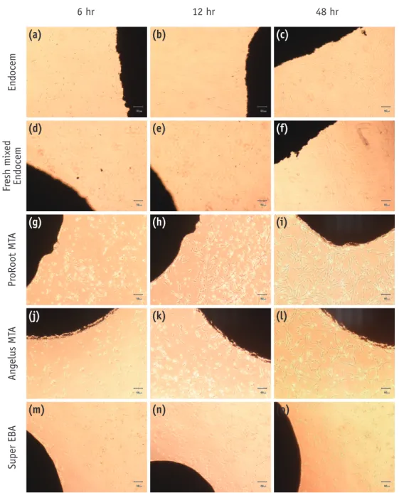
Cell morphology and cell density around the border of each material were evaluated by using a light microscope. As shown in Figure 1, in most groups, cells were attached on the plate about 6 hours after initial plating except for the fresh mixed Endocem and Super EBA groups. MTA and Angelus MTA groups showed the high cell density from the initial phase even at the material borders (Figures 1g and 1j). Endocem group did not show any cell attachment
Figure 1. Morphological changes of periodontal ligament cells in contact with each experimental material. (a - c) Endocem; (d - f) fresh mixed Endocem; (g - i) ProRoot MTA; (j - l) Angelus MTA; (m - o) Super EBA at 6, 12, and 48 hours.
6 hr 12 hr 48 hr En docem Fr esh mix ed En docem Pr oRoot MT A An gelus MT A Super EB A (a) (b) (c) (d) (e) (f) (g) (h) (i) (j) (k) (l) (m) (n) (o)
immediately after plating. However, after 6 hours, some irregular and less dense attachments were observed and as time passed further, pattern similar to that of both MTA and Angelus MTA groups was found (Figures 1a - 1c). In both fresh mixed Endocem and Super EBA groups, after 6 hours, relatively round and unattached cells were observed around the border of materials (Figures 1d and 1m). However, over time up to 48 hours, cell density of fresh mixed Endocem group has increased on the material border and periphery as well. In super EBA groups, it was hard to find any attached cell that was in contact with material as time passed. Seventy-two hours after plating, cells stained with trypan blue existed only at the Super EBA group. Cell numbers were not significantly different between groups except the Super EBA group (Table 1). Super EBA group showed significantly less viable cells than any other groups (p < 0.05).
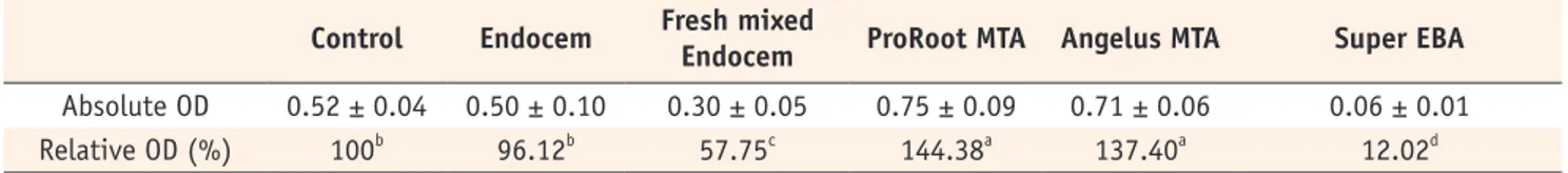
As the results of WST-1 assay, the relative proportions of cell viability in comparing the 5 groups were not equivalent (p < 0.05, Table 2). Compared with untreated control group, there was no significantly different cell viability of the Endocem group, however, the fresh mixed Endocem group had significantly less cell viability. The cells exposed to ProRoot MTA and Angelus MTA showed the highest viability, whereas the cells exposed to Super EBA displayed the lowest viability.
Discussion
The cytotoxicity of MTA has been investigated in many studies. The results have shown that ProRoot MTA and Angelus MTA are both biocompatible and nontoxic to surrounding cells. These studies have been performed both in vivo and in vitro.9,13,15
In this study, we evaluated
and compared the cytotoxicity of some root-end filling materials such as ProRoot MTA, Angelus MTA and Super EBA with the new pozzolan cement.
After endodontic microsurgery, ideal healing includes the regeneration of alveolar bone and regrowth of PDL along the resected root surface.16 Thus, how PDL cells behave and react as a result of direct contact with the root-end filling materials is important. Also, human cells can be conveniently cultured with a low number of passages resulting in minimal cell changes due to cell culture manipulation and choosing human PDL cells presents the additional advantage of reducing bias concerning species origin and non-tissue specific cell lines.17-18 Therefore, we chose the human PDL cell from extracted tooth to simulate the clinical environment, primary PDL cells of the 4th passage were used in this study to obtain the reliable results.19
In the present study, as a criterion for the evaluation of the biocompatibility of the materials, we observed the morphology of the PDL cells which had the direct contact with the root-end filling materials, and conducted WST-1 assay to evaluate the cytotoxicity of the materials. Attachment of cell to material is essential to survive and proliferate.20 Therefore, many studies evaluated the cytotoxicity of the materials with the morphologic analysis of cells.21-23 In vitro cytotoxicity assays with cultured cells are widely used in the sensitivity testing of dental materials because they are rapid, economical and reproducible. Among various cytotoxicity tests, MTT assay has been used widely as an indicator of cell viability. MTT (3-(4,5-dimethylthiazol-2-yl)-2,5-diphenyl tetrazolium bromide) is reduced in metabolically active cells by a mitochondrial enzyme to form insoluble purple formazan crystals, which are dissolved by the addition of a
Table 1. The number of cell counted based on trypan blue dye exclusion technique
Control Endocem Fresh mixed Endocem ProRoot MTA Angelus MTA Super EBA
Cell number (x 104) 21.33 ± 2.52 23.67 ± 0.58 20.33 ± 2.31 24.00 ± 3.46 22.33 ± 2.52 8.67 ± 5.13* Asterisk indicates a statistically significant difference between groups (p < 0.05).
Table 2. The absolute and relative value of optical density based on WST-1 assay
Control Endocem Fresh mixed Endocem ProRoot MTA Angelus MTA Super EBA
Absolute OD 0.52 ± 0.04 0.50 ± 0.10 0.30 ± 0.05 0.75 ± 0.09 0.71 ± 0.06 0.06 ± 0.01
Relative OD (%) 100b 96.12b 57.75c 144.38a 137.40a 12.02d
The same letters indicate mean values with no statistically significant difference among the groups. OD, optical density.
detergent. However, the insoluble purple formazan crystals can be cytotoxic and other required detergent also can complicate the assay.24 The WST-1 assay works similarly to the MTT by reacting with the mitochondrial succinate-tetrazolium reductase and forming the formazan dye. The WST-1 reagent produces a water-soluble formazan, which is stable in solution for more than 24 hours and is not cytotoxic. Furthermore, it is reported that the WST-1 assay has a sensitivity that is three times greater than that of the MTT assay.25
In the direct contact test, ProRoot MTA and Angelus MTA groups showed the cell attachment immediately after plating and also the most active attachment and proliferation. Cells exposed to set Endocem showed spindle shape of PDL which was similar to the control group. When compared with the cells exposed to ProRoot MTA and Angelus MTA, the set Endocem group showed difference in the cell shape and numbers at 6 hours and 12 hours after the plating, but at 48 hours after the plating, it showed the similar cell morphology and distribution as other groups. On the contrary, cells that were in direct contact with the fresh mixed Endocem showed round shape with the low density even after 6 hours since the plating, which means the cytotoxicity of the initial mixing. This can be explained by a high pH and heat of the cement surface that were produced at the initial mixing in the same manner as the Portland cement. The high pH and the heat can directly damage the cells by apoptosis and/ or necrosis and indirectly damage the cells by denature of culture medium proteins.26-28 The initial cytotoxicity had a tendency to decrease over time as the fresh mixed Endocem group showed the cell attachment and proliferation with the spindle shape after 48 hours since the plating. Distinctively, Super EBA did not show any cell attachment or proliferation even after 48 hours which can be explained by the cytotoxicity of the eugenol. Lin et al. reported as amount of the eugenol increased, cell survival rate was decreased and the eugenol was released even after hardening of the cement.29
In the present study, we performed the cell counting with the trypan blue and the WST-1 assay to evaluate the cell viability. At 72 hours after plating, all cells except Super EBA group showed attachment on the experimental materials. In the Super EBA group, cells appeared to be floating, small and round even after 3 days. The Super EBA group also showed the lowest survival cell numbers (Table 1), which agreed with the result of the previous direct contact test and WST-1 assay.
Contrary to the result of cell counting, WST-1 assay resulted that relative OD values of ProRoot MTA and Angelus MTA groups (144.38% and 137.40%, respectively) were higher than the relative OD value of Endocem group with significant difference. There was no significant difference between ProRoot MTA and Angelus MTA
groups. Recently, it is reported that cements such as MTA makes some influences to a genetic factor that induces cell division of PDL cells.30 Also, in a study about MTA’s influence on the pulpal cell’s apoptosis and differentiation, Moghaddame-jafari et al. reported that MTA stimulated differentiation of surrounding cells.31 In a cell cycle, the highest cell ration occurs in S phase and G2 phase which is the DNA synthesis phase. In the present study, the high relative OD values of ProRoot MTA and Angelus MTA groups are related to cell divisions and possible indirect cell proliferation. Endocem group did not show any significantly different OD result compared to the control group. This WST-1 assay result agreed with the cell counting and we can conclude that the cytotoxicity of Endocem is merely low. Even though relatively low cell viability was seen in the fresh mixed Endocem group, the viability is expected to be improved over time.
Conclusions
Within the limitation of this study, the cytotoxicity of the pozzolan cement (Endocem) was comparable with MTA and Angelus MTA, therefore, Endocem can be used as the alternative of retrofilling material.
Acknowledgement
This study was supported by a faculty research grant of Yonsei University College of Dentistry for 2013 (6-2013-0080).
Conflict of Interest: No potential conflict of interest relevant to this article was reported.
References
1. Torabinejad M, Hong CU, Pitt Ford TR, Kaiyawasam SP. Tissue reaction to implanted super-EBA and mineral trioxide aggregate in the mandible of guinea pigs: a preliminary report. J Endod 1995;21:569-571.
2. Al-Sa’eed OR, Al-Hiyasat AS, Darmani H. The effects of six root-end filling materials and their leachable components on cell viability. J Endod 2008;34:1410-1414.
3. Bondra DL, Hartwell GR, MacPherson MG, Portell FR. Leakage in vitro with IRM, high copper amalgam, and EBA cement as retrofilling materials. J Endod 1989; 15:157-160.
4. Samara A, Sarri Y, Stravopodis D, Tzanetakis GN, Kontakiotis EG, Anastasiadou E. A comparative study of the effects of three root-end filling materials on proliferation and adherence of human periodontal ligament fibroblasts. J Endod 2011;37:865-870.
5. Bodrumlu E. Biocompatibility of retrograde root filling materials: a review. Aust Endod J 2008;34:30-35. 6. Asgary S, Eghbal MJ, Parirokh M, Ghoddusi J, Kheirieh
S, Brink F. Comparison of mineral trioxide aggregate’s composition with Portland cements and a new endodontic cement. J Endod 2009;35:243-250.
7. Dorn SO, Gartner AH. Retrograde filling materials: A retrospective success-failure study of amalgam, EBA, and IRM. J Endod 1990;16:391-393.
8. Torabinejad M, Hong CU, McDonald F, Pitt Ford TR. Physical and chemical properties of a new root-end filling material. J Endod 1995;21:349-353.
9. Torabinejad M, Hong CU, Lee SJ, Monsef M, Pitt Ford TR. Investigation of mineral trioxide aggregate for root-end filling in dogs. J Endod 1995;21:603-608.
10. Rubinstein RA, Kim S. Long-term follow-up of cases considered healed one year after apical microsurgery. J Endod 2002;28:378-383.
11. Taschieri S, Del Fabbro M, Testori T, Weinstein R. Endoscopic periradicular surgery: a prospective clinical study. Br J Oral Maxillofac Surg 2007;45:242-244. 12. Enkel B, Dupas C, Armengol V, Akpe Adou J, Bosco
J, Daculsi G, Jean A, Laboux O, LeGeros RZ, Weiss P. Bioactive materials in endodontics. Expert Rev Med Devices 2008;5:475-494.
13. Parirokh M, Torabinejad M. Mineral trioxide aggregate: a comprehensive literature review-Part I: chemical, physical, and antibacterial properties. J Endod 2010;36: 16-27.
14. Parirokh M, Torabinejad M. Mineral trioxide aggregate: a comprehensive literature review-Part III: Clinical applications, drawbacks, and mechanism of action. J Endod 2010;36:400-413.
15. Torabinejad M, Parirokh M. Mineral trioxide aggregate: a comprehensive literature review-part II: leakage and biocompatibility investigations. J Endod 2010;36:190-202.
16. Song M, Kim SG, Shin SJ, Kim HC, Kim E. The influence of bone tissue deficiency on the outcome of endodontic microsurgery: a prospective study. J Endod 2013;39:1341-1345.
17. Karimjee CK, Koka S, Rallis DM, Gound TG. Cellular toxicity of mineral trioxide aggregate mixed with an alternative delivery vehicle. Oral Surg Oral Med Oral Pathol Oral Radiol Endod 2006;102:e115-120.
18. Huang FM, Chang YC. Cytotoxicity of resin-based restorative materials on human pulp cell cultures. Oral Surg Oral Med Oral Pathol Oral Radiol Endod 2002; 94:361-365.
19. Geurtsen W, Lehmann F, Spahl W, Leyhausen G. Cytotoxicity of 35 dental resin composite monomers/ additives in permanent 3T3 and three human primary
fibroblast cultures. J Biomed Mater Res 1998;41:474-480.
20. Pérez AL, Spears R, Gutmann JL, Opperman LA. Osteoblasts and MG-63 osteosarcoma cells behave differently when in contact with ProRoot MTA and White MTA. Int Endod J 2003;36:564-570.
21. Abdullah D, Ford TR, Papaioannou S, Nicholson J, McDonald F. An evaluation of accelerated Portland cement as a restorative material. Biomaterials 2002;23: 4001-4010.
22. Balto HA. Attachment and morphological behavior of human periodontal ligament fibroblasts to mineral trioxide aggregate: a scanning electron microscope study. J Endod 2004;30:25-29.
23. Zhu Q, Haglund R, Safavi KE, Spangberg LS. Adhesion of human osteoblasts on root-end filling materials. J Endod 2000;26:404-406.
24. Ishiyama M, Tominaga H, Shiga M, Sasamoto K, Ohkura Y, Ueno K. A combined assay of cell viability and in vitro cytotoxicity with a highly water-soluble tetrazolium salt, neutral red and crystal violet. Biol Pharm Bull 1996;19:1518-1520.
25. Ishiyama M, Shiga M, Sasamoto K, Mizoguchi M, He PG. A new sulfonated tetrazolium salt that produces a highly water-soluble formazan dye. Chem Pharm Bull 1993;41:1118-1122.
26. De Deus G, Ximenes R, Gurgel-Filho ED, Plotkowski MC, Coutinho-Filho T. Cytotoxicity of MTA and Portland cement on human ECV 304 endothelial cells. Int Endod J 2005;38:604-609.
27. Saidon J, He J, Zhu Q, Safavi K, Spångberg LS. Cell and tissue reactions to mineral trioxide aggregate and Portland cement. Oral Surg Oral Med Oral Pathol Oral Radiol Endod 2003;95:483-489.
28. Shin SJ. In vitro studies addressing celluar mechanisms underlying the bone and dentin inductive property of mineral trioxide aggregate (MTA). Master thesis in oral Biology. University of Pennsylvania; 2004. p75.
29. Lin CP, Chen YJ, Lee YL, Wang JS, Chang MC, Lan WH, Chang HH, Chao WM, Tai TF, Lee MY, Lin BR, Jeng JH. Effects of root-end filling materials and eugenol on mitochondrial dehydrogenase activity and cytotoxicity to human periodontal ligament fibroblasts. J Biomed Mater Res B Appl Biomater 2004;71:429-440.
30. Bonson S, Jeansonne BG, Lallier TE. Root-end filling materials alter fibroblast differentiation. J Dent Res 2004;83:408-413.
31. Moghaddame-Jafari S, Mantellini MG, Botero TM, McDonald NJ, Nör JE. Effect of ProRoot MTA on pulp cell apoptosis and proliferation in vitro. J Endod 2005;31: 387-391.