Proximate, Phytochemical, and In Vitro Antimicrobial Properties
of Dried Leaves from Ocimum gratissimum
Justina Y Talabi1 and Solomon Akinremi Makanjuola2
1Department of Human Nutrition and Dietetics, Afe Babalola University, Ado Ekiti 360001, Nigeria 2BloomMak Scientific Services, Lagos 234001, Nigeria
Prev. Nutr. Food Sci. 2017;22(3):191-194
https://doi.org/10.3746/pnf.2017.22.3.191 pISSN 2287-1098ㆍeISSN 2287-8602
Received 30 May 2017; Accepted 10 August 2017; Published online 30 September 2017
Correspondence to Solomon Akinremi Makanjuola, Tel: +234-803-468-4871, E-mail: [email protected]
Copyright © 2017 by The Korean Society of Food Science and Nutrition. All rights Reserved.
This is an Open Access article distributed under the terms of the Creative Commons Attribution Non-Commercial License (http://creativecommons.org/licenses/by-nc/4.0) which permits unrestricted non-commercial use, distribution, and reproduction in any medium, provided the original work is properly cited.
ABSTRACT: Ocimum gratissimum is a common plant in the tropics and has been used in food and medicine. Its usage in food and medicine could be attributed to its phtyochemical and antimicrobial properties. In this study we investigated the proximate, phytochemical, and antimicrobial attributes of air dried leaves of O. gratissimum. The aqueous extract was found to contain phtyochemicals with alkaloid and saponin present in appreciable amounts. The proximate analysis (crude tein and crude fibre content were 15.075% and 17.365%, respectively) showed that the leaf could be a good source of pro-tein and fibre. The aqueous ethanolic extract of the leaf exhibited activity against a wider range of organisms when com-pared to the aqueous extract at the investigated concentrations. Aqueous ethanolic extracts of O. gratissimum leaf was ac-tive against Escherichia coli, Pseudomonas aeruginosa, Staphylococcus aureus, and Bacillus cereus and the aqueous extract of the leaf was active against P. aeruginosa.
Keywords: Ocimum gratissimum, phytochemical, proximate analysis, antimicrobial activity
INTRODUCTION
The use of plants as medicine is an ancient practice com-mon to all societies especially the African society (1). Ocimum gratissimum grows in the tropics and subtropics especially in tropical Africa and India (2). O. gratissimum has found usage in food and medicine. Its application in food includes the use as flavourings and nutraceuticals. In Nigeria, the leaf is used as a condiment in the prepa-ration of dishes such as ‘pepper soup’, ‘jollof rice’, and veg-etable soups. It was initially used in the preparation of these dishes to enhance their flavour. However, their us-age in the preparation of these dishes is gaining increased acceptance due to the perceived nutraceutical benefit. The extract from the leaves of O. gratissimum possesses good antioxidant potential, which may be attributed to its phytochemical constituents (3). O. gratissimum is also used in traditional medicine for the treatment of several ailments such as urinary tract, wound, skin, and gastro-intestinal infections, and this practice continues to exist in the developing nations (4). The steam distillation ex-tract of the leaf has also been reported to have inhibitory effects on some selected bacteria that cause diarrhoea (5). The ethanolic extract of the leaf has been reported to inhibit the growth of Proteus mirabilis, Staphylococcus
aur-eus, Pseudomonas aeruginosa, and Candida albicans (4). Tra-ditionally, in Nigeria, fresh leaves are usually harvested, rinsed, and squeezed in cold water for 3 to 5 min. The squeezing in cold water is repeated three times, and the extracts are collected and served for drinking immediate-ly. However, with increased urbanisation, access to fresh-ly harvested leaves has decreased. Drying of the fresh leaves provides an option for preserving the leaves and making it available in the urban centers. Thus this study sought to investigate the qualitative and quantitative phytochemical, proximate and antimicrobial properties of dried leaves from O. gratissimum.
MATERIALS AND METHODS
Dry leaf preparation
The leaves were obtained from a garden in Ado Ekiti, Nigeria. Leaves were sorted and gently rinsed. The leaves were then spread on paper inside a room for 5 days to dry and then ground using a blender.
Phytochemical screening
Leave powder was soaked in water for 24 h at room tem-perature and then filtered. Chemicals tests were carried
192 Talabi and Makanjuola
Table 1. Phytochemical screening of Ocimum gratissimum leaf Parameters Alkaloids +++ Saponins +++ Tannins + Phlobatannins + Glycosides ++ Phenols ++ Anthraquinones + Cardenolides − Steroids − Terpenes − Flavonoids − Chalcones −
+++, appreciable amount; ++, moderate amount; +, a minute or trace amount; −, completely absent.
Table 2. Proximate and phytoquantitative analysis of Ocimum gratissimum leaf Parameters % Composition Proximate analysis Crude protein 15.08±0.29 Crude fat 5.10±0.014 Crude fibre 17.37±0.021 Ash 9.79±0.035 Moisture 8.32±0.042 Carbohydrate 44.36±0.25 Phytoquantitative analysis Phytate 0.13±0.0014 Oxalate 0.11±0.0014 Tannin 0.012±0.0014 Saponin 0.23±0.0021 Alkaloid 0.29±0.0021 Phlobatannin 0.006±0.0014 Phenol 0.030±0.0021 Glycoside 0.11±0.0021
out on the extract using standard procedure to identify the constituents as described by Sofowora (6), Trease and Evans (7) and Harborne (8). Phytochemicals screened were: tannin, phlobatannin, saponin, flavonoid, steroid, terpenoid, glycoside, cardenolide, alkaloids, anthraqui-none, chalcones, and phenols.
Proximate analysis
Proximate analysis was assayed as described in Associa-tion of Official Analytical Chemists (AOAC) (9). The leaf powder was analysed for crude protein, crude fat, crude fibre, ash, and moisture, and carbohydrate was calculated by difference.
Phytochemical quantification
Analyses were carried out in the aqueous extract. Alka-loids were measured as described in Soetan (10). Tannins were measured using the method of AOAC (11). Sapo-nins were determined using the method of Brunner (12). Glycosides were determined as described by Sofowora (6). Phenols were measured using the method of Mako (13) and phlobatannins were assayed as described by Salau (14).
Microbial inhibition study
Extract preparation: Fifty grams of the ground sample was soaked with 250 mL of sterile water for 24 h. The mix-ture was filtered with Whatman No 1 filter paper. The filtrate was concentrated to 1/10 of its original volume using a rotary evaporator. The influence of using aqueous ethanolic solvent as medium for extraction on the anti-microbial property of the leaf extract was also investi-gated. The same procedure as the aqueous extraction was used for the ethanolic extract, but 80% ethanol was used instead of water.
Microbial assay: The antimicrobial activity of the ethanolic and aqueous extracts was evaluated by the agar well
dif-fusion method (15). Inocula of test bacterial isolates were prepared by inoculating a loopful of test bacteria from a stock culture into freshly prepared nutrient both and in-cubated at 37oC for 24 h. Absorbance of the grown cul-ture was read at 530 nm after adjustment with sterile distilled water to match that of 0.5 M McFarland stand-ard solution which is equivalent to between 1.0×10⁶∼ 1.0×10⁷ CFU/mL. One milliliter of the bacterial suspen-sion was spread on Mueller-Hinton agar. The plates were allowed to stand for 1.5 h for the test bacterial isolates to be fully embedded and properly established in the seeded medium. With a sterile cork borer (No 4 Gallen-kamp), wells of equal depth of 0.5 cm (Δ=5 mm diame-ter) were dug inside the agar. Each well was aseptically filled up with 0.5 mL of the respective extracts while avoiding splashes and overfilling. The sensitivity of the test organisms to the different extracts was indicated by clearing around each well. The halo’s diameter as an in-dex of the degree of sensitivity was measured with a transparent plastic ruler. Isolates tested were Escherichia coli, Pseudomonas aeruginosa, Staphylococcus aureus, and Ba-cillus cereus.
RESULTS
Alkaloids and saponins were present in appreciable amounts. Glycosides and phenols were present in moder-ate amounts while tannins, phlobatannins, and anthra-quinones were present in minute quantities (Table 1). Cardenolides, steroids, terpenes, flavonoids, and chal-cones were absent.
The proximate analysis indicated that the leaf powder had a high carbohydrate content 44.36% (Table 2). The crude protein and crude fibre content were 15.08% and
Properties of Dried Leaves from Ocimum gratissimum 193
Table 3. Diameters of inhibition zones of aqueous and aqueous ethanolic extracts Organisms Inhibition zone (mm) 0.1 mg/mL 0.2 mg/mL 0.3 mg/mL 0.4 mg/mL 0.5 mg/mL Aqueous extract Escherichia coli 0 0 0 0 0 Pseudomonas aeruginosa 9.43 9.93 10.9 12 13.1 Staphylococcus aureus 0.5 1.1 1.57 2.43 3.03 Bacillus cereus 0 0 0 0 0
Aqueous ethanolic extract
Escherichia coli 7.03 8.01 9 10.03 10.1
Pseudomonas aeruginosa 11.03 11.83 11.97 12.9 14.97
Staphylococcus aureus 6.5 7.03 8.1 9.1 10.07
Bacillus cereus 9.47 10.07 10.97 11.97 12.97
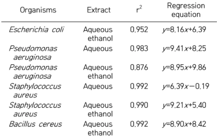
Table 4. Coefficient of determination of the relationship be-tween extract concentration and inhibition zone
Organisms Extract r2 Regression
equation
Escherichia coli Aqueous
ethanol 0.952 y =8.16x+6.39 Pseudomonas aeruginosa Aqueous 0.983 y =9.41x+8.25 Pseudomonas aeruginosa Aqueous ethanol 0.876 y =8.95x+9.86 Staphylococcus aureus Aqueous 0.992 y =6.39x−0.19 Staphylococcus aureus Aqueous ethanol 0.990 y =9.21x+5.40
Bacillus cereus Aqueous
ethanol
0.992 y =8.90x+8.42
r2: coefficients of determination.
y , inhibition zone; x, extract concentration.
17.37%, respectively. The crude fat, ash, and moisture contents had the lowest percentages of 5.10%, 9.79%, and 8.32%, respectively (Table 2).
As observed in the phytochemical screening, quantita-tive analysis also indicated that alkaloids (0.2875%) and saponins (0.225%) had the highest concentrations com-pared to the other phytochemicals that were quantified (Table 2). Phytates, oxalates, and glycosides were also present in moderate quantities with a concentration of 0.127%, 0.106%, and 0.106%, respectively. The phyto-chemical with the lowest concentrations was phlobatan-nin with a concentration of 0.006% (Table 2).
The aqueous extract exhibited antimicrobial activity against P. aeroginosa, and to a lesser extent against S. aur-eus (Table 3). The aqueous extract showed no microbial activity against E. coli and B. cereus. The aqueous ethanolic leaf powder extract showed antimicrobial activity against E. coli, P. aeroginosa, S. aureus, and B. cereus (Table 3). The aqueous and aqueous ethanolic extracts showed the highest inhibition against P. aeruginosa (13.10±0.17 mm and 14.97±0.06 mm, respectively) at the highest extract concentration of 0.5 mg/mL compared with the other organisms tested. The increase in diameter of the
inhibi-tion zones was found to have a linear relainhibi-tionship with the concentration of the extracts. The regression equa-tions and coefficients of determination (r2) are shown in Table 4.
DISCUSSION
Akinmoladun et al. (3) and Nweze and Eze (4) carried out the phytochemical screening of O. gratissimum; how-ever, quantitative analysis of the individual phytochem-ical components were not studied. Also, their study did not screen for phenols, cardenolides, and chalcones. Akinmoladun et al. (3) also reported the absence of alka-loids in the aqueous extracts of the leaf, while this study confirmed the presence of alkaloids in the aqueous ex-tracts of the leaf. Akinmoladun et al. (3) confirmed the presence of steroids, terpenoids, and flavonoids in the aqueous and ethanolic leaf extracts; however, these phy-tochemicals were found to be absent in this current in-vestigation. Flavonoids were also reported to be absent in the ethanolic extract of the leaf (4). Variation in the phytochemical content of the leaf extract could be due to planting location, seasonal variation, and extraction varia-bles (temperature, time, concentration, and particle size).
This study showed that the O. gratissimum leaf has a crude fat content of 5.1%. Essential oils that have been extracted from the oil of the leaf include eugenol, thymol, citral, geraniol, and linalool (16). Adebolu and Oladimeji (5) suggested that only the oil from the leaves had anti-bacterial activity against some selected diarrhoea causing bacteria. Ethanol could improve the extraction of esstial oils and this might have been responsible for the en-hanced activity of the aqueous ethanolic extract against the tested bacteria compared to the aqueous extract. Ac-cording to Lahlou (16), essential oils are poorly soluble in water, and the use of various solvents (such as acetone and ethanol) in the dilution of essential oils has been rec-ommended.
194 Talabi and Makanjuola
This suggests that the aqueous ethanolic leaf extract was active against E. coli, P. aeruginosa, S. aureus, and B. cereus while the aqueous extract was only active against P. aeru-ginosa at the concentrations studied. The antimicrobial activity of the leaf extract could be attributed to the phy-tochemical content of the leaf.
P. aeruginosa has become an important cause of Gram- negative infection, especially in patients with compro-mised host defense mechanisms. It is the most common pathogen isolated from patients who have been hospital-ised longer than one week and a frequent cause of noso-comial infection (19). Also, three of the organisms in this investigation (E. coli, S. aureus, and B. cereus) have been implicated in food borne diseases (20,21). According to the FDA, there are 48 million cases of foodborne illness annually, and each year, these illnesses result in an esti-mated 128,000 hospitalizations and 3,000 deaths (22). This study suggests that the aqueous and aqueous etha-nolic extracts of O. gratissimum could be potent therapeu-tic agents in treating some opportunistherapeu-tic infections and food borne illnesses caused by these bacteria.
While the leaf extract is useful in the inactivation of pathogenic microorganisms, its usage should be balanced with respect to its effect on beneficial microorganisms in the intestinal microflora. This brings to fore the impor-tance of dosage in the use of the leaf extract of O. gratis-simum.
Aqueous ethanolic extracts of the O. gratissimum leaf were active against E. coli, P. aeruginosa, S. aureus, and B. cereus, and the aqueous extract of the leaf was active against P. aeruginosa at the investigated concentrations. This brings to fore the role of solvent type in influencing the activity of O. gratissimum against microbes. Further investigation is required to understand how the phyto-chemical contents of O. gratissimum extracts could be af-fected by planting conditions and other processing varia-bles such as variation in particle size, drying method, ex-traction temperature and exex-traction time.
AUTHOR DISCLOSURE STATEMENT
The authors declare no conflict of interest.
REFERENCES
1. Usman H, Osuji JC. 2007. Phytochemical and in vitro antimi-crobial assay of the leaf extract of Newbouldia laevis. Afr J Tradit Complement Altern Med 4: 476-480.
2. Aruna K, Sivaramakrishnan VM. 1990. Plant products as
pro-tective agents against cancer. Indian J Exp Biol 28: 1008-1011. 3. Akinmoladun AC, Ibukun EO, Afor E, Obuotor EM, Farombi
EO. 2007. Phytochemical constituent and antioxidant activ-ity of extract from the leaves of Ocimum gratissimum. Sci Res Essays 2: 163-166.
4. Nweze EI, Eze EE. 2009. Justification for the use of Ocimum gratissimum L in herbal medicine and its interaction with disc antibiotics. BMC Complementary Altern Med 9: 37.
5. Adebolu TT, Oladimeji SA. 2005. Antimicrobial activity of leaf extracts of Ocimum gratissimum on selected diarrhoea caus-ing bacteria in southwestern Nigeria. Afr J Biotechnol 4: 682- 684.
6. Sofowora A. 1993. Medicinal plants and traditional medicines in Africa. John Wiley and Sons Ltd., New York, NY, USA. p 256. 7. Trease GE, Evans WC. 1989. Trease and Evans’ pharmacognosy.
13th ed. Bailliere Tindall, London, UK. p 53.
8. Harborne JB. 1973. Phytochemical methods: a guide to modern tech-niques of plant analysis. Chapman and Hall, London, UK. p 279. 9. AOAC. 2005. Official methods 988.05, 2003.06, 958.06,
942.05, and 967.08. In Official Methods of Analysis of the Associ-ation of Analytical Chemists InternAssoci-ational. 18th ed. Gathersburg, MD, USA.
10. Soetan KO. 2012. Comparative evaluation of phytochemicals in the raw and aqueous crude extracts from seeds of three Lablab purpureus varieties. Afr J Plant Sci 6: 410-415. 11. Edeogu CO, Ezeonu FC, Okaka ANC, Ekuma CE, EIom SO.
2007. Antinutrients evaluation of staple food in Ebonyi State, South-Eastern, Nigeria. J Appl Sci 7: 2293-2229.
12. Brunner JH. 1984. Direct spectrophotometer determination of saponin. Anal Chem 34: 1314-1326.
13. Mako AA. 2013. Performance of West African Dwarf goats fed graded levels of sun-cured water hyacinth (Eichhornia cras-sipes Mart. Solms-Laubach) replacing Guinea grass. Livestock Res Rural Dev 25: 127.
14. Salau AK, Yakubu MT, Oladiji AT. 2013. Cytotoxic activity of aqueous extracts of Anogeissus leiocarpus and Terminalia avicen-nioides root barks against Ehrlich ascites carcinoma cells. Indian J Pharmacol 45: 381-385.
15. Nair R, Chanda SV. 2004. Antibacterial activity of some me-dicinal plants of Saurashtra region. J Tissue Res 4:117-120. 16. Sulistiarini D. 1999. Ocimum gratissimum L. In Plant Resources
of South East Asia. No.19: Essential-Oils Plants. Oyen LPA, Dung NX, eds. ProSea, Bogor, Indonesia. p 140-142. 17. Lahlou M. 2004. Methods to study the phytochemistry and
bioactivity of essential oils. Phytother Res 18: 435-448. 18. Usman H, Haruna AK, Akpulu IN, Ilyas M, Ahmadu AA,
Musa YM. 2005. Phytochemical and antimicrobial screenings of the leaf extracts of Celtis integrifolia Lam. J Trop Biosci 5: 72- 76.
19. Friedrich M, Cunha BA, Lessnau KD, Lazo KG. Pseudomonas aeruginosa Infections. http://emedicine.medscape.com/arti-cle/226748-overview (accessed Apr 2017).
20. Le Loir Y, Baron F, Gautier M. 2003. Staphylococcus aureus and food poisoning. Genet Mol Res 2: 63-76.
21. Biswas B, Rogers K, McLaughlin F, Daniels D, Yadav A. 2013. Antimicrobial activities of leaf extracts of guava (Psidi-um guajava L.) on two Gram-negative and Gram-positive bac-teria. Int J Microbiol 2013: 746165.
22. FDA. Foodborne Illnesses: What You Need to Know. http:// www.fda.gov/Food/FoodborneIllnessContaminants/Food borneIllnessesNeedToKnow/default.htm (accessed Jun 2016).