35
-Vol. 14, No. 1(June), 2017 The Journal of Medicine and Life Science
Diffuse alveolar hemorrhage (DAH) is a rare and life-threatening complication in children with systemic lupus erythematosus (SLE). Despite recent advances in medical therapy, DAH could drive into catastrophic respiratory failure and hemodynamic unstability1).
The extracorporeal life support (ECLS) or extracorporeal membrane oxygenation (ECMO) means the use of extracorporeal cardiopulmonary bypass in patients with acute and reversible cardiac or respiratory failure2). When assessing children with acute respiratory failure for ECMO, it is significant to establish that their illnesses are potentially reversible3).
We present a case of a 9-year-old girl with lupus nephritis who had rapidly aggravating dyspnea and massive pulmonary hemorrhage. She was diagnosed as having DAH
with acute respiratory distress syndrome and was successfully managed with combined treatment of venovenous ECMO, plasmapheresis, and continuous renal replacement therapy (CRRT).
A previously healthy 9-year-old girl was admitted to our hospital with 2-week history of foamy urine and with 1-month history of weight gain of 2kg and generalized edema on December 13, 2016. One month prior to admission, she presented a skin rash on both arms and legs. She had a family, in which her mother and three aunts were suffering from autoimmune disease, including SLE and mixed connective tissue disease (Fig.1).
When she was admitted, she was ill looking and generally edematous. Initial vital signs were stable, with a blood pressure (BP) of 99/73 mmHg, pulse rate of 102 beats/min, respiratory rate of 20 breaths/min, and body temperature of 36.9℃. A neurological assessment showed her Glasgow Coma Scale score of 12 (E3, V4, M5). Her body weight was 37.5 kg increased 2 kg beyond usual weight. The pupil reflex
The combination therapy with extracorporeal membrane oxygenation,
plasmapheresis and continuous renal replacement therapy for massive
diffuse alveolar hemorrhage in a child with systemic lupus erythematosus
Su Wan Kim
1, Kyoung Hee Han
21Department of Thoracic and Cardiovascular surgery, Jeju National University School of Medicine, Jeju, Korea 2Department of Pediatrics, Jeju National University School of Medicine, Jeju, Korea
(Received May 15, 2017; Revised May 22, 2017; Accepted May 29, 2017)
Diffuse alveolar hemorrhage (DAH) is a rare and life-threatening complication in children with systemic lupus erythematosus (SLE). Despite recent advances in medical therapy, DAH could drive into catastrophic respiratory failure and hemodynamic unstability. The authors present a case of a 9-year-old girl with lupus nephritis who had rapidly aggravating dyspnea and massive pulmonary hemorrhage. She was diagnosed as having DAH with acute respiratory distress syndrome and was successfully managed with combined treatment of venovenous extracorporeal membrane oxygenation (ECMO), plasmapheresis, and continuous renal replacement therapy (CRRT). We propose to introduce ECMO and plasmapheresisas early as possible as a rescue therapy in children with SLE complicated to DAH, who are irresponsive to conventional treatment. (J Med Life Sci 2017;6(1):35-38)
Key Words
: Systemic lupus erythematosus (SLE) ; Diffuse alveolar hemorrhage (DAH) ; Extracorporeal membrane oxygenation(ECMO) ; Plasmapheresis ; Continuous renal replacement therapy (CRRT) ; Child
Introduction
Corresponding Author : Kyoung Hee Han
Department of Pediatrics, Jeju National University School of Medicine, 15, Aran 13gil, Jeju-si, Jeju Special self–governing province, 63241, Republic of Korea
E-mail : [email protected]
Abstract
Case report
의과학 논문집 14권-1호 4차시안_8호제주대학교논문-수정3 17. 07. 04 오전 11:08 페이지 3536
-Su Wan Kim, Kyoung Hee Han
was prompt and both conjunctivae were not anemic. Her abdomen was distended, but she had no specific tenderness. Her extremities were edematous, and she had pitting edema, and purpura on the 4-extremities. Laboratory findings on admission were as follows: white blood cell (WBC), 5,100 /L (segmental neutrophil 72.4%, lymphocyte 20.0%); hemoglobin, 11.8 g/dL; hematocrit, 34.9%, platelets (PLT) 213,000 /L; albumin, 1.5 g/dL; total protein, 3.4 g/dL; total bilirubin, 0.4 mg/dL; aspartate aminotransferase, 33 IU/L, alanine aminotransferase, 14 IU/L; total cholesterol, 297 mg/dL; blood urea nitrogen (BUN), 24.7 mg/dL; creatinine (Cr), 0.6 mg/dL; sodium, 139 mmol/L; potassium, 4.8 mmol/L; calcium, 7.6 mg/dL; phosphorus, 4.5 mg/dL; and uric acid, 6.8 mg/dL. C-reactive protein (CRP) was 0.10 mg/dL and erythrocyte sedimentation rate (ESR) was 17 mm/hr. IgG was 456 mg/dL (normal range, 680 ~ 1620 mg/dL), IgA 30 mg/dL (normal range, 84 ~ 438 mg/dL), IgM 75 mg/dL (normal range, 57 ~ 288 mg/dL), C3 24 mg/dL (normal range, 65 ~ 135 mg/dL), C4 5 mg/dL (normal range, 13 ~ 35 mg/dL), and CH50 <5 IU/mL. Antinuclear antibody was positive in a speckled pattern by 1:320X titer. HBsAg, anti-neutrophil cytoplasmic antibody (ANCA) and anti-streptolysin O (ASO) were all negative.
Urinalysis was as followed: specific gravity, 1.020; pH, 5.5; protein, 4+, red blood cells, many/high-power field (HPF); and WBC many/HPF. Urine protein to Cr ratio increased to 11.64 mg/mg Cr. Chest X-ray showed minimal amount of bilateral pleural effusion (Fig. 2A).
Initially, we suspected lupus nephritis. On hospital day (HD) 3, a renal biopsy was performed, and diffuse lupus nephritis, ISN/RPS Class IV-G (A) with active lesions and diffuse global proliferative lupus nephritis was noted (Fig. 3). On HD 4, high-dose intravenous methylprednisolone (MP) pulse therapy was administered at a dose of 30 mg/kg/day with a maximum
of 1 g/day for 3 consecutive days. After the second dose of MP, she complained of fever and severe abdominal pain. Under the presumptive diagnosis of bacterial peritonitis, she was treated with intravenous cefotaxime and vancomycin. Blood culture was confirmed to the pneumococcal bacteremia. On HD 8, she experienced tachypnea, orthopnea, hemoptysis and oliguria. She had an acutely ill appearance. Her respiratory rates increased to 30/min, and SpO2was 86% on room air. A laboratory examination was as follows: WBC, 11,200 /L (segmental neutrophil 90.3%); hemoglobin, 5.9 g/dL; hematocrit, 17.7%, PLT 88,000 /L; BUN, 45.4 mg/dL; Cr, 1.6 mg/dL mg/dL; sodium, 146 mmol/L; potassium, 3.8 mmol/L; calcium, 7.9 mg/dL; phosphorus, 8.1 mg/dL; and uric acid, 9.6 mg/dL. Chest x-ray showed aggravation of bilateral pleural effusion and pulmonary congestion on both lungs (Fig. 2B). She was treated with plasmapheresis and emergency dialysis for the management of flare-up of SLE and acute kidney injury. While preparing to board an aircraft indwelling with endotracheal tube to transfer to another hospital in Seoul, she developed worsening hemoptysis and desaturation. Copious bright red blood was seen upon endotracheal tube indicative of massive pulmonary hemorrhage. She was emergently taken to intensive care unit and we performed cardiopulmonary resuscitation (CPR) in our hospital. She was emergently cannulated for venovenous ECMO. Chest x-ray finding was aggravated to total white lung opacity in 8 hours (Fig. 2C). Following cannulation, she was maintained on she was maintained on venovenous ECMO, CRRT, plasmapheresis and mechanical ventilator with broad spectrum antibiotics due to acute respiratory distress syndrome and renal failure. To avoid further bleeding, platelets and fresh frozen plasma was transfused to keep PLT counts counts >100,000 /L and fibrinogen >100 mg/dL. Anticoagulation therapy with nafamostat mesilate was given during CRRT and ECMO.
On HD 9, chest tubes were placed in both lungs for bilateral hemothorax. On HD 19, her urine output gradually increased. Plasmapheresis discontinued and oral prednisolone began to administer at a dose of 1 mg/kg/day. On HD 22, tracheostomy was done. She was successfully decannulated from ECMO on HD 28.
On HD 34, the blood culture was reported to be positive for Candida albicansand antifungal therapy was commenced with intravenous fluconazole, but repeated blood cultures on HD 37, 38 and 41 were all positive for C. albicans. The patient was transitioned to caspofungin therapy from the intravenous fluconazole for the persistent ongoing Candidal septicaemia on HD 43. After 2 days of caspofungin therapy, Figure 1. Family history of autoimmune disease in this
case.
37
-The combination therapy with extracorporeal membrane oxygenation, plasmapheresis and continuous renal replacement therapy for massive diffuse alveolar hemorrhage in a child with systemic lupus erythematosus
blood culture was negative for C. albicans and she received antifungal agents for 2 weeks from then.
On HD 50, BUN and Cr improved to 21.4 mg/dL and 0.6 mg/dL, respectively, and CRRT was stopped. Tacrolimus was given to her at a dose of 0.2 mg/kg/day on HD 60. She could be weaned from the ventilator on HD 63. The dose of oral prednisolone was increased to 2 mg/kg/day for the
control of heavy proteinuria related with lupus nephritis on HD 67. Tracheostomy tube was removed on HD 70. She was discharged in a stable condition breathing room air on HD 85. Predischarge chest X-ray showed linear atelectasis in both lungs (Fig. 2D).
Figure 2. Follow-up chest X-rays in this case.
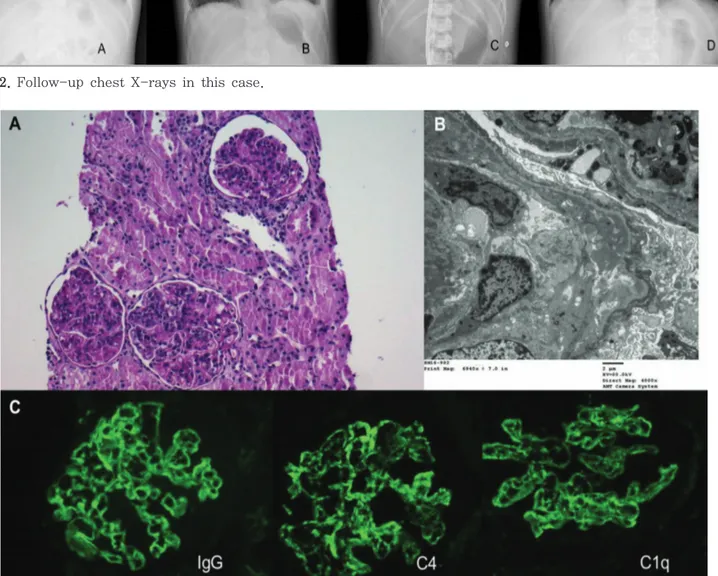
Figure 3. Renal biopsy revealed markedly hypercellular glomeruli involving endocapillary and mesangial cells on
microspy. Ultrastructural examination disclosed diffuse numerous subepithelial deposits and moderate amounts of subendothelial and mesangial deposits. The fluorescence microscopy showed a diffuse granular peripheral staining of IgG, IgM, IgA, C3, C4 and C1q(A: Periodic acid-Schiff staining, x10; B: electron microscopy image, x4000; C: fluorescence microscopy, x260)
38
-Su Wan Kim, Kyoung Hee Han
DAH denotes bleeding into the alveolar spaces caused by a broken alveolar-capillary basement membrane4). There are varied causes related with the development of DAH, but it can be divided into three major categories in the histopathologic pattern: pulmonary capillaritis, bland pulmonary hemorrhage, and diffuse alveolar damage4,5). Pulmonary capillaritis are characteristic into neutrophilic infiltration of the alveolar wall with many of those cells undergoing fragmentation4). Causes of pulmonary capillaritis include the systemic vasculitides, rheumatic diseases, certain drugs, and idiopathic pulmonary hemosiderosis. Bland pulmonary hemorrhage is pathologically seen in which the alveolar spaces are filled with red blood cells without disruption of the alveolar septa4). Occasionally, anti-GBM disease or SLE can be associated with bland pulmonary hemorrhage. Diffuse alveolar damage can occur in acute respiratory distress syndrome, in which the alveolar septa are swollen and hyaline membrane are lining the alveoli. Diffuse alveolar damage can lead to DAH4).
Treatment of DAH in children has not been established, but there are some reports for the management of DAH in children with SLE based upon the severity of disease1,6). Children with life-threatening manifestations of SLE secondary to major organ involvement require intensive immunosuppressants such as high doses of systemic glucocorticoids, intravenous cyclophosphamide or rituximab as an induction therapy for disease controll7). In our case, an initial treatment for lupus nephritis was difficult due to the complication of peritonitis caused by pneumococcal bacteremia. Induction therapy for SLE combined with treatment for infection control did not achieve the remission and progress the disease flare into DAH.
In recent years, prompt institution of ECMO and plasmapheresis as a rescue therapy are reported for management of children with DAH caused by SLE1,8). Although exposed to severe pneumococcal bacteremia and Candidal septicaemia, emergent institution of ECMO and
plasmapheresis followed by immunosuppressive agents were responsive for the disease control in terms of reemergence of urination within 20 days of renal replacement therapy and breathing without assistance after 2 months of ventilator therapy in this case.
In conclusion, we propose to introduce ECMO, plasmapheresisas and CRRT early as possible as a rescue therapy in children with SLE complicated to DAH who are irresponsive to conventional treatment.
1) Kimura D, Shah S, Briceno-Medina M, Sathanandam S, Haberman B, Zhang J, et al. Management of massive diffuse alveolar hemorrhage in a child with systemic lupus erythematosus. J Intensive care 2015;3:10.
2) Skinner SC, Hirschl RB, Bartlett RH. Extracorporeal life support. Semin Pediatr Surg 2006;15:242-50.
3) Rambaud J, Guilbert J, Guellec I, Jean S, Durandy A, Demoulin M, et al. Extracorporeal membrane oxygenation in critically ill neonates and children. Arch Pediatr 2017[Epub ahead of print]
4) Lara AR FS, Schwarz MI. Diffuse alveolar hemorrhage. In: Interstitial Lung Disease: People's Medical Publishing House, Shelton, CT, USA, 2011.
5) Franks TJ, Koss MN. Pulmonary capillaritis. Curr Opin Pulm Med 2000;6:430-5.
6) Prasun Giri P, Sinha R, Pal P, Sarkar B. Therapeutic plasma exchange in paediatric SLE: a case series from India. Lupus 2015;24:889-91.
7) Lehman TJ, Singh C, Ramanathan A, Alperin R, Adams A, Barinstein L, et al. Prolonged improvement of childhood onset systemic lupus erythematosus following systematic administration of rituximab and cyclophosphamide. Pediatr Rheumatol Online J 2014;12:3. 8) Lger P, Ponthier L, Rambaud J, Guilbert J, Hallalel F, Chevalier J, et al. Extracorporeal membrane oxygenationin immunocompromised children. Pediatr Crit Care Med 2014;15:28-34.