저작자표시-비영리-변경금지 2.0 대한민국 이용자는 아래의 조건을 따르는 경우에 한하여 자유롭게 l 이 저작물을 복제, 배포, 전송, 전시, 공연 및 방송할 수 있습니다. 다음과 같은 조건을 따라야 합니다: l 귀하는, 이 저작물의 재이용이나 배포의 경우, 이 저작물에 적용된 이용허락조건 을 명확하게 나타내어야 합니다. l 저작권자로부터 별도의 허가를 받으면 이러한 조건들은 적용되지 않습니다. 저작권법에 따른 이용자의 권리는 위의 내용에 의하여 영향을 받지 않습니다. 이것은 이용허락규약(Legal Code)을 이해하기 쉽게 요약한 것입니다. Disclaimer 저작자표시. 귀하는 원저작자를 표시하여야 합니다. 비영리. 귀하는 이 저작물을 영리 목적으로 이용할 수 없습니다. 변경금지. 귀하는 이 저작물을 개작, 변형 또는 가공할 수 없습니다.
A Thesis for the Degree of Doctor of Philosophy
Characterization, Genomic Analysis, and Application of
Salmonella Typhimurium-targeting Bacteriophages
Salmonella Typhimurium을 숙주로 하는 박테리오파지의
특성 분석, 유전체분석과 생물방제제 활용에 관한 연구
August, 2017
Jaewoo Bai
Department of Agricultural Biotechnology
College of Agriculture and Life Sciences
농학박사학위논문
Characterization, Genomic Analysis, and Application of
Salmonella Typhimurium-targeting Bacteriophages
Salmonella Typhimurium을 숙주로 하는 박테리오파지의
특성 분석, 유전체분석과 생물방제제 활용에 관한 연구
지도교수 유 상 렬
이 논문을 농학박사학위논문으로 제출함
2017년 7월
서울대학교 대학원
농생명공학부
배 재 우
배재우의 박사학위논문을 인준함
2017년 7월
위 원 장 __________________ (인)
부위원장 __________________ (인)
위 원 __________________ (인)
위 원 __________________ (인)
위 원 __________________ (인)
I
Abstract
Bai, Jaewoo Department of Agricultural Biotechnology The Graduate School Seoul National University
Salmonella is a Gram-negative, rod-shaped, and flagellated bacteria which can
be found in all warm-blooded animals including human and in the environment. Up to date, more than 2,500 serotypes of Salmonella have been discovered and it is noticed to be one of the most important pathogens associated with various foodborne illnesses all over the world. Salmonella infections can cause gastroenteritis with symptoms including nausea, vomiting, abdominal cramps, diarrhea, fever, and headache. In most cases, symptoms of salmonellosis are relatively mild to healthy people and recovered in a few days without specific treatments. However, in some cases of young and elderly patients, Salmonella infection can become severe and even cause death. Recently, infections by antibiotics resistant non-typhoid
Salmonella have emerged as one of the important problems by pathogenic bacteria.
Due to the emergence of multidrug-resistant Salmonella, Salmonella-infecting bacteriophages have been considered as promising alternative biocontrol agents to antibiotics.
To develop a novel biocontrol agent against S. Typhimurium, 26 new bacteriophages targeting S. Typhimurium were newly isolated and characterized. Host receptor analysis identified five different cell wall receptors including flagella,
II
O-antigen, BtuB, LPS core oligosaccharide (OS) region, and OmpC which are utilized by S. Typhimurium phages. For further understanding of the host-phage interactions, whole genomes of selected phages were sequenced and analyzed. Comparative genomic analysis among the phages showed that phage tail and tail fiber structures are important to determine the host ranges as well as the host receptor.
Based on the receptor study, three phages (BSPM4, BSP101 and BSP22A) target different receptors including flagella, O-antigen, and BtuB, respectively were selected. Genome sequence analysis results revealed that all three phages neither have lysogen module nor toxin genes, supporting that they are strictly virulent and safe to be developed as biocontrol agents. A phage cocktail comprised of three phages was designed and its antimicrobial efficiency was evaluated. In-vitro treatment of the phage cocktail showed a significant reduction in the development of bacterial resistance to phage infection. Since a significant number of foodborne outbreaks and sporadic infections of Salmonella are mediated by contaminated fresh produces, the antimicrobial efficiency of the phage cocktail was evaluated using two fresh produces, lettuce and cucumber, as food models. The phage cocktail significantly inhibited S. Typhimurium growth in fresh produces for 12 h. These results suggest that the phage cocktail composed of phages targeting three different host receptors would be a useful material for developing a novel biocontrol agent against S. Typhimurium to ensure the safety of fresh produces.
Bacteriophage endolysin, a peptidoglycan hydrolase encoded by phage genomes, are synthesized at the end of the phage life cycle and play important roles in the host cell lysis after phage replication and propagation. Since the endolysins show specific
III
activities only to the peptidoglycan layer generally found in bacteria, they have been considered as safe to humans. Therefore, they have also been suggested as a novel biocontrol agent as well as a natural food preservative to control food-borne pathogens in foods. However, the use of endolysins are still limited to control of positive bacteria because of the presence of the outer membrane in Gram-negative bacteria which prevent endolysin assessment to the peptidoglycan substrate. On this account, studies of endolysins targeting Gram-negative bacteria are still in the beginning stage. Therefore, further studies of endolysins from Gram-negative bacteria targeting phages are required to develop endolysin-based novel antimicrobial agents against Gram-negative bacteria.
For this purpose, a novel endolysin designated M4LysA was newly identified from the phage BSPM4 genome and characterized. Bioinformatics analysis revealed that M4LysA was not homologous to the previously known endolysins. However, when M4LysA was induced in E. coli cell, rapid cell lysis was observed, suggesting that M4LysA is a host cell lysis protein. Indeed, colorimetric assay revealed that M4LysA have endopeptidase activity. Domain analysis results showed that M4LysA is a membrane protein having an apparent transmembrane domain (TMD). By deletion of the C-terminal TMD from M4LysA, solubility was increased while the peptidoglycan lysis activity still remained, suggesting that M4LysA cause cell lysis by degrading the peptidoglycan. Since M4LysA contains unusual membrane domain in C-terminal region, it was revealed to be secreted Sec-translocase pathway independently. Instead, TMD of C-terminal region seemed to be important for its translocation to the periplasm. In addition, the host ranges of M4LysA were broader
IV
than those of the parental phage BSPM4, supporting its potential use as a novel antimicrobial agent against Gram-negative bacteria.
Despite the advantages of endolysins as biocontrol agents, their applications to the Gram-negative bacteria still have limitations because of the outer membrane barrier. In order to overcome this problem, newly purified endolysin BSP16Lys which is revealed to have N-acetylmuramonyl-L-alanine amidase activity was encapsulated into the lipid vesicles and its antimicrobial activity was evaluated. Without outer membrane permeabilizers addition, the amount of S. Typhimurium was successfully reduced (3-log CFU/mL) within 1 h at room temperature (25ºC) by treating BSP16Lys endolysin-encapsulated liposome. In addition, a liposome containing commercial lysozyme also showed antimicrobial activity without any other membrane permeabilizers. The results suggested the promising use of peptidoglycan hydrolases-encapsulated liposomes as antimicrobial agents against Gram-negative bacteria.
In this study, I suggested novel approaches to control S. Typhimurium by utilizing and maximizing the advantage of bacteriophages and endolysins as biocontrol agents. These results will provide not only deep insight into the phage biology but also advanced application strategies of the phages and endolysins as novel antimicrobial agents.
Key words: Salmonella Typhimurium, bacteriophage, genome analysis, endolysin,
biocontrol
V
Contents
Abstract………..………...………..…………..I
Contents……….………...V
List of Figures………..………...………..XVI
List of Tables………..…..………..…...……XIX
Chapter I. General Introduction……….…….…....1
I-1. Bacteriophage..……..……….………....….2
I-1-1. General features and phylogeny .……….………...….2
I-1-2. Phage therapy ..……….……….………...….3
I-1-3. Phages as biocontrol agents ..….……….……….………...….5
I-1-4. Challenges in phage application ……….……….………...….6
I-1-5. Phage endolysins …….….….….….………….……….………...….8
I-2. Salmonella, a foodborne pathogen ………..……….…10
I-3. Purpose of this study………...………...……13
VI
Chapter II. Characterization of S. Typhimurium-infecting
bacteriophages and their genomes………....………..20
II-1. Introduction……….………...…….……21
II-2. Materials and Methods………...……23
II-2-1. Bacterial strains, growth conditions, and mutant construction...23
II-2-2. Bacteriophage isolation and propagation ………....…....…..27
II-2-3. Bacterial challenge assay………...…..…...……..…..30
II-2-4. Bacteriophage host range determination ……….….…....….30
II-2-5. Transmission electron microscopy (TEM)………...…..……....30
II-2-6. Receptor analysis using various mutants………..….…….31
II-2-7. Adsorption assay………..…..……….31
II-2-8. Extraction of bacteriophage genomic DNA……….…...…….32
II-2-9. Full-genome sequencing and bioinformatics analysis ………32
II-2-10. Proteomic analysis of phage BSPM1 and BSPM2…..………….…33
II-2-11. LC-MS/MS analysis ………...………...33
II-2-12. Database searching………...………..……...……….…...……34
VII
II-3. Results and Discussion………...…….……….36
II-3-1. Isolation of bacteriophages and their receptor determination ….…....36
II-3-2. Host range determination ……….………..40
II-3-3. Bacterial challenge assay..………..……….44
II-3-4. Morphological analysis ………..………48
II-3-5. Full genome sequence analysis of bacteriophages………..51
II-3-5-1. Phage BSP3…………..………..………...……..51
II-3-5-2. Comparative genome analysis of BSP3 and Felix-O1 phages ...54
II-3-5-3. Phage BSP16……….…..57
II-3-5-4. Phage BSP25……….…..60
II-3-5-5. Comparative genome analysis of BSP25 and related phages .…..64
II-3-5-6. Phage BSP64……….…..67
II-3-5-7. Phages BSPM1 and BSPM2 ……….…..71
II-3-5-8. Comparative genome analysis of BSPM1 and BSPM2 phages..74
II-3-5-9. Comparative proteomic analysis of BSPM1 and BSPM2 phages.. ……….. 78
II-3-5-10. Tail fiber mutant construction in phage BSPM2 ………….…..81
VIII
Chapter III. Enhanced inhibition of Salmonella Typhimurium by a phage
cocktail
targeting
different
host
receptors
…...….………91
III-1. Introduction………...……….92
III-2. Materials and Methods………..………96
III-2-1. Bacterial strains, plasmids, media, and growth conditions…...…….96 III-2-2. Bacteriophage isolation, purification and propagation ……….…...100 III-2-3. Receptor analysis and complementation……….…….101 III-2-4. Transmission electron microscopy (TEM) analysis………….……104 III-2-5. Extraction of bacteriophage genomic DNA ………...…….104 III-2-6. Genome sequencing and bioinformatics analysis ...…………..…..104 III-2-7. Bacteriophage host range determination ……….105 III-2-8. Bacterial challenge assay………..….…..105 III-2-9. Motility assay ………....…...106 III-2-10. The frequency of bacteriophage-insensitive mutants ....……...….106 III-2-11. Food application……….………...….107
III-3. Results and Discussion……….………….…..….109
IX
receptors...109
III-3-2. Morphological analysis..………...……...112
III-3-3. Analysis of genome sequences of the BSPM4, BSP101, and BSP22A phages..……...……….………114
III-3-4. Host range analysis ……...……..………...…..………120
III-3-5. Bacterial challenge assay ……….………...………122
III-3-6. Frequencies of BIM development and phage susceptibility in BIMs …...……….126
III-3-7. Efficacy validation of the three-phage cocktail in fresh produce spiked with Salmonella.………...………131
III-4. Conclusion……….………...………...……135
III-5. References……….………...………...……136
Chapter IV. Characterization of a Novel Bacteriophage Lysis Protein and
Its Possible Host Lysis Mechanism………..147
IV-1. Introduction……….……..148
IV-2. Materials and Methods………...……..…151
X
IV-2-2. Cloning and expression of the lysis proteins …..……….…153
IV-2-3. In silico analysis ……...……….…………...…..155
IV-2-4. Overexpression and purification of M4LysAΔTMD protein………155
IV-2-5. Activity and the host range of M4LysAΔTMD ….…...………156
IV-2-6. Enzymatic activity confirmation….……...………..157
IV-2-7. Functional analysis of M4LysA …..……...………..157
IV-3. Results and discussion………...…………...………159
IV-3-1. Identification of putative lysis protein in BSPM4 phage ...…...….159
IV-3-2. M4LysA sequence is conserved in Chi-like phages……….161
IV-3-3. Domain analysis of M4LysA and its lytic activity ….…..……..….164
IV-3-4. M4LysA is a Sec translocase-independent membrane protein....….172
IV-3-5. C-terminal TMD are required for M4LysA activity....………..176
IV-4. Conclusion……….………...………...……180
IV-5. References……….………...………...….…182
Chapter V. Liposome-mediated delivery of phage endolysins to penetrate
Gram-negative bacteria cell wall……….187
XI
V-1. Introduction………..………….……..188
V-2. Materials and Methods………...……..…189
V-2-1. Bacterial strains and growth conditions……….….………...190
V-2-2. Cloning, expression, and purification of BSP16Lys endolysin ….…192 V-2-3. Lysis activities of BSP16Lys endolysin …..……….…………...…..193
V-2-4. Effects of pH, temperature, and NaCl concentrations on the enzymatic activities of BSP16Lys endolysin………193
V-2-5. Liposome preparation ………...….…...………194
V-2-6. Dynamic light scattering (DLS)…………...………..195
V-2-7. Transmission electron microscopy (TEM) ...………..195
V-2-8. Entrapment efficiency ………..…………...………..196
V-2-9. Antimicrobial activity of liposome-encapsulated BSP16Lys endolysin. ………..…197
V-3. Results and discussion………...…………...…..……198
V-3-1. Purification and antibacterial activities of BSP16Lys endolysin.….198 V-3-2. Optimal pH, temperature and NaCl concentrations for enzyme activity……….202
XII
V-3-4. Antimicrobial activity of liposomes…………...….213
V-4. References……….………...………...……217
Appendix 1 : A study of Cronobacter Sakazakii-targeting phage CR5...223
VI-1. Abstract….………..………….……..224
VI-2. Introduction…..………..………….……..226
VI-3. Materials and Methods..………...……..…229
VI-3-1. Bacterial strains and growth conditions..…….….………...229
VI-3-2. Bacteriophage isolation and purification………....….…232
VI-3-3. Transmission electron microscopy.…..……….………....…..233
VI-3-4. Host range analysis……….…..……….………....…..233
VI-3-5. Bacterial challenge test.………….…..……….………….…....…..234
VI-3-6. Mutant construction and complementation for identification of host receptor……….………….…..……….………….…...…..234
VI-3-7. Bacteriophage DNA isolation and purification.………….…....…..237
VI-3-8. Bacteriophage genome sequencing and bioinformatics analysis...237
XIII
VI-3-10. Food application………...………...239
VI-3-11. Nucleotide sequence accession number….……...………...239
VI-4. Results ………...………...…………...…..……240
VI-4-1. Isolation of bacteriophage CR5………...….240
VI-4-2. Host range analysis and bacterial challenge test.………....….242
VI-4-3. Identification of the host receptor.………...….244
VI-4-4. Genome sequence analysis……..………...….246
VI-4-5. Proteomic analysis of the phage structural proteins.…………...….251
VI-4-6. Food application………..…………...….254
VI-5. Discussion...………...……….……...…………...…..……258
VI-6. References………….………...………...…..…...262
Appendix 2 : A Review for the Bacteriophages and Endolysins based
Applications……….…269
VII-1. Abstract...………..………….……..270
VII-2. Introduction....………..………….……..272
XIV
VII-3-1. General features and phylogeny……….….………...274
VII-3-2. Phage therapy………....….…275
VII-3-3. Food applications…..………....….…277
VII-4. Biocontrol of food-borne pathogens using phages and endolysins..278
VII-4-1. Phage applications…….………....….…278
VII-4-1-1. E. coli O157:H7………282
VII-4-1-2. Salmonella enterica ………..………283
VII-4-1-3. Campylobacter jejuni..………..………285
VII-4-1-4. Listeria monocytogenes..………..………286
VII-4-1-5. Staphylococcus aureus..………..……..………287
VII-4-1-6. Cronobacter sakazakii and Vibrio spp..……..……..………288
VII-4-2. Phage endolysin...…….………....….…289
VII-4-2-1. General features………291
VII-4-2-2. Endolysin applications………292
VII-5. Phage Rapid Detection of Food-borne Pathogens in Foods………..295
VII-5-1. CBD-based rapid detection in foods..………....….…295
XV
VII-6. Conclusion………..………..303
VII-7. References………....…...………..305
XVI
List of Figures
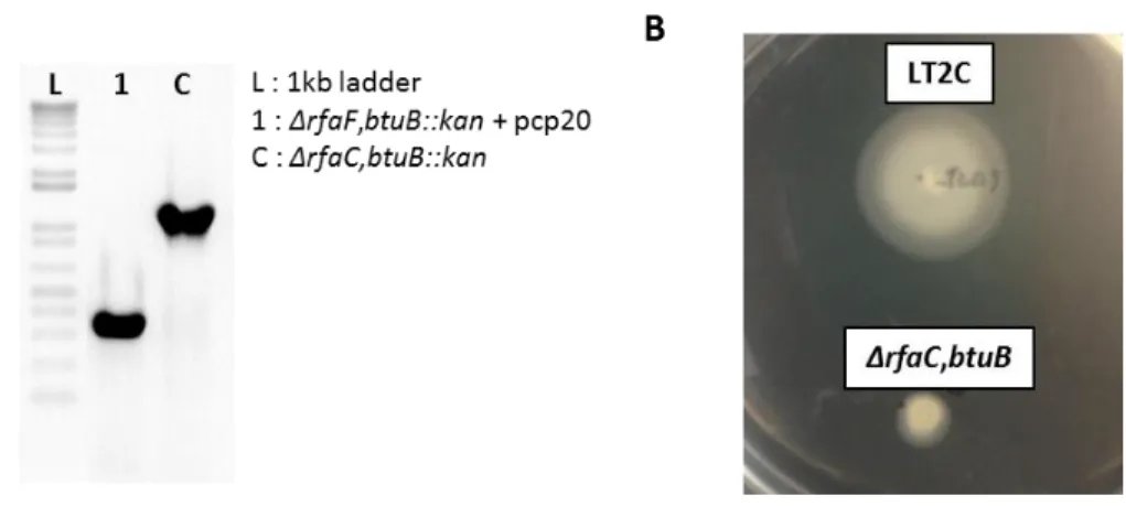
Fig. 2.1. Construction of LT2C-ΔrfaCbtuB mutant and phenotype confirmation...39
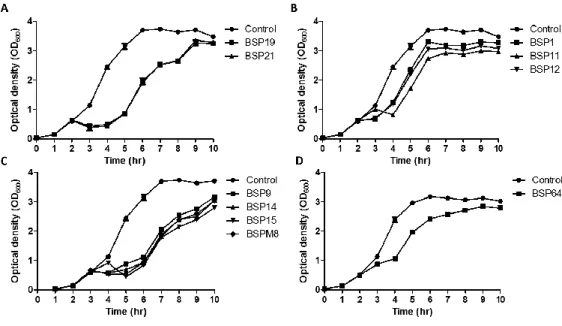
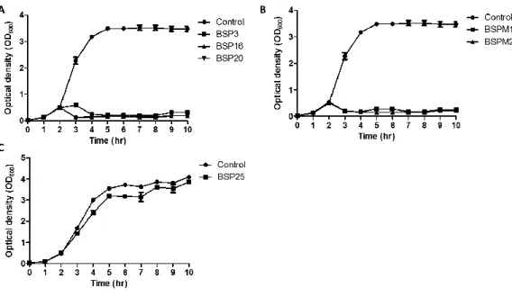
Fig. 2.2. Inhibition assays of LT2C (wild type) with each phage. …..…….………..46
Fig. 2.3. Inhibition assays of LT2C rfbP mutant (rough type) with each phage..….47
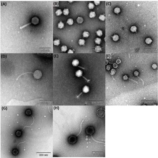
Fig. 2.4. Transmission electron micrograph images of the phages……...………..49
Fig. 2.5. Genome map of S. Typhimurium phage BSP3……….………..52
Fig. 2.6. Comparative genomic analysis of BSP3 and Felix-O1. .……….………..55
Fig. 2.7. Genome map of S. Typhimurium phage BSP16...………….…………...58
Fig. 2.8. Genome map of bacteriophage BSP25………...62
Fig. 2.9. Comparative genomic analysis of BSP25, BSP101 and SFP10 phages…..65
Fig. 2.10. Genome map of the BSP64 phage………..………..69
Fig. 2.11. Genome maps of (A) BSPM1 and (B) BSPM2 phages..………..72
Fig. 2.12. Comparative genomic analysis of BSPM1 and BSPM2 phages….……..75
Fig. 2.13. In vitro adsorption assay of BSPM1 and BSPM2 phages…….….……..77
Fig. 2.14. Comparison of phage structural proteins by LC-MS/MS analysis ….…..79
XVII
Fig. 2.16. Confirmation of mutated region in BSPM2 phage tail fiber genes.….…..84
Fig. 3.1. Identification of S. Typhimurium host receptor………...……111
Fig. 3.2. TEM image of phages isolated….………..………...…….…...113
Fig. 3.3. Genome map of phages…..…..………...…………117
Fig. 3.4. Bacterial challenge test of phage BSPM4, BSP101, BSP22A and cocktail
of three phages with S. Typhimurium LT2C..………..………124
Fig. 3.5. Susceptibility of obtained BIMs to other receptor-targeting phages..….130
Fig. 3.6. Food application of phage cocktail………...133
Fig. 4.1. In-silico prediction and lysis activity of putative lysis proteins of phage
BSPM4………..……….…..160
Fig. 4.2. Amino acid sequence alignments of M4LysA with other endolysins…..162
Fig. 4.3. Domain prediction of M4LysA and its activity………...167
Fig. 4.4. Enzymatic activity of M4LysA ΔTMD……....………...169
Fig. 4.5. The cell lysis kinetics in the presence of translocase inhibitor and functional
analysis of M4LysA TMD...174
XVIII
Fig. 5.1. Purification of BSP16Lys endolysin and its antibacterial activities....…..200
Fig. 5.2. Antimicrobial activities of BSP16Lys endolysin………..201
Fig. 5.3. The optimum conditions for the enzymatic activity of BSPLys endolysin
………203
Fig. 5.4. DLS number distribution of DPPC liposomes...208
Fig. 5.5. Morphological analysis of liposomes………...212
Fig. 5.6. Antimicrobial activities of endolysin and lysozyme containing
liposomes...215
Fig. 6.1. TEM image of phage CR5………...…..241
Fig. 6.2. Bacterial challenge test of phage CR5 with C. sakazakii ATCC 29544..243
Fig. 6.3. Identification of the C. sakazakii host receptor by deletion and
complementation of the flgK gene………245
Fig. 6.4. Genome map of phage CR5………...248
Fig. 6.5. Comparative sequence analysis of RNA polymerase beta/beta’ subunits.250
Fig. 6.6. Proteomic analysis of the structural proteins of phage CR5……….252
Fig. 6.7. Food application of phage CR5………...……….256
XIX
List of Tables
Table 2.1. Bacterial strains and plasmids used in chapter II …..…...………24
Table 2.2. Primers used in in chapter II………...………26
Table 2.3. Environmental sample lists used in chapter II….………...…29
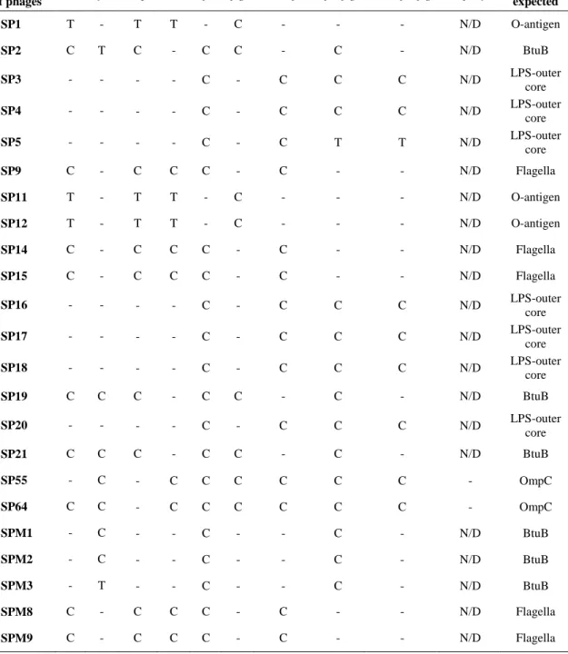
Table 2.4. Receptor determination of isolated phages in chapter II..………..……38
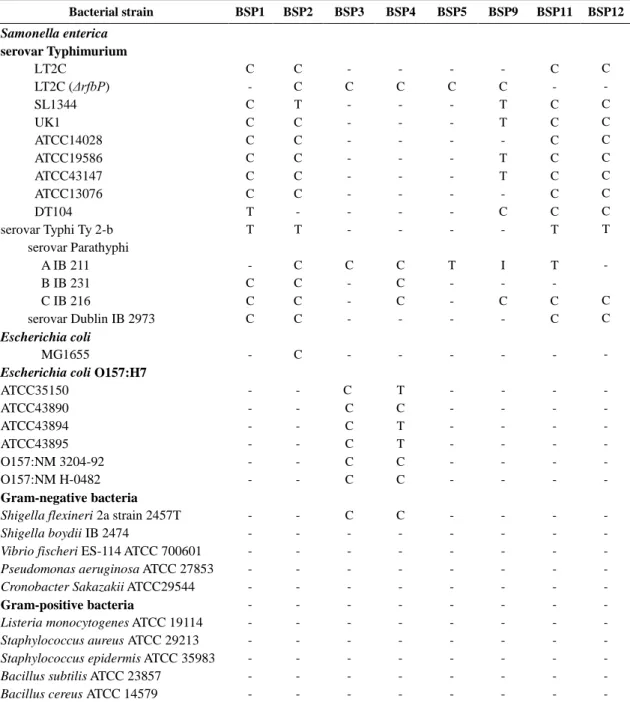
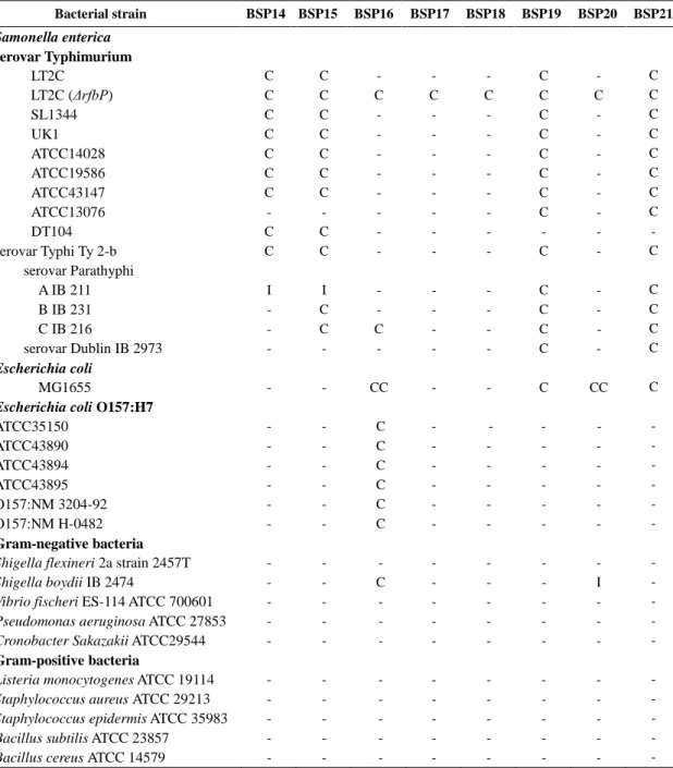
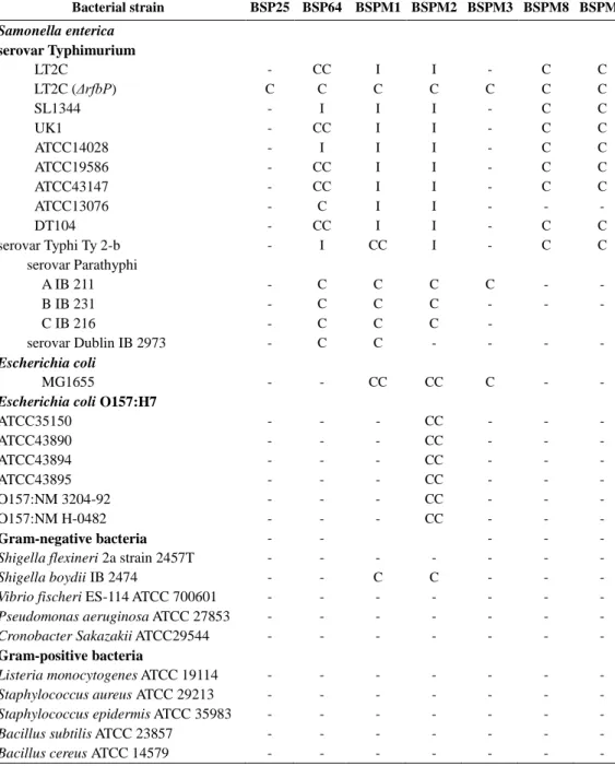
Table 2.5. The host range of 23 phages………...…………..…………..…41
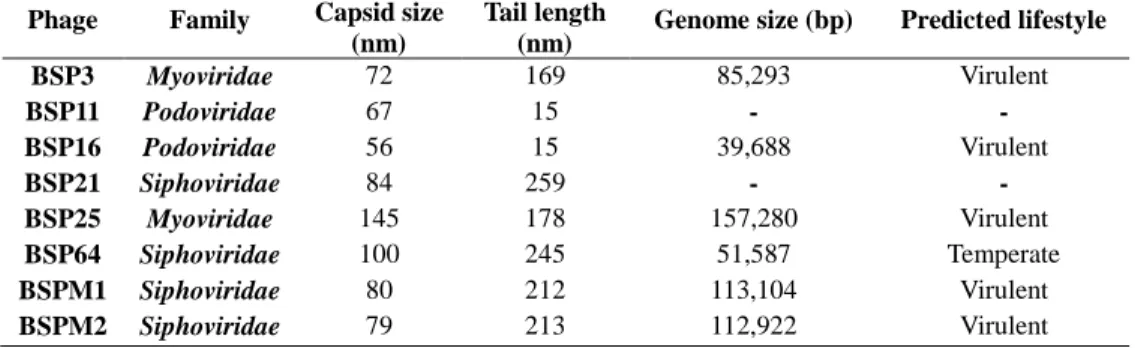
Table 2.6. Morphological features of S. Typhimurium phages ……...………...…50
Table 2.7. The host range comparison of BSP3 and Felix-O1 phages ………...…56
Table 3.1. Bacterial strains and plasmids used in chapter III……..………97
Table 3.2. Primers used in chapter III………...………..…..103
Table 3.3. Receptor screening of newly isolated phages……….….110
Table 3.4. Comparative analysis of genes related to the host receptor …..….….119
Table 3.5. Host range analysis of three phages……….…..….….121
XX
Table 4.1. Bacterial strains and plasmids used in chapter IV.……….……..……152
Table 4.2. Primers used in chapter IV……….…..154
Table 4.3. Host range analysis of M4LysAΔTMD..………..…171
Table 5.1. Bacterial strains and plasmids used in chapter V..………191
Table 5.2. Physicochemical properties of liposomes ……….…..211
Table 6.1. Host range of C. sakazakii phage CR5…………..……….……..……230
Table 6.2. Primers used for construction of C. sakazakii mutants.………….…..236
Table 7.1. Examples of phages used in studies related to pathogen reduction in
foods………...279
Table 7.2. Examples of endolysins used in studies related to pathogen reduction in
1
Chapter I.
2
I-1. Bacteriophage
I-1-1. General features and phylogeny
Bacteriophages (phages) are the most abundant microorganisms on Earth, and also have the ability to infect bacteria. Basic structure of phages in the order
Caudovirales consists of two parts: phage head and its tail. The phage head contains
a genetic material in a form of DNA or RNA [1]. Linked to the phage head, the phage tail generally plays roles in recognition and adsorption of the specific bacterial host receptor [2]. After binding to the host bacterium, phage injects its genetic material into the host cytosol via tail structure by diffusion, osmotic pressure, or transport by specific protein [3, 4]. The injected genetic material undergoes host genome integration for lysogenic cycle or replication for lytic cycle. During the lytic cycle, structural proteins are produced from encoded genes in the phage genome. After replication of the genetic material and production of structural proteins, progeny phages are assembled with them and released from the host bacterium [5, 6].
Since the first phage was discovered and characterized by Felix d’Herelle in 1917 [7], tailed phages in the order Caudovirales are the most abundant (about 96% of all phages). This order consists of three major families including
Siphoviridae, Myoviridae, and Podoviridae with different morphological
characteristics. Among the reported phages to date, phages in the Siphoviridae family are the most abundant (61.6% of all phages) with long flexible noncontractile tails ranged from 79 to 539 nm. The phages in the Myoviridae family are the second most abundant (24.5%) and they have larger heads ranged from 53 to 160 nm in
3
comparison to those of other two families. Moreover, contractile tails give
Myoviridae its unique characteristics. The phages in the Podoviridae family (13.9%)
have a distinctly short non-contractile tail ranged from 3 to 40 nm [8].
I-1-2. Phage therapy
The first clinical study using phages was a direct phage injection in six patients with staphylococcal boils in 1921 [9]. Since then, phages have been used to cure various diseases caused by bacterial infections for several decades in Eastern Europe. However, antibiotics have been widely used for the same purpose in other parts of the world and this resulted in the emergence of antibiotic-resistant bacteria. Therefore, it has been a big issue how to control these antibiotic-resistant bacteria. Because recently, phage has attracted the public attention due to its high host specificity and efficient host lysis, phage therapy has been revisited to control these problematic bacteria in Western Europe [1].
To date, numerous clinical phage trials have been reported against various pathogens including Escherichia coli, Klebsiella pneumoniae, Staphylococcus aureus,
Pseudomonas aeroginosa, and Salmonella Typhimurium. As an example, in Poland,
550 patients with gastrointestinal, head, neck, and skin infections caused by these pathogens were successfully treated and symptoms of 506 patients (92%) were relieved [6]. In addition, in Russia, 1,340 patients with conjunctivitis, dermatitis, pharyngitis, and rhinitis were divided into three groups for different treatment regimens: phage treatment (360 patients), antibiotics treatment (404 patients), and combination (576 patients) [6]. Interestingly, the phage-treated and the combination
4
groups were clinically improved up to 86% and 83%, respectively. However, the antibiotics-treated group showed minor improvement up to 48%, suggesting that phage therapy may be effective to control these pathogens but combination of phages and antibiotics did not show synergistic effect. In Ireland, ten methicillin-resistant S.
aureus (MRSA) DPC5246-infected human hands were soaked in a solution
containing 108 PFU/ml of a single phage K, revealing 2 log reduction, suggesting
that MRSA can be controlled using a specific phage [10, 11]. Based on the clinical studies, many commercial phage therapy products were developed and produced in Eastern Europe, including “Phagestaph”(JSC Biochimpharm, Tbilisi, Georgia), “E.
coli bacteriophage” (Microgen, Moscow, Russia), and “Complex pyobacteriophage”
(Microgen). In addition, other countries have many phage therapy-related companies producing commercial products: USA (Elanco Food Solutions, Gangagen Inc., Intralytix, Neurophage Pharmaceuticals, New Horizons Diagnostics, OmniLytics Inc., Phage International, Targanta Therapeutics, Viridax ), UK (AmpliPhi Biosciences Corporation, Blaze Venture Technologies, BigDNA, Novolytics, Phico), Georgia (Biopharm Ltd., JSC Biochimpharm, Phage Therapy Center), Australia (Special Phage Services Pty, Ltd.), Canada (Biophage Pharma Inc.), Germany (Hexal Genentech), India (Gangagen Biotechnologies PVT Ltd.), Ireland (Phage Works), Israel (Phage Biotech Ltd.), Portugal (Innophage), South Korea (CJ CheilJedang Corporation) and the Netherlands (EBI Food Safety) [12]. Consequently, phage therapy would provide novel insights and approaches to overcome the limitations of antibiotics and biocontrol of various antibiotics-resistant bacteria without any side effect in humans.
5
I-1-3. Phages as biocontrol agents
Food safety issues has become one of the major concerns for consumers because it threatens human health. Every year in the United States, about 9.4 million cases of foodborne illness with about 56,000 hospitalizations and 1,300 deaths caused by major food-borne pathogens including Salmonella, Clostridium
perfringens, Listeria monocytogenes, and Campylobacter have been reported [13].
Because of food contaminations by pathogens, about 25% of their food productions were lost in food industries every year [14]. In general, control of these food-borne pathogens has been continuing by using various natural or chemical food preservatives. Natural preservatives such as organic acids, bacteriocins, chitosan, and lactoferrin have tendency to exhibit weak and limited antimicrobial activities [15]. Meanwhile, consumers generally do not prefer chemical preservatives due to their side effects [16]. Furthermore, while antibiotics have strong and stable antimicrobial activities, they are not allowed to use in foods.
Phages are bacterial viruses which have host specificity and lysis activities, indicating that they can infect and lyse the specific host bacteria for their replication and propagation [5]. Phages have been suggested as natural biocontrol agents against food-borne pathogens because phages are specific to bacteria cell and safe to human [17]. Therefore, phages could be applied as promising alternative biocontrol agents, especially in food industry. ListSheild (Intralytix, Inc., Baltimore, MD, USA), a cocktail of six phages targeting L. monocytogenes, was first approved by the United States Food and Drug Administration (FDA) and U.S. Department of Agriculture (USDA)’s Food Safety and Inspection Service (FSIS) for food additives in 2006 and
6
re-approved as GRAS status by FDA in 2014. In addition, EcoShield (Intralytix), a cocktail of three phages (ECP-100) targeting E. coli O157:H7, was also approved by FDA and FSIS for food applications in 2011. Listex P100 (Micreos Food Safety, Wageningen, The Netherlands), a single phage targeting L. monocytogenes, was also approved as GRAS status by FDA in 2006. Recently, SalmoFresh (Intralytix), a cocktail consisted of six Salmonella-targeting phages, was also approved as GRAS status by FDA in 2013 [18]. These phage products were allowed to use as food preservatives to control specific food-borne pathogens in foods. Furthermore, phages can be also used as sanitizers to prevent cross contamination in food-contact materials as well as food processing facilities [19].
I-1-4. Challenges in phage application
Compare to antibiotics, the therapeutic use of phages have some advantages as follows; i) natural product, ii) high host specificity, iii) low inherent toxicity, iv) relatively low cost, etc [20]. However, though phages have a number of advantages as antibacterial agents those can be used as alternatives to chemical antibiotics, there still remain some problems and concerns associated with phage application.
First, not all phages are able to be used as biocontrol agents. To be used as a biocontrol agent, phage should be obligatory lytic without any transduction of bacterial toxin genes or virulence factors after the host cell lysis. Since not all isolated phages are totally lytic but have properties of temperate phages, it is essential to check their life cycles after the phage isolation. However, since these selection processes need too much time and labor, new methods for selecting useful
7
phages need to be developed. It has become much easier to select virulent phages based on genomic data due to the bioinformatics tools and genome sequencing analysis have been developed. However, most open reading frames (ORFs) from phages are predicted as hypothetical proteins with unknown functions and has not yet fully studied. Therefore, more studies are required for better understanding the phage genomes.
Second, bacteria have developed various strategies against phage infection; alterations or modifications of host cell receptors against phage adsorption, prevention of phage DNA entry by cutting or blocking, and defense systems by abortive phage infection targeting critical steps of phage multiplication [21]. Among them, prevention of phage adsorption by altering or modifying the host receptors is one of the common strategies developed by host bacteria. To date, many studies about modifications or hiding of host receptors such as LPS, membrane proteins, and flagella against phages infection have been reported [22-24]. Although many approaches to construct phage cocktails have been studied and suggested, only a few have been approved to be used [25]. Therefore, more studies are needed to develop efficient phage cocktails and their safety verifications are also required.
Third, unfamiliarity of the Western medical establishment with therapeutic phage applications is one of the greatest hurdles to overcome. Moreover, though some phage products have been approved by Food and Drug Administration (FDA) as GRAS (generally recognized as safe) or allowed by United States Department of Agriculture (USDA), more efforts are still required for the public acceptance [26]. Since phages are one of “virus” particles, they can be misinterpreted by the public
8
with confusion equivalent to “human viruses”. Therefore, researches about phage applications as well as educations and promotions to the public are need to be invigorated.
I-1-5. Phage endolysins
Endolysins are phage-encoded enzymes which are synthesized at the final step of the phage multiplication cycle. After replication and propagation of the
Caudovirales phages in the host bacterial cells, assembled phages are released upon
breakdown of bacterial cell wall caused by phage encoded endolysins. Additionally, endolysin has a specific activity to hydrolyze peptidoglycan of the cell wall and holin are known to help endolysin to cross the bacterial membrane to reach cell wall [27]. Therefore, endolysins are promising biocontrol agents which could be applied in food biotechnology. Especially, endolysin can lyse Gram-positive food-borne pathogens from outside of the cells as they do not have outer membrane in their cell wall structure. In contrast, Gram-negative bacterial cells which possess the outer membrane that prevents access of hydrophilic enzymes become insensitive to external peptidoglycan hydrolase treatment.
Generally, endolysins from Gram-positive bacteria-targeting phages have two conserved protein domains, enzymatic activity domain (EAD, also called as catalytic domain, located in N-terminal region) and cell wall binding domain (CBD, located in C-terminal region) [28]. Meanwhile, the majority of endolysins (~ 94%) purified from Gram-negative bacteria-targeting phages contain one catalytic domain and form a globular structure [29]. It has been reported that there are five types of
9
EADs according to the peptidoglycan cleavage sites: N-acetylmuramidases (lysozymes), N-acetyl-β-D-glucosaminidases (glycosidases), N-acetylmuramoyl-L-alanine amidases, L-alanoyl-D-glutamate endopeptidases, and interpeptide bridge-specific endopeptidases [30].
Since the endolysins specifically target the peptidoglycan layer in the bacteria, they have been considered safe for humans without any immunological responses [31]. Furthermore, no studies on the emergence of endolysin resistance strains have been reported up to date [32-35]. Therefore, endolysins could be good candidates for biocontrol agents to control food-borne pathogens in foods without harming humans. However, most of endolysins control bacteria limited to Gram-positives and the studies for Gram-negative bacteria-targeting endolysins are still at the preliminary stage. Therefore, further studies are needed to develop Gram-negative bacteria-targeting endolysins as novel antibacterial agents.
10
I-2. Salmonella, a foodborne pathogen
Salmonella is a Gram-negative, rod-shaped, facultative anaerobic, and
flagellated food-borne pathogen causing illness called salmonellosis. Salmonellosis is one of major causes of bacterial enteric disease with symptoms, mild diarrhea, common gastroenteritis, enteric fever, and ulceration in both humans and warm-blooded animals. Up to date, more than 2,600 serotypes (serovars) of Salmonella have been isolated and reported [36].
According to the current Centers for Disease Control and Prevention (CDC) system, the Salmonella genus contains two distinct species: Salmonella enterica and
Salmonella bongori. Based on biochemical properties and genomic relations, S. enterica is divided into six subspecies: S. enterica subsp. enterica, S. enterica subsp. salamae, S. enterica subsp. arizonae, S. enterica subsp. diarizonae, S. enterica subsp. houtenae, and S. enterica subsp. indica [37, 38]. All isolated serotypes are
considered as potential pathogens and the majority serotypes can infect different hosts.
Depend on the symptoms and target hosts, Salmonella serovars can be divided into two groups: invasive typhoidal serotypes and non-typhoidal Salmonella (NTS) serovars. Despite they have high genetic similarity, two serovars trigger different illnesses and distinct immune responses in humans. The former group comprised of Salmonella Typhi, Salmonella Paratyphi, and Salmonella Sendai and they are highly adapted to human host in specific. They usually cause enteric fever especially in the developing country where clean water and adequate sanitation are
11
limited. Enteric fever is a systemic disease with high a mortality, resulting in more than 200,000 deaths annually over the world [39, 40]. During recent years, the occurrences of serovar Paratyphi A infections have risen, particularly in South–East Asia [41]. The latter group includes Salmonella Enteritidis and Salmonella Typhimurium. In contrast to typhoidal infection which is common in the developing countries, NTS salmonellosis outbreaks are worldwide. Every year, approximately 93.8 million cases of gastroenteritis are caused by NTS infection with more than 155,000 deaths and about 80.3 million cases were foodborne [42]. Food-mediated NTS infection is generally caused by contaminated animal-derived products including poultry, eggs, and dairy products. In addition, NTS infection is also caused by the consumption of contaminated fresh produces such as sprouts, tomatoes, peanuts, and spinach. Moreover, NTS transmission also can be resulted by person to person contact or by contact with animals like cats, dogs, reptiles, or amphibians [43]. In general, NTS mediated infection predominantly causes self-limiting diarrheal illness, bloodstream, or focal infection. Actually, it is rarely shown in high-income countries, however, in Africa, bloodstream infection caused by NTS are commonly shown to both adults and children, presenting fever, and about 25% of them are associated with a case fatality [36]. Moreover, two major changes in the epidemiology of non-typhoid salmonellosis occurred in the 20th century in Europe and the United States: one is the emergence of foodborne human infections by S. Enteritidis and the other one is emergence of multidrug-resistant strains of S. Typhimurium. Moreover, non-typhoid Salmonella resistant strains against fluoroquinolones and third-generation cephalosporin were increased in the 21th
12
century [44, 45]. As described above, Salmonella infection has been a notable burden in both developing and developed countries. Although various food preservatives have been used to prevent Salmonella contaminations, they are not specific for
Salmonella and even worse not that effective. Accordingly, efforts to reduce the
spread of Salmonella by food and other routes must be carried out all over the world. In addition, better strategies to prevent the emergence of antibiotics-resistant
13
I-3. Purpose of this study
S. Typhimurium is a common intracellular pathogen that cause a number of
foodborne outbreaks all over the world. Because of its strong persistence and survival in the environment such as food matrices, it is urgent to control Salmonella in foods. Because of the increasing concern about the possible hazards and limitations of chemical and physical treatments, bacteriophages and endolysins have been considered to be developed as novel and safe biocontrol agents. However, studies about phage receptors, phage genome, and biological applications of phages and endolysins are not sufficient yet. In this respect, it would be important not only to isolate novel phages but also to characterize them in detail from biology to genomes and apply them to the foods in order to examine their capabilities in field test. Moreover, there is pressing need to purify and characterize new endolysins, and their novel application method are needed to be developed.
Therefore, this study aims to (i) isolate and characterize S. Typhimurium-targeting phages, (ii) construct a phage cocktail which is composed of various phages using different host receptors to enhance its antibacterial activity, and (iii) purify, characterize new endolysins, and develop a novel endolysin application method to control S. Typhimurium.
14
I-4. References
1.
Clark JR, March JB. 2006. Bacteriophages and biotechnology:
vaccines, gene therapy and antibacterials. Trends Biotechnol
24:212-218.
2.
Bertin A, de Frutos M, Letellier L. 2011. Bacteriophage-host
interactions leading to genome internalization. Curr Opin Microbiol
14:492-496.
3.
Grayson P, Molineux IJ. 2007. Is phage DNA 'injected' into
cells-biologists and physicists can agree. Current Opinion in Microbiology
10:401-409.
4.
Ming HX, Zhu L, Zhang Y. 2011. Rapid quantification of infectious
enterovirus from surface water in Bohai Bay, China using an
integrated cell culture-qPCR assay. Mar Pollut Bull 62:2047-54.
5.
Kutter E, Sulakvelidze A. 2005. Bacteriophages : biology and
applications. CRC Press, Boca Raton, FL.
6.
Inal JM. 2003. Phage therapy: a reappraisal of bacteriophages as
antibiotics. Archivum Immunologiae Et Therapiae Experimentalis
51:237-244.
7.
Duckworth DH. 1976. "Who discovered bacteriophage?". Bacteriol
Rev 40:793-802.
8.
Ackermann HW. 1998. Tailed bacteriophages: the order caudovirales.
Adv Virus Res 51:135-201.
15
Clinical Infectious Diseases 48:1096-1101.
10.
O'Flaherty S, Ross RP, Meaney W, Fitzgerald GF, Elbreki MF,
Coffey A. 2005. Potential of the polyvalent anti-Staphylococcus
bacteriophage K for control of antibiotic-resistant staphylococci from
hospitals. Applied and Environmental Microbiology 71:1836-1842.
11.
Li D, Gu AZ, Yang W, He M, Hu XH, Shi HC. 2010. An integrated
cell culture and reverse transcription quantitative PCR assay for
detection of infectious rotaviruses in environmental waters. J
Microbiol Methods 82:59-63.
12.
Endersen L, O'Mahony J, Hill C, Ross RP, McAuliffe O, Coffey
A. 2014. Phage therapy in the food industry. Annu Rev Food Sci
Technol 5:327-349.
13.
Scallan E, Hoekstra RM, Angulo FJ, Tauxe RV, Widdowson MA,
Roy SL, Jones JL, Griffin PM. 2011. Foodborne Illness Acquired in
the United States-Major Pathogens. Emerging Infectious Diseases
17:7-15.
14.
Sarhan WA, Azzazy HME. 2015. Phage approved in food, why not
as a therapeutic? Expert Review of Anti-Infective Therapy 13:91-101.
15.
Juneja VK, Dwivedi HP, Yan X. 2012. Novel natural food
antimicrobials. Annu Rev Food Sci Technol 3:381-403.
16.
Pawlowska AM, Zannini E, Coffey A, Arendt EK. 2012. "Green
preservatives": combating fungi in the food and feed industry by
applying antifungal lactic acid bacteria. Adv Food Nutr Res
66:217-238.
16
17.
McCallin S, Alam Sarker S, Barretto C, Sultana S, Berger B, Huq
S, Krause L, Bibiloni R, Schmitt B, Reuteler G, Brussow H. 2013.
Safety analysis of a Russian phage cocktail: from metagenomic
analysis to oral application in healthy human subjects. Virology
443:187-96.
18.
Sharma M. 2013. Lytic bacteriophages: Potential interventions
against enteric bacterial pathogens on produce. Bacteriophage
3:e25518.
19.
Sulakvelidze A. 2013. Using lytic bacteriophages to eliminate or
significantly reduce contamination of food by foodborne bacterial
pathogens. Journal of the Science of Food and Agriculture
93:3137-3146.
20.
Loc-Carrillo C, Abedon ST. 2011. Pros and cons of phage therapy.
Bacteriophage 1:111-114.
21.
Labrie SJ, Samson JE, Moineau S. 2010. Bacteriophage resistance
mechanisms. Nat Rev Microbiol 8:317-327.
22.
Sun Q, Lan R, Wang Y, Wang J, Luo X, Zhang S, Li P, Wang Y,
Ye C, Jing H, Xu J. 2011. Genesis of a novel Shigella flexneri
serotype
by
sequential
infection
of
serotype-converting
bacteriophages SfX and SfI. BMC Microbiol 11:269.
23.
Riede I, Eschbach ML. 1986. Evidence That Trat Interacts with
Ompa of Escherichia-Coli. Febs Letters 205:241-245.
24.
Silverman MR, Simon MI. 1972. Flagellar Assembly Mutants in
Escherichia-Coli. Journal of Bacteriology 112:986-993.
17
25.
Bai J, Kim YT, Ryu S, Lee JH. 2016. Biocontrol and Rapid
Detection of Food-Borne Pathogens Using Bacteriophages and
Endolysins. Frontiers in Microbiology 7.
26.
Kutter E, De Vos D, Gvasalia G, Alavidze Z, Gogokhia L, Kuhl S,
Abedon ST. 2010. Phage therapy in clinical practice: treatment of
human infections. Curr Pharm Biotechnol 11:69-86.
27.
Young R, Blasi U. 1995. Holins - Form and Function in
Bacteriophage Lysis. Fems Microbiology Reviews 17:191-205.
28.
Chang Y, Ryu S. 2017. Characterization of a novel cell wall binding
domain-containing Staphylococcus aureus endolysin LysSA97. Appl
Microbiol Biotechnol 101:147-158.
29.
Briers Y, Lavigne R. 2015. Breaking barriers: expansion of the use
of endolysins as novel antibacterials against Gram-negative bacteria.
Future Microbiology 10:377-390.
30.
Borysowski
J,
Weber-Dabrowska
B,
Gorski
A.
2006.
Bacteriophage endolysins as a novel class of antibacterial agents.
Experimental Biology and Medicine 231:366-377.
31.
Loessner MJ. 2005. Bacteriophage endolysins - current state of
research and applications. Current Opinion in Microbiology
8:480-487.
32.
Schmelcher M, Donovan DM, Loessner MJ. 2012. Bacteriophage
endolysins as novel antimicrobials. Future Microbiology 7:1147-1171.
33.
Callewaert L, Walmagh M, Michiels CW, Lavigne R. 2011. Food
18
22:164-171.
34.
Hermoso JA, Garcia JL, Garcia P. 2007. Taking aim on bacterial
pathogens: from phage therapy to enzybiotics. Curr Opin Microbiol
10:461-472.
35.
Rodriguez-Rubio L, Martinez B, Rodriguez A, Donovan DM,
Gotz F, Garcia P. 2013. The phage lytic proteins from the
Staphylococcus aureus bacteriophage vB_SauS-phiIPLA88 display
multiple active catalytic domains and do not trigger staphylococcal
resistance. PLoS One 8:e64671.
36.
Feasey NA, Dougan G, Kingsley RA, Heyderman RS, Gordon MA.
2012. Invasive non-typhoidal salmonella disease: an emerging and
neglected tropical disease in Africa. Lancet 379:2489-2499.
37.
Popoff M, Le Minor L. 1997. Antigenic formulas of the Salmonella
serovars 7th revision: World Health Organization Collaborating
Centre for Reference and Research on Salmonella. Pasteur Institute,
Paris, France.
38.
Brenner FW, Villar RG, Angulo FJ, Tauxe R, Swaminathan B.
2000. Salmonella nomenclature. J Clin Microbiol 38:2465-2467.
39.
Crump JA, Luby SP, Mintz ED. 2004. The global burden of typhoid
fever. Bulletin of the World Health Organization 82:346-353.
40.
Buckle GC, Walker CL, Black RE. 2012. Typhoid fever and
paratyphoid fever: Systematic review to estimate global morbidity
and mortality for 2010. J Glob Health 2:010401.
19
Bhattacharya SK, Agtini M, Deen JL, Wain J, Kim DR, Ali M,
Acosta CJ, Jodar L, Clemens JD. 2005. Salmonella paratyphi A
rates, Asia. Emerg Infect Dis 11:1764-1766.
42.
Majowicz SE, Musto J, Scallan E, Angulo FJ, Kirk M, O'Brien SJ,
Jones TF, Fazil A, Hoekstra RM, International Collaboration on
Enteric Disease 'Burden of Illness S. 2010. The global burden of
nontyphoidal Salmonella gastroenteritis. Clin Infect Dis 50:882-889.
43.
Gal-Mor O, Boyle EC, Grassl GA. 2014. Same species, different
diseases: how and why typhoidal and non-typhoidal Salmonella
enterica serovars differ. Front Microbiol 5:391.
44.
Rabsch W, Tschape H, Baumler AJ. 2001. Non-typhoidal
salmonellosis: emerging problems. Microbes Infect 3:237-247.
45.
Chen HM, Wang Y, Su LH, Chiu CH. 2013. Nontyphoid Salmonella
infection: microbiology, clinical features, and antimicrobial therapy.
Pediatr Neonatol 54:147-52.
20
Chapter II.
Characterization of S. Typhimurium-infecting
bacteriophages and their genomes
21
II-1. Introduction
Salmonella can cause non-typhoidal salmonellosis, the most common
food-borne disease with symptoms like common gastroenteritis, enteric fever, and ulceration (1). Even though Salmonella has been widely detected in various animal-based foods (2), it is very difficult to control in the foods. Natural and chemical food preservatives have been used for prevention of food-borne pathogen contaminations, however, they are not specific for Salmonella. Therefore, the development of novel type antimicrobial agents has been required all over the world.
Bacteriophages (also called as phages), natural antibacterial agents, are bacterial viruses that lyse specific bacterial host strains (3). Phages are the most numerous particles (approximately ~1032 particles) on earth, outnumbering their host
bacteria by about 10-fold (4). They are considered to coexist with their host bacterial cells and consequently they have been expected to play an important role in microbial ecology (5). The initial step of phage infection begins with recognition and adsorption, the step binding to a specific target site on the host bacterial surface. A phage adsorption is mediated by receptor-binding proteins (RBPs) and tail fibers those are localized on the tip of phage tail structure. Phages recognize their hosts by interacting with surface molecules on host bacteria using those structures. Many studies have revealed that various surface structures of negative and Gram-positive bacteria are used as phage receptors, including outer membrane proteins such as OmpA, OmpC, OmpF, OmpT, OmpX, LamB, FhuA, BtuB, lipopolisaccharides (LPS), capsular and slime polysaccharides, flagella, pili, and
22
wall teichoic acids (6, 7). Since phages utilize diverse surface molecules on host cell as receptors, adsorption step renders high host specificity to individual phages.
Since phages have been approved as novel type of food preservatives by the US Food and Drug Administration (FDA) (8), they have been recognized as interesting subjects for biocontrol agents of pathogenic bacteria including
Salmonella (9-12). Therefore, more detailed characterizations of newly isolated
phages are required to understand phage biology and their genomic features for the development and application of phage-based biocontrol agents near future.
In this study, 23 new bacteriophages targeting S. Typhimurium were isolated and characterized using various Salmonella mutant strains as hosts. Host receptor analysis results revealed five different cell wall receptors including BtuB, flagella, O-antigen, outer-core region of LPS, and OmpC. Comparative genomic analysis between phages within the similar morphology group revealed that phage tail compositions are important to determine the host ranges as well as host receptors. These results will provide useful information for further understanding about the S. Typhimurium targeting phages including general features, genomic characteristics and phage-host interactions.
23
II-2. Materials and Methods
II-2-1. Bacterial strains, growth conditions, and mutant construction
The bacterial strains used in this study are listed in Table 2.1. All of the strains were aerobically grown in Luria-Bertani (LB) broth or LB agar medium supplemented with 1.5% Bacto agar (Difco, Detroit, MI, USA) at 37°C or 30°C. Soft top agar containing LB was prepared with 0.4% agar for phage plaque confirmation. For phage isolation, prophage-cured S. Typhimurium LT2 strain (designated as LT2C) from Cancer Research Centerand its various mutants were used as host bacterial strains. LT2C-derived mutants were constructed by specific gene knock-out method as previously described (13) and primers used for mutant construction are listed in Table 2.2. When appropriate, antibiotics were added as follow (final concentrations): kanamycin (50 μg/ml), ampicillin (50 μg/ml), and chloramphenicol (25 μg/ml).24
Table 2.1. Bacterial strains and plasmids used in chapter II
Strains (description) Source or referencesa Strains (description) Source or referencesa
Samonella enterica Salmonella Typhimurium LT2C mutants
serovar Typhimurium ΔrfaC (14)
SL1344 NTCT ΔrfaCbtuB This study
UK1 (15) ΔbtuB (16)
LT2 (17) ΔrfbP (16)
LT2C [prophage-cured LT2 strain] (18) ΔflgK Park (2014)
ATCC14028 ATCC ΔbtuBrfbP (16)
ATCC19586 ATCC ΔflgKrfbP (14)
ATCC43147 ATCC ΔflgKbtuB This study
ATCC13076 ATCC ΔbtuBrfbPflgK Park (2014)
DT104 ΔompC Kim (2010)
serovar Typhi Ty 2-b IVI ΔompCrfbP (14)
serovar Parathyphi
A IB 211 IVI
B IB 231 IVI Escherichia coli O157:H7
C IB 2973 IVI ATCC35150 ATCC
serovar Dublin IB 2973 IVI ATCC43890 ATCC
ATCC43894 ATCC
Escherichia coli ATCC43895 ATCC
K-12 MG1655 Wild type O157:NM 3204-92 IVI
DH5α Invitrogen O157:NM H-0482 IVI
25
Table 2.1. Bacterial strains and plasmids used in chapter II (continued)
Strains (description) Source or
referencesa
Strains (description) Source or
referencesa
Gram-negative bacteria Plasmid
Shigella flexineri 2a strain 2457T IVI pKD46 (13)
Shigella boydii IB 2474 IVI [PBAD-gam-beta-exo oriR101 repA101ts; AmpR]
Vibrio fischeri ES-114 ATCC 700601 IVI pKD13 (13)
Pseudomonas aeruginosa ATCC 27853 ATCC [R6Kγ ori, AmpR-FRT, and KmR-FRT]
Cronobacter Sakazakii ATCC29544 ATCC pCP20 (13)
[pSC101 (Ts) ori, AmpR, CmR, cI857, and λP
Rflp]
Gram-positive bacteria pUHE21-2 lacIq (19)
Listeria monocytogenes ATCC 19114 ATCC [reppMB1 lacIq; AmpR]
Staphylococcus aureus ATCC 29213 ATCC pACYC184 (20)
Staphylococcus epidermis ATCC 35983 ATCC [repp15A CmR TetR]
Bacillus subtilis ATCC 23857 ATCC pCas9 (21)
Bacillus cereus ATCC 14579 ATCC pCRISPR (21)
26
Table 2.2. Primers used in chapter II
Primer Nucleotide sequences [5`→ 3`]a
btuB-RED-F-LT2 ATG ATT AAA AAA GCT ACG CTG CTG ACG GCG TTC TCC GTC ATG TAG GCT GGA GCT GCT TCG
btuB-RED-R-LT2 TAA TGG CGT ATC GGT AAT CGC ATT ACG CGC ATC AAC GTA A AT TCC GGG GAT CCG TCG ACC
btuB-Di-F CAT CAT CGC GTA CTA TCG AT btuB-Di-R GAT GTG AGG TGA CCG GAT AT
K1 CAG TCA TAG CCG AAT AGC CT
K2 TTG TCA AGA CCG ACC TGT CC
BSPM2_158_guide1_di_F GCA TCT ATT AAT AGC ATT AAT GAA TCT BSPM2_158_guied2_di_F GCT GTT GTT CAA GCT ATG AAA GTA CAG pCRISPR_di_R ATC AGT GAT AGA GAT TGA CAT CCC pCas9_di_R ATG GAC GAT CAC ACT ACT CTT C BSPM2_158_di_F CAG AAC TTC CAT CAC CCC ACA AAA BSPM2_158_di_R AGC AGA AGC TGA TGC TGA TGT TGG
27
II-2-2. Bacteriophage isolation and propagation
Twenty-three samples were collected from diverse environments including sewage, sludge, soil, pond water, fountain water, animal feces, swine mucin samples, and etc. as summarized in Table 2.3. All samples were used for bacteriophage isolation as described previously with slight modifications (22).Briefly, 25 g of each sample was mixed with 225 ml of sterile Butterfield’s phosphate-buffered dilution water (0.25 M KH2PO4 adjusted to pH 7.2 with NaOH) in sterile bags and then
homogenized for 90 s using a BagMixer 400 blender (Interscience Laboratory Inc.,
St. Nom, France). Twenty-five milliliter of each homogenized sample was then mixed with same volume of 2X LB broth and incubated at 37°C for 12 h with vigorous shaking (220 rpm). After incubation, the samples were centrifuged at 10,000 x g for 10 min and filtrated using 0.22-μm pore sized filter (Millipore, Billerica, MA) to remove residual bacterial cells. Ten-milliliter of each supernatant was added to 40 ml LB broth containing proper Salmonella strains (1%, final concentration) and then incubated at 37°C for 12 h with shaking (220 rpm). The culture was centrifuged at 10,000 × g for 10 min and the supernatant was filtrated using 0.22-μm pore sized filter (Millipore). To confirm the presence of phages, 10 µL of each serially diluted phage was spotted onto LB plates covered with 0.4% soft agar containing 1% (final concentration) Salmonella strain. Plates were incubated at 37°C for 12 h and plaque formation was observed.
To purify the phage, a single plaque was picked with sterile tip and eluted in sodium chloride-magnesium sulfate (SM) buffer (50 mM Tris-HCl, pH 7.5, 100
28
mM NaCl, 10 mM MgSO4) and the overlaid onto LB plate.Purification step was
repeated at least three times for purification.
For phage propagation, the lysate of a single phage was added at a multiplicity of infection (MOI) of 1 to the culture of host Salmonella strain (2 h, OD600 = 0.5) and the mixture was incubated at 37°C for 3 h.This phage propagation
steps were serially performed with three different culture volumes (3, 50, and 200 ml culture) to obtain enough phages, accompanied with centrifugation and filtration as described above. To prepare a high-titer phage, the phage particles were precipitated using polyethylene glycol (PEG) 6000 (Junsei, Japan) and concentrated using CsCl gradient (step density = 1.3, 1.45, 1.5 and 1.7 g/ml) ultracentrifugation (himac CP 100β, Hitachi, Japan) at 78,500 × g for 2 h at 4°C. Finally, the phage band fraction was collected and purified with dialysis for 1 h, repeated twice in standard dialysis buffer (5 M NaCl, 1 M MgCl2 and 1 M Tris∙HCl at pH 8.0) using Spectra/Por
dialysis membrane (molecular weight cutoff, 12,000 to 14,000; Spectrum Laboratories, Inc.). Purified phage was stored in glass vial at 4°C.
29
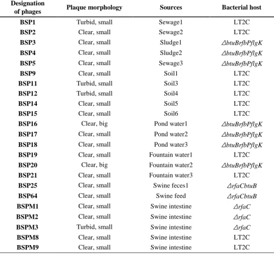
Table 2.3. Environmental sample lists used in chapter II
Designation
of phages Plaque morphology Sources Bacterial host BSP1 Turbid, small Sewage1 LT2C
BSP2 Clear, small Sewage2 LT2C
BSP3 Clear, small Sludge1 ΔbtuBrfbPflgK BSP4 Clear, small Sludge2 ΔbtuBrfbPflgK BSP5 Clear, small Sewage3 ΔbtuBrfbPflgK BSP9 Clear, small Soil1 LT2C
BSP11 Turbid, small Soil3 LT2C
BSP12 Turbid, small Soil4 LT2C
BSP14 Clear, small Soil5 LT2C
BSP15 Clear, small Soil6 LT2C
BSP16 Clear, big Pond water1 ΔbtuBrfbPflgK BSP17 Clear, small Pond water2 ΔbtuBrfbPflgK BSP18 Clear, small Pond water3 ΔbtuBrfbPflgK BSP19 Clear, small Fountain water1 LT2C
BSP20 Clear, big Fountain water2 ΔbtuBrfbPflgK BSP21 Clear, small Fountain water3 LT2C
BSP25 Clear, small Swine feces1 ΔrfaCbtuB BSP64 Clear, small Swine feed ΔrfaCbtuB BSPM1 Clear, small Swine intestine ΔrfaC BSPM2 Clear, small Swine intestine ΔrfaC BSPM3 Turbid, small Swine intestine ΔrfaC BSPM8 Clear, small Swine intestine LT2C
30
II-2-3. Bacterial challenge assay
Fifty milliliters of LB broth was sub-inoculated with appropriate S. Typhimurium strains (1%, final concentration) and the culture was incubated at 37°C with shaking (220 rpm) until it reached the early exponential phase (OD600 = 0.5). At
that time, the culture was infected with the phage at a MOI of 1. Only SM buffer was added into the bacterial culture as a negative control. After phage infection, bacterial samples were collected every 1 h and OD600 was measured. Samples were serially
diluted if necessary. All experiments were performed in triplicate.
II-2-4. Bacteriophage host range determination
Bacterial strains used for phage host range analysis were listed in Table 2.1. Each bacterial strain was incubated overnight at 37°C with shaking and then a 100 μl of each bacterial culture was added to 5 mL of the 0.4% molten LB soft agar and mixed by slight vortexing. Then the mixture was overlaid on the 1.5% LB agar plate. Subsequently, serially diluted phage lysates were spotted onto the prepared bacterial lawns and incubated at 37°C for 12 h. After incubation, the formation of a single phage plaque or the inhibition zone was observed to determine the sensitivity of each bacterium.
II-2-5. Transmission electron microscopy (TEM)
Morphological analysis of the phages was performed with transmission electron microscopy (TEM). Each phage stock diluent (approximately 109 to 1010