Human adipose-derived mesenchymal
stem cells improve neuropathic pain and
enhance nerve regeneration in a
neuropathic pain model
Hye Yeong Lee
Department of Medical Science
The Graduate School, Yonsei University
Human adipose-derived mesenchymal
stem cells improve neuropathic pain and
enhance nerve regeneration in a
neuropathic pain model
Directed by Professor Do Heum Yoon
The Master’s Thesis Submitted to the Department of Medical
Science, the Graduate School of Yonsei University in partial
fulfillment of the requirements for the degree of Master of
Medical Science
Hye Yeong Lee
December 2013
This certifies that the Master’s Thesis
of Hye Yeong Lee is approved
_____________________
Thesis Supervisor: Do Heum Yoon
_____________________
Thesis committee Member#1: Dong Ah Shin
_____________________
Thesis committee Member#2: Phil Hyu Lee
The Graduate School
Yonsei University
ACKNOWKEDGEMENTS
이 논문을 완성함에 있어 정성 어린 지도와 많은 격려를 해 주신 윤도흠 교수님께 감사의 말씀을 드리며 좋은 방향으로 연구를 진행 할 수 있도록 해주신 신동아 교수님, 이필휴 교수님께도 감사의 마음을 바칩니다. 또한 석사과정 중 많은 도움을 주신 하윤 교수님, 김긍년교수님, 이성 교수님께도 감사를 드립니다. 제가 신경외과 교실에서 겪은 많은 경험은 앞으로 더욱 발전할 수 있는 계기가 될 것이라고 생각합니다. 끝으로 늘 저를 항상 믿고 응원해 주신 부모님과, 언니 옆에서 늘 든든하게 지지해 주는 동생에게 감사의 마음을 보내며 이 글을 마칩니다.Table of Contents
Abstract ∙∙∙∙∙∙∙∙∙∙∙∙∙∙∙∙∙∙∙∙∙∙∙∙∙∙∙∙∙∙∙∙∙∙∙∙∙∙∙∙∙∙∙∙∙∙∙∙∙∙∙∙∙∙∙∙∙∙∙∙∙∙∙∙∙∙∙∙∙∙∙∙∙∙∙∙∙∙∙∙∙∙∙∙∙∙∙∙∙∙∙∙∙∙∙∙∙∙∙∙∙∙∙∙∙∙∙∙ 1
I. Introduction ∙∙∙∙∙∙∙∙∙∙∙∙∙∙∙∙∙∙∙∙∙∙∙∙∙∙∙∙∙∙∙∙∙∙∙∙∙∙∙∙∙∙∙∙∙∙∙∙∙∙∙∙∙∙∙∙∙∙∙∙∙∙∙∙∙∙∙∙∙∙∙∙∙∙∙∙∙∙∙∙∙∙∙∙∙∙∙∙∙∙∙∙∙∙∙∙ 3
II. Materials and Method
1. Human adipose-derived mesenchymal stem cells culture and
characterization ∙∙∙∙∙∙∙∙∙∙∙∙∙∙∙∙∙∙∙∙∙∙∙∙∙∙∙∙∙∙∙∙∙∙∙∙∙∙∙∙∙∙∙∙∙∙∙∙∙∙∙∙∙∙∙∙∙∙∙∙∙∙∙∙∙∙∙∙∙∙∙∙∙∙∙∙∙∙∙∙∙∙∙∙∙∙ 5
2. Neuronal differentiation ∙∙∙∙∙∙∙∙∙∙∙∙∙∙∙∙∙∙∙∙∙∙∙∙∙∙∙∙∙∙∙∙∙∙∙∙∙∙∙∙∙∙∙∙∙∙∙∙∙∙∙∙∙∙∙∙∙∙∙∙∙∙∙∙∙∙∙∙∙∙∙∙∙ 5
3. Establish of a neuropathic pain animal model ∙∙∙∙∙∙∙∙∙∙∙∙∙∙∙∙∙∙∙∙∙∙∙∙∙∙∙∙∙∙∙∙∙∙∙∙∙∙∙ 6
4. Dynamic plantar aesthesiometer ∙∙∙∙∙∙∙∙∙∙∙∙∙∙∙∙∙∙∙∙∙∙∙∙∙∙∙∙∙∙∙∙∙∙∙∙∙∙∙∙∙∙∙∙∙∙∙∙∙∙∙∙∙∙∙∙∙∙∙∙ 7
5. Sciatic functional index using foot print analysis ∙∙∙∙∙∙∙∙∙∙∙∙∙∙∙∙∙∙∙∙∙∙∙∙∙∙∙∙∙∙∙∙∙∙ 8
6. Evoked potential ∙∙∙∙∙∙∙∙∙∙∙∙∙∙∙∙∙∙∙∙∙∙∙∙∙∙∙∙∙∙∙∙∙∙∙∙∙∙∙∙∙∙∙∙∙∙∙∙∙∙∙∙∙∙∙∙∙∙∙∙∙∙∙∙∙∙∙∙∙∙∙∙∙∙∙∙∙∙∙∙∙∙∙∙ 8
7. Immunohistochemistry ∙∙∙∙∙∙∙∙∙∙∙∙∙∙∙∙∙∙∙∙∙∙∙∙∙∙∙∙∙∙∙∙∙∙∙∙∙∙∙∙∙∙∙∙∙∙∙∙∙∙∙∙∙∙∙∙∙∙∙∙∙∙∙∙∙∙∙∙∙∙∙∙ 11
8. Luxol fast blue staining, toluidine blue staining and electron microscopy
∙∙∙∙∙∙∙∙∙∙∙∙∙∙∙∙∙∙∙∙∙∙∙∙∙∙∙∙∙∙∙∙∙∙∙∙∙∙∙∙∙∙∙∙∙∙∙∙∙∙∙∙∙∙∙∙∙∙∙∙∙∙∙∙∙∙∙∙∙∙∙∙∙∙∙∙∙∙∙∙∙∙∙∙∙∙∙∙∙∙∙∙∙∙∙∙∙∙∙∙∙∙∙∙∙∙∙∙∙∙∙∙∙ 15
III. Result
1. Human adipose-derived mesenchymal stem cells(h-ADMSCs) cultures
∙∙∙∙∙∙∙∙∙∙∙∙∙∙∙∙∙∙∙∙∙∙∙∙∙∙∙∙∙∙∙∙∙∙∙∙∙∙∙∙∙∙∙∙∙∙∙∙∙∙∙∙∙∙∙∙∙∙∙∙∙∙∙∙∙∙∙∙∙∙∙∙∙∙∙∙∙∙∙∙∙∙∙∙∙∙∙∙∙∙∙∙∙∙∙∙∙∙∙∙∙∙∙∙∙∙∙∙∙∙∙∙∙ 16
2. Differentiation to a neuronal cell phenotype using two different methods
∙∙∙∙∙∙∙∙∙∙∙∙∙∙∙∙∙∙∙∙∙∙∙∙∙∙∙∙∙∙∙∙∙∙∙∙∙∙∙∙∙∙∙∙∙∙∙∙∙∙∙∙∙∙∙∙∙∙∙∙∙∙∙∙∙∙∙∙∙∙∙∙∙∙∙∙∙∙∙∙∙∙∙∙∙∙∙∙∙∙∙∙∙∙∙∙∙∙∙∙∙∙∙∙∙∙∙∙∙∙∙∙∙ 18
3. h-ADMSCs relieve nociception and pain ∙∙∙∙∙∙∙∙∙∙∙∙∙∙∙∙∙∙∙∙∙∙∙∙∙∙∙∙∙∙∙∙∙∙∙∙∙∙∙∙∙∙∙∙ 21
4. Assessment of allodynia using dynamic plantar aesthesiometer ∙∙∙∙∙∙∙∙∙ 24
5. Electrophysiological assessment ∙∙∙∙∙∙∙∙∙∙∙∙∙∙∙∙∙∙∙∙∙∙∙∙∙∙∙∙∙∙∙∙∙∙∙∙∙∙∙∙∙∙∙∙∙∙∙∙∙∙∙∙∙∙∙∙∙∙ 27
6. Transplanted h-ADMSCs were differentiated into a neuronal lineage 32
7. Transplanted h-ADMSCs promoted the remyelination of the injured nerve
∙∙∙∙∙∙∙∙∙∙∙∙∙∙∙∙∙∙∙∙∙∙∙∙∙∙∙∙∙∙∙∙∙∙∙∙∙∙∙∙∙∙∙∙∙∙∙∙∙∙∙∙∙∙∙∙∙∙∙∙∙∙∙∙∙∙∙∙∙∙∙∙∙∙∙∙∙∙∙∙∙∙∙∙∙∙∙∙∙∙∙∙∙∙∙∙∙∙∙∙∙∙∙∙∙∙∙∙∙∙∙∙∙ 34
IV. Discussion ∙∙∙∙∙∙∙∙∙∙∙∙∙∙∙∙∙∙∙∙∙∙∙∙∙∙∙∙∙∙∙∙∙∙∙∙∙∙∙∙∙∙∙∙∙∙∙∙∙∙∙∙∙∙∙∙∙∙∙∙∙∙∙∙∙∙∙∙∙∙∙∙∙∙∙∙∙∙∙∙∙∙∙∙∙∙∙∙∙∙∙∙∙∙∙∙∙∙∙∙∙ 38
V. Conclusion ∙∙∙∙∙∙∙∙∙∙∙∙∙∙∙∙∙∙∙∙∙∙∙∙∙∙∙∙∙∙∙∙∙∙∙∙∙∙∙∙∙∙∙∙∙∙∙∙∙∙∙∙∙∙∙∙∙∙∙∙∙∙∙∙∙∙∙∙∙∙∙∙∙∙∙∙∙∙∙∙∙∙∙∙∙∙∙∙∙∙∙∙∙∙∙∙∙∙∙∙ 41
References ∙∙∙∙∙∙∙∙∙∙∙∙∙∙∙∙∙∙∙∙∙∙∙∙∙∙∙∙∙∙∙∙∙∙∙∙∙∙∙∙∙∙∙∙∙∙∙∙∙∙∙∙∙∙∙∙∙∙∙∙∙∙∙∙∙∙∙∙∙∙∙∙∙∙∙∙∙∙∙∙∙∙∙∙∙∙∙∙∙∙∙∙∙∙∙∙∙∙∙∙∙∙∙∙∙∙∙ 42
List of Figures
Figure Ⅰ. Point of measurement for electrophysiological assessment
∙∙∙∙∙∙∙∙∙∙∙∙∙∙∙∙∙∙∙∙∙∙∙∙∙∙∙∙∙∙∙∙∙∙∙∙∙∙∙∙∙∙∙∙∙∙∙∙∙∙∙∙∙∙∙∙∙∙∙∙∙∙∙∙∙∙∙∙∙∙∙∙∙∙∙∙∙∙∙∙∙∙∙∙∙∙∙∙∙∙∙∙∙∙∙∙∙∙∙∙∙∙∙∙∙∙∙∙ 10
Figure Ⅱ. Operative view ∙∙∙∙∙∙∙∙∙∙∙∙∙∙∙∙∙∙∙∙∙∙∙∙∙∙∙∙∙∙∙∙∙∙∙∙∙∙∙∙∙∙∙∙∙∙∙∙∙∙∙∙∙∙∙∙∙∙∙∙∙∙∙ 12
Figure Ⅲ. Characterization of h-ADMSCs ∙∙∙∙∙∙∙∙∙∙∙∙∙∙∙∙∙∙∙∙∙∙∙∙∙∙∙∙∙∙∙∙∙∙∙∙∙ 17
Figure Ⅳ. h-ADMSCs differentiation to neuronal cell phenotype. 19
Figure Ⅴ. Pain reducing effect of h-ADMSCs on mechanical
allodynia ∙∙∙∙∙∙∙∙∙∙∙∙∙∙∙∙∙∙∙∙∙∙∙∙∙∙∙∙∙∙∙∙∙∙∙∙∙∙∙∙∙∙∙∙∙∙∙∙∙∙∙∙∙∙∙∙∙∙∙∙∙∙∙∙∙∙∙∙∙∙∙∙∙∙∙∙∙∙∙∙∙∙∙∙∙∙∙∙∙∙∙∙ 22
Figure Ⅵ. Improved motor function by h-ADMSCs ∙∙∙∙∙∙∙∙∙∙∙∙∙∙∙∙∙∙∙∙∙∙ 25
Figure Ⅶ. Electrophysiological studies after transplantation of
h-ADMSCs ∙∙∙∙∙∙∙∙∙∙∙∙∙∙∙∙∙∙∙∙∙∙∙∙∙∙∙∙∙∙∙∙∙∙∙∙∙∙∙∙∙∙∙∙∙∙∙∙∙∙∙∙∙∙∙∙∙∙∙∙∙∙∙∙∙∙∙∙∙∙∙∙∙∙∙∙∙∙∙∙∙∙∙∙∙∙∙∙∙∙ 28
Figure Ⅷ. Immunohistochemistry showing recovery of neuronal
regeneration ∙∙∙∙∙∙∙∙∙∙∙∙∙∙∙∙∙∙∙∙∙∙∙∙∙∙∙∙∙∙∙∙∙∙∙∙∙∙∙∙∙∙∙∙∙∙∙∙∙∙∙∙∙∙∙∙∙∙∙∙∙∙∙∙∙∙∙∙∙∙∙∙∙∙∙∙∙∙∙∙∙∙∙∙∙∙∙ 33
Figure Ⅸ. Microscopic findings showing the remyelination of the
List of Table
Table Ⅰ. Antibodies used in the experiments ∙∙∙∙∙∙∙∙∙∙∙∙∙∙∙∙∙∙∙∙∙∙∙∙∙∙∙∙∙∙∙∙∙ 13
Table Ⅱ. Motor evoked potential(MEP) in rat treated with
h-ADMSCs 4 weeks after transplantation. ∙∙∙∙∙∙∙∙∙∙∙∙∙∙∙∙∙∙∙∙∙∙∙∙∙∙∙∙∙∙∙∙∙∙∙∙∙∙∙∙∙∙∙ 30
Table Ⅲ. Somatosensory evoked potential(SSEP) in rat treated with
1
Abstract
Human adipose-derived mesenchymal stem cells improve neuropathic pain and enhance nerve regeneration in a neuropathic pain model
Hye Yeong Lee
Department of Medical Science
The Graduate School, Yonsei University
(Directed by Professor Do Heum Yoon)
Adipose derived mesnchymal stem cell(ADMSC) has been a promising source of cell therapy due to its capability of autologous transplantation, availabily of easy acquisition ,and potency to cross differentiation. While researchers has vigorously investigated on the efficacy of ADMSCs on nervous diseases, it is still not clear whether positive results are coming from its neural differentiation or anti-inflammatory effects. Currently, direct transplantation has been accepted as the standard method of stem cell delivery. However, a fragile lesion may be further damaged by a small volume of transplanting cell. Therefore, we evaluated the neuronal differentiation of ADMSCs and compared indirect transplantation with direct injection of h-ADMSCs in a rat model of sciatic nerve crush injury. In our result, h-ADMSCs significantly improved mechanical allodynia and neuronal regeneration compared with control group (p=8). Epineural injection was superior to intraneural injection in terms of less mechanical allodynia and better neuronal regeneration (p=8). It is assumed that secondary injury by needling and
2
volume expansion was avoided by less-invasive epineural soaking. In conclusion, h-ADMSCs transplantation with epineural soaking may be a good alternative for the treatment of peripheral nerve injury and neuropathic pain.
Key words: Neuropathic pain, human adipose mesenchymal stem cell, differentiation, cell therapy
3
Human adipose-derived mesenchymal stem cells improve neuropathic pain and enhance nerve regeneration in a neuropathic pain model
Hye Yeong Lee
Department of Medical Science
The Graduate School, Yonsei University
(Directed by Professor Do Heum Yoon)
Ⅰ. Introduction
Neuropathic pain is a result of neural injury or dysfunction. The nervous system has inherent regenerative potential after damage. Schwann cells and macrophages rapidly remove myelin debris and produce inflammatory cytokines, chemokines, extracellular matrix molecules, and proteolytic enzymes as well as a number of growth factors that play critical roles in neuronal survival and axonal regeneration. However, nerve regeneration is often limited by incomplete and non-specific regeneration, axonal loss, variable clinical results, or the development of chronic neuropathic pain disorders1. Recently, cell transplantation has become a challenging field of interest in neuro-degeneration; in experimental nerve gap models, the local implantation of neural stem cells or bone marrow mesenchymal stem cells has been shown to improve peripheral nerve regeneration through multiple mechanisms: trophic factor secretion, synthesis of extracellular matrix molecule, microenvironment stabilization, and immune modulation2.
4
Human adipose-derived mesenchymal stem cells (h-ADMSCs) influenced relevant immune modulatory activity both in vivo and in vitro in a broad range of immune cells. Moreover, several research groups have found evidence for neural and glial trans differentiation of h-ADMSCs in vitro and in vivo. Finally, h-ADMSCs secrete a variety of neurotrophins and cytokines, which might positively impact neural cell survival and neural degeneration3,4.
The intrinsic limitation of the general method is the additional trauma necessary in reaching the injection site as well as the difficulty in treating multiple or diffused sites of injury. In such circumstances, a systemic administration of cells capable of reaching the damaged PNS would be desirable.
The effects of h-ADMSCs treatment were evaluated in terms of functional recovery and nerve regeneration, assessing the ability of injected cells to integrate into the injured peripheral nerve, to modulate the immune response, and to influence the local micro environment through the release of neurotrophic molecules5.
5 Ⅱ. Methods
1. Human adipose-derived mesenchymal stem cells culture and characterization
The cell were cultured at 1x10⁴ cells in a 25 cm2 flask in alpha MEM (Gibco, Grand Island, NY), Fetal bovine serum (10%; Gibco, Grand Island, NY), penicillin streptomycin (1%; Gibco, Grand Island, NY), at 37℃ in a 5% CO2 atmosphere. When
70~80% confluent adhesion cells were trypsinized with 0.5% trypsin/EDTA (Gibco, Grand Island, NY) they were expanded in 75 cm2 flask. The medium was replaced every 3~4 days with fresh subculture6.
To confirm the presence isolation of h-ADMSCs, immunocytochemical analysis was conducted. h-ADMSCs were specified by the expression of CD44 and CD90 and absence of CD14 and CD19 (All primary antibodies purchased for Millipore, Billerica, MA).
For all experiments in this study, cells at passage 5~6 were used.
2. Neuronal differentiation
To induce neuronal differentiation for h-ADMSCs, to methods were used: serum-free cytokine induction, and butylated hydroxyanisole chemical induction.
Serum-free induction. h-ADMSCs were cultured in suspension using a bacterial dish
containing sphere medium composed of neurobasal medium (Gibco, Grand Island, NY) with B27 (2%; Gibco, Grand Island, NY), L-glutamine (2 mM; Gibco, Grand Island, NY), EGF (20 ng/ml; R&D system, Minneapolis, MN), and bFGF (20 ng/ml; R&D system, Minneapolis, MN). After 4~7 days, h-ADMSCs formed spheroid aggregate (i.e., neurospheres). Neurospheres were plated on Matrigel (BD bioscience, San Jose, CA)-coated coverslip in neurobasal medium containing the 2% B27, 2 mM L-glutamine, 10
6
ng/ml recombinant human-BDNF (Peprotech, Rocky Hill, NJ), and 1% penicillin streptomycin. The medium was replaced every 3 days up to 10~14 days.7,8
Chemical induction. There are also other ways to induce neuronal differentiation in
h-ADMSCs. Cells were plated on Matrigel-coated coverslips in DMEM (Gibco, Grand Island, NY) containing 20% FBS for 1 day. Then, the medium was changed to DMEM with butylated hydroxyanisole (200 μM; Sigma, St. Louis, MO), KCl (5 mM; Sigma, St. Louis, MO), valproic acid (2 μM; Sigma, St. Louis, MO), forskolin (10 μM; Sigma, St. Louis, MO), hydrocortisone (1 μM; Sigma, St. Louis, MO), and insulin (5 μg/ml; Sigma, St. Louis, MO) for 3 days.7
Immunocytochemistry. To detect changes in cell morphology following neuronal
induction, we analyzed of h-ADMSCs culture from 4~14 days following neuronal induction. Images were viewed using bright field microscopy. For immunocytochemistry, we grew cells on cover slips, fixed h-ADMSCs with 4% paraformaldehyde, and washed two times in wash buffer with BSA (0.1%; Sigma, St. Louis, MO) in 1xPBS (Gibco, Grand Island, NY). These were then incubated with normal donkey serum (10%; Jackson, Bar harbor, ME) and Triton® X-100 (0.3%; Sigma, St. Louis, MO) in 1xPBS blocking buffer to prevent non-specific antibody binding. Primary was antibodies were allowed to react for 16 hours at 4℃. Subsequently, the cover slips were rinsed three times with PBS containing 0.1% BSA. Next, the samples were treated with a secondary antibody at room temperature for 1 hour. In addition to the nuclear staining, the cells were mounted with DAPI (4’, 6-diamidino-2-phenylindole)-conjugated mounting medium.9
3. Establish of a neuropathic pain animal model
Adult male Sprague Dawley rats (200±20 g) were housed in a facility accredited by the Association for Assessment and Accreditation of laboratory Animal Care (AAALAC). Animals were randomized into four groups of eight animals each: a PBS epineural
7
soaking group, a phosphate-buffered saline (PBS) intraneural injection group, a h-ADMSCs epineural soaking group, and a h-h-ADMSCs intraneural injection group. All animals were anaesthetized with Rompun (10 mg/kg; Bayer Korea, Seoul, South Korea) and Zoletil 50 (25 mg/kg; Virbac Laboratories, Carros, France). The left hind leg was shaved and the biceps femoris incised to expose the sciatic nerve. The crush injury was performed 10 mm proximal to its trifurcation using a 60-g force vascular clip (Micro Serrefines; Fine Science tools, North Vancouver, BC, Canada) for 2 minutes. The nerve were kept moist with 37℃ sterile saline solution throughout the surgical procedure. After the injury, the muscles were approximated, and the wound was closed by suturing the skin with 4/0 interrupted nylon sutures.3
5x10⁵ h-ADMSCs were administered directly or indirectly 7 days after the surgery in 3ul PBS. The animals were sacrificed by CO2 chamber euthanasia at 35days after the
crush, and the sciatic nerves were dissected. Sciatic nerves stored at -80℃ for histological and immunohistochemical analyses.10,11
After surgery, cefazolin (25 mg/kg; Chong Kun Dang, Seoul, south Korea) injection twice daily for 5 days and cyclosporine (20 mg/kg; Chong Kun Dang, Seoul, South Korea) injection until the end of examination after transplantation of cells.
4. Dynamic plantar aesthesiometer
To estimate rat hind paw mechanical threshold, rats were adapt to the plastic box with a metal wire mesh table for 15 minutes. The mechanical stimulus was delivered to the plantar surface of the hind paw from below the floor of the test chamber by an automated testing device (Dynamic Plantar Aesthesiometer; Ugo Basile, Comerio, Varese, Italy).12-14 A 0.5 mm-diameter steel rod of was pushed against the hind paw with ascending force of 0 to 50 g over a period of 20 seconds at a rate of 2.5 g/s. When the animal withdrew its hind paw, the mechanical stimulus automatically stopped, and the force at which the
8
animal withdrew its paw was recorded to the nearest 0.1 g. Animals were subjected to three consecutive trials with at least 5–6 minutes between the trials and averaged to select animals.
5. Sciatic functional index using foot print analysis
Walking track analysis was performed the day before the surgery and then weekly on the 7th, 14th, 21st, 28th, and 35th days post-operatively. Briefly, the plantar surface of the hind paws was dipped with red ink, and the rats were allowed to ambulate down a standard walking trace on which a strip of graph paper (60x15 cm) was displayed. Several measurements were taken from the footprints: (I) distance from the heel to the third toe, the print length (PL); (II) distance from the first to the fifth toe, the toe spread (TS); and (III) distance from the second to the fourth toe, the intermediary toe spread (ITS). All three measurements were taken from the experimental (E) and normal (N) sides.15-17
( ) ( ) ( ) Measurements were made according to the Sciatic Functional Index (SFI), where 0 corresponds to normal function, and -100 corresponds to a total functional impairment of one side.
6. Evoked potential
Somatosensory evoked potentials (SSEPs) (n = 6 in each group) and motor evoked potentials (MEPs) (n = 6 in each group) were measured 5 weeks after transplantation. The rats were randomly assigned to the recording of SSEPs or MEPs without any indication of the extent of the lesion.10,18,19 The animals were anesthetized with Rompun and Zoletil 50 Rompun (10 mg/kg, 25 mg/kg) before electrophysiological examinations.
9
A pulse generator (DG2A, Digitimer, Hertfordshire, England) was used to generate trigger pulses required for repetitive stimulation. It makes a constant output pulse, and a current stimulator (DS3, Digitimer, Hertfordshire, England) added amplitude to the signal.20 A signal with desired pulsed current was given to the animal. For SSEP recording, left sciatic nerve was exposed and bipolar stimulating electrodes (730338, Harvard apparatus, Holliston, MA) was placed using a stereotaxic frame (WPI, Sarasota, FL) on the sensorimotor cortex at 2mm posterior to bregma and 2mm lateral to sagittal suture after craniectomy.21 For MEP recording, the stimulating electrode was fixed at the temporalis muscle and the recording electrode was inserted in the gastrocnemius muscle (with the injured sciatic nerve). The signal was amplified by a bio amplifier (axoclamp 900A, Molecular Devices, Union city, CA) with the specific parameters. The amplified wave was analyzed by software (qCLAMP 10, Molecular devices, Union city, CA)
10
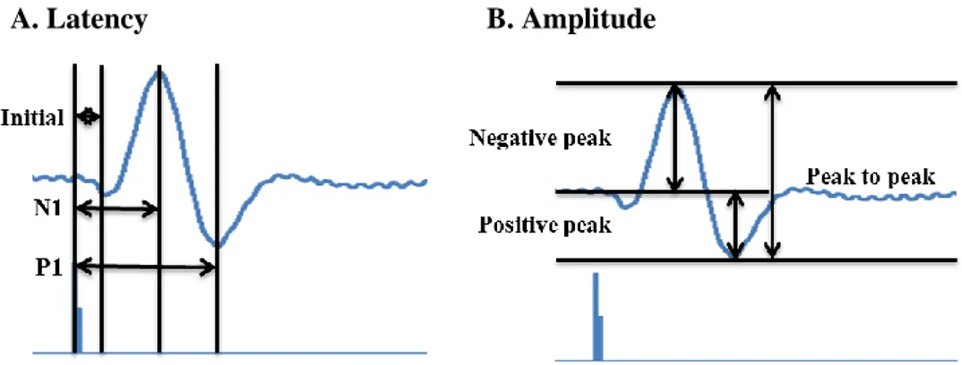
A. Latency B. Amplitude
Figure 1. Point of measurement for electrophysiological assessment
A) The latency of the evoked potentials was measured from the onset of the first negative deflection (initial latency), from the peak of the first negative deflection (N1 latency), and from the peak of the positive deflection (P1 latency). B) The amplitudes was also measured from the peak of the first negative deflection to the baseline (negative peak amplitude), from the peak of the first positive deflection to the baseline (positive peak amplitude), and from the peak of the first negative deflection to the peak of the positive deflection (peak-to-peak amplitude)
11
7. Immunohistochemistry
The animals were sacrificed at 2 weeks and 5 weeks. For immunohistochemistry, L4-L6 DRGs and sciatic nerves were removed, fixed in 4% paraformaldehyde in PBS overnight, cryoprotected in 10, 20, and 30% sucrose, and cryostat-cut into 10um-thick section.
Sections were incubated overnight at 4℃ with a primary antibody with incubation buffer (1% bovine serum albumin, 1% normal donkey serum, 0.3% Trion® X-100, and 0.01% sodium azide in PBS) and followed by secondary species-specific fluorescent antibodies for 1 hour at room temperature. Then, sections were mounted with mounting solution (UltraCruzTM Mounting Medium, Santa Cruz Biotechnology, Dallas, Texas)22,23
12
A.
B.
C.
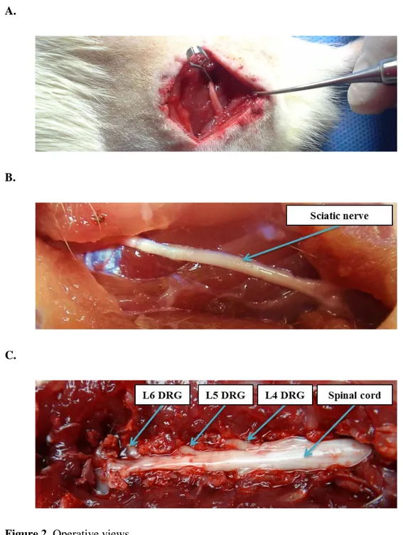
Figure 2. Operative views.
A) Sciatic nerve crush injury model B) Sciatic nerve of rat treated with h-ADMSCs 4 weeks after transplantation. C) L4, L5, L6 dorsal root ganglion of rat treats treated with h-ADMSCs 4 weeks after transplantation.
13
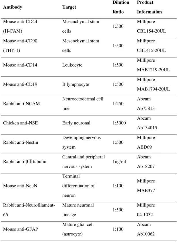
Table 1. Antibodies used in the experiments
Antibody Target Dilution Ratio Product Information Mouse anti-CD44 (H-CAM) Mesenchymal stem cells 1:500 Millipore CBL154-20UL Mouse anti-CD90 (THY-1) Mesenchymal stem cells 1:500 Millipore CBL415-20UL Mouse anti-CD14 Leukocyte 1:500
Millipore
MAB1219-20UL Mouse anti-CD19 B lymphocyte 1:500
Millipore MAB1794-20UL Rabbit anti-NCAM Neuroectodermal cell line 1:250 Abcam Ab75813 Chicken anti-NSE Early neuronal 1:5000
Abcam Ab134015 Rabbit anti-Nestin Developing nervous system 1:500 Millipore ABD69 Rabbit anti-βⅢtubulin
Central and peripheral nervous system 1ug/ml Abcam Ab18207 Mouse anti-NeuN Terminal differentiation of neuron 1:100 Millipore MAB377 Rabbit anti-Neurofilament-66 Mature neuronal lineage 1:500 Millipore 04-1032 Mouse anti-GFAP
Mature glial cell (astrocyte)
1:100
Abcam Ab10062
14 Rabbit anti-S100 Astrocyte, Shwann cell 1:40 Abcam Ab52642 Mouse anti-CNPase Oligodendrocyte 1:200
Abcam Ab6319 Chicken anti-vimentin Peripheral nerve (Schwann) 1:5000 Millipore AB5733 Mouse anti-human nuclei
Nuclei of all human cell type
1:200
Millipore MAB1281 Mouse anti-CGRP Inflammation marker 1:100
Abcam Ab81887 FITC-Donkey anti-mouse IgG(H+L) Mouse primary antibody 1:100 Jackson 751-095-151 CyTM3-Donkey anti-mouse IgG(H+L) Mouse primary antibody 1:500 Jackson 715-164-151 FITC-Donkey anti-rabbit IgG(H+L) Rabbit primary antibody 1:100 Jackson 711-095-152 CyTM3-Donkey anti-rabbit IgG(H+L) Rabbit primary antibody 1:500 Jackson 711-165-152 CyTM3-Donkey
anti-chicken IgG(H+L) Chicken primary antibody 1:500 Jackson 703-165-155
15
8. Luxol fast blue staining, toluidine blue staining and electron microscopy
Luxol Fast Blue Staining. The sectioned tissue was stained by Luxol Fast Blue (LFB)
to examine general morphology and the extent of demyelination. At first, the sections hydrated and then were stained with LFB solution (Luxol® fast blue solution, L0294) at 60℃ overnight. Using lithium carbonate solution and 70% alcohol, excess staining and permount were removed.24
Electron microscopy and toluidine blue staining. The regenerated nerves were
harvested and immersion-fixed in Karnovsky’s solution (2% Glutaraldehyde, 2% Paraformaldehyde, 0.1M Phosphate buffer) overnight at 4℃. The specimens had post fixation in 1% OsO4 solution, dehydrated with an ascending series of ethanol followed by
infiltration with propylene oxide. Specimens were then embedded in mixture solution including EPON mixture (EPON 812, MNA, DDSA, DMP30) and propylene oxide. Semithin sections of 250~300nm were cut then stained with 0.5% toluidine blue. Thin section for transmission electron microscopy were placed on glider thin bar mesh grids and double stained with uranyl acetate and lead citrate.25
16 Ⅲ. Results
1. Human adipose-derived mesenchymal stem cells cultures
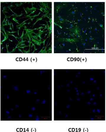
The isolated human adipose tissue cells, which were cultured in alpha-MEM medium, were positive for CD44 and CD 90, but negative for CD14 and CD19. It tested using immunofluorescence staining (Fig. 3).
17
Figure 3. Characterization of h-ADMSCs
Isolated h-ADMSCs approached with confluency, showing spindle shape-like fibroblastic morphology. In addition, they have mesenchymal stem cell markers CD44 and CD99 and were not reactive at CD14 and CD19.
18
2. Differentiation to a neuronal cell phenotype using two different methods
In our study, we used two protocols for confirming neuronal differentiation of h-ADMSCs. The early neural marker Tuj1 was positively expressed by cells by serum-free induction method. However, astrocyte markers, GFAP and oligoendrocyte markers, and CNPase were positively expressed (Fig. 4A,B). On the other hand, early neural markers Tuj1 and Nestin were positively expressed by chemical induction method. In addition, astrocyte markers, S100 oligoendrocyte markers, CNPase, and peripheral nerve markers vimentin were positively expressed (Fig. 4C). These two method show that h-ADMSCs have a neuronal differentiation ability.
19 A
B
20
Figure 4. h-ADMSCs differentiate to neuronal cell phenotype.
A) Scheme of neuronal differentiation in serum free induction. B) Inflected h-ADMSCs differentiated into Tuj1-positive neuron (red) and NF-positive mature neuron (green), but were negative for CNPase and GFAP. C) Inducted h-ADMSCs differentiated into NCAM positive neuroectodermal cell line (green), NSE positive early neuronal cell line (red), Nestine, Tuj1 positive neuron (green), S100 positive astrocyte (red), CNPse positive oligodendrocyte (green), and vimentine positive Schwann cell (red)
21
3. h-ADMSCs relieve mechanical allodynia
When tested with dynamic plantar aesthesiometer, mean paw withdrawal threshold of the PBS epineural soaking group was 13.12±0.72, 16.13±1.81, 16.94±1.24, 19.77±1.34, and 21.18±1.90g; PBS intraneural injection group was 13.12±0.72, 14.19±1.31, 16.09±1.16, 16.42±1.45, and 18.94±1.94; h-ADMSCs epineural soaking group was 13.12±0.72, 17.76±1.60, 17.98±1.29, 22.97±1.06, and 24.05±1.69; and h-ADMSCs intraneural injection group was 13.12±0.72, 14.92±1.02, 17.02±1.66, and 21.53±1.76g. Paw withdrawal thresholds were recorded at various time intervals, with values ranging from 31.37±0.37g to 34.73±0.32 g in sham operated animals. h-ADMSCs epineural soaking transplantation showed the best convalescence, while there was no significant difference between the h-ADMSCs intraneural injection and PBS epineural soaking groups (Fig. 5A).
22 A.
*
*
*
*
*
*
*
*
*
23
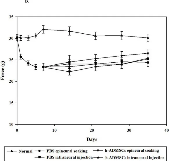
B.
Figure 5. Pain reducing effect of h-ADMSCs on mechanical allodynia
A) Result of dynamic plantar aesthesiometer test for left leg. B) Result of dynamic plantar aesthesiometer test for right leg. (*P<0.05 vs PBS intraneural injection)
24
4. Assessment of allodynia using dynamic plantar aesthesiometer
The data show each group’s SFI score at each time point. One week later, the SFI score was -51.98±2.51 in the injured group and -6.25±1.22 in the normal group. This showed a difference 1 week later after transplantation (PBS epineural soaking: -32.41±1.42; PBS intraneural injection: -33.05±1.27; ADMSCs epineural soaking: -27.14±1.26; and h-ADMSCs intraneural injection: -30.52±1.53), which was maintained until the end of experimentation (PBS epineural soaking: 13.29±2.05; PBS intraneural injection: -18.10±0.78; h-ADMSCs epineural soaking: -9.18±1.36; and h-ADMSCs intraneural injection: -14.16±2.11). Sciatic nerve damage led to a decrease of TS and IT length and increase of PI length by release of paw flexion. After 35 days following injury, the h-ADMSCs epineural soaking transplantation group showed recovery matching the normal group (Fig. 6).
25 A. B.
*
*
*
*
26
Figure 6. Motor functional recovery is accelerated after h-ADMSCs treatment.
A,B) The disease curve was measured weekly by SFI in each group. Improved plantar flexion and toe spread are visible in track obtained from the h-ADMSCs epineural soaking group as compared with the PBS intraneural injected group 2 weeks after injury. (*P<0.05 vs PBS intraneural injection)
27
5. Electrophysiological assessment
SSEPs and MEPs were recorded 4 weeks after transplantation. The curves of MEPs have patterns of negative-negative potentials, and SSEPs have positive-negative-positive potentials (Fig. 7A,B). Sciatic nerve crush injury rat transplanted with h-ADMSCs epineural soaking group showed shortened initial latency compared with the intraneural injection group. However, it did not show any difference with the PBS epineural soaking group, though amplitude of MEP in the h-ADMSCs epineural soaking group show increased amplitude compared with other groups (Table. 2). The h-ADMSCs epineural soaking group showed shortened initial latency and N1 and P1 latency of SSEPs. Overall, the amplitude showed a clear tendency to be increased in other groups (Table. 3).
28
A. Motor evoked potential (MEP)
29
Figure 7. Electrophysiological studies after transplantation of h-ADMSCs.
A) Sciatic nerve crush injury rat transplanted with h-ADMSCs epineural soaking group showed shortened initial latency similar to that in the normal group, though N1 latency and P1 latency did not. In addition, amplitude of MEP significantly increased in the h-ADMSCs epineural soaking group. B) The h-h-ADMSCs epineural soaking group showed shortened initial latency and N1and P1 latency. Overall, the amplitude showed a clear tendency to increase in h-ADMSCs epineural soaking group the other groups.
30
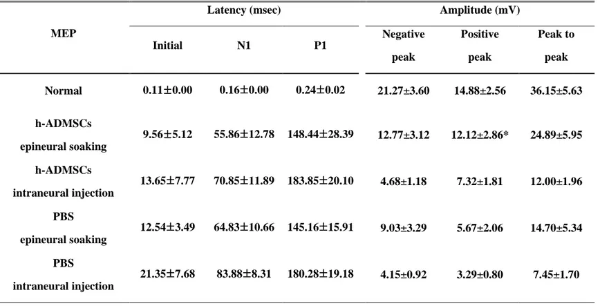
Table 2. Motor evoked potential in rat treated with h-ADMSCs 4 weeks after transplantation.
Values are mean ±SE. Input power is 20mA and the number of experiments is 8 MEP, Motor evoked potential; h-ADMSCs, human adipose derived mesenchymal stem cells; PBS, phosphate-buffered saline. (*P<0.05 vs PBS intraneural injection)
MEP
Latency (msec) Amplitude (mV)
Initial N1 P1 Negative peak Positive peak Peak to peak Normal 0.11±0.00 0.16±0.00 0.24±0.02 21.27±3.60 14.88±2.56 36.15±5.63 h-ADMSCs epineural soaking 9.56±5.12 55.86±12.78 148.44±28.39 12.77±3.12 12.12±2.86* 24.89±5.95 h-ADMSCs intraneural injection 13.65±7.77 70.85±11.89 183.85±20.10 4.68±1.18 7.32±1.81 12.00±1.96 PBS epineural soaking 12.54±3.49 64.83±10.66 145.16±15.91 9.03±3.29 5.67±2.06 14.70±5.34 PBS intraneural injection 21.35±7.68 83.88±8.31 180.28±19.18 4.15±0.92 3.29±0.80 7.45±1.70
31
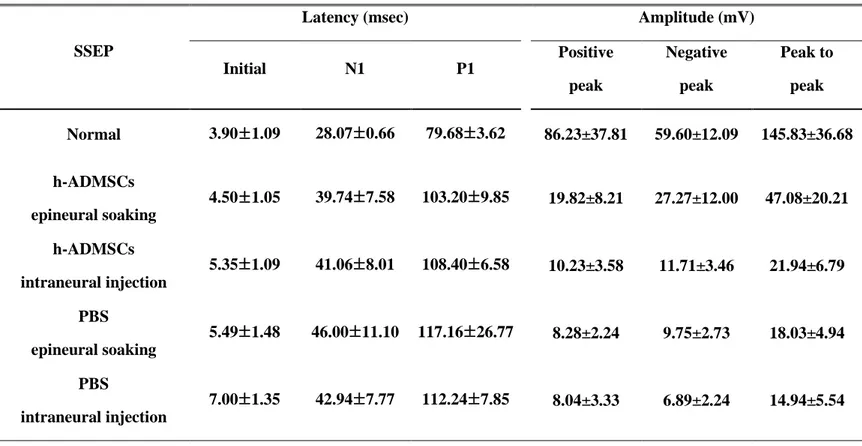
Table 3. Somatosensory evoked potential in rat treated with h-ADMSCs 4 weeks after transplantation.
SSEP
Latency (msec) Amplitude (mV)
Initial N1 P1 Positive peak Negative peak Peak to peak Normal 3.90±1.09 28.07±0.66 79.68±3.62 86.23±37.81 59.60±12.09 145.83±36.68 h-ADMSCs epineural soaking 4.50±1.05 39.74±7.58 103.20±9.85 19.82±8.21 27.27±12.00 47.08±20.21 h-ADMSCs intraneural injection 5.35±1.09 41.06±8.01 108.40±6.58 10.23±3.58 11.71±3.46 21.94±6.79 PBS epineural soaking 5.49±1.48 46.00±11.10 117.16±26.77 8.28±2.24 9.75±2.73 18.03±4.94 PBS intraneural injection 7.00±1.35 42.94±7.77 112.24±7.85 8.04±3.33 6.89±2.24 14.94±5.54
Values are mean ±SE. Input power is 3mA, and the number of experiment experiments number is 8 SSEP, somatosensory evoked potential; h-ADMSCs, human adipose derived mesenchymal stem cells; PBS, phosphate-buffered saline. (*P<0.05 vs PBS intraneural injection)
32
6. Transplanted h-ADMSCs were differentiated to a neuronal lineage
Transplanted cells derived from h-ADMSCs were confirmed by human nuclei antibody. Double immunoreactivity for human nuclei and neuronal markers showed that transplanted cells differentiated into neurons and oligodendrocytes. However, definite astrocyte marker GFAP was not seen. In the epineural soaking group, cells were almost differentiated from Schwann cells; in the intraneural injected group, cells were differentiated from neurons. (Fig. 8)
33
Figure 8. Immunohistochemistry showing recovery of neuronal regeneration
Immunostaining images 4 weeks after transplant of h-ADMSCs. Sciatic nerve sections were stained with anti-human nuclei (green), anti-Tuj1 of neuron (red), anti-NF of mature neuron (red), and anti-vimentin of Schwann cells (red).
34
7. Transplanted h-ADMSCs promote the remyelination of the injured nerve
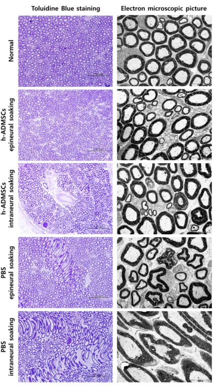
In LFB staining, morphological changes were observed following the h-ADMSCs transplanted group. In the control group, the PBS was not repaired after 5 weeks. In the h-ADMSCs transplanted group, tissue recovery and remyelination occurred. However, in the h-ADMSCs intraneural injecting group, there remained a needle track that was not restored (Fig. 9A). In toluidine blue staining, the normal nerve showed numerous large well-myelinated axons. The h-ADMSCs transplanted group showed almost perfectly recovery similar to normal tissue, unlike with the PBS group, which showed abnormal shape of nerves as circular bundles (Fig. 9B). In transmission electron microscopic examination, transplantation of the h-ADMSCs enhanced structural maturation and organization of the injured sciatic nerve, characterized by better myelination and less fibrosis, which corroborates the findings of the toluidine blue staining. In the PBS groups with nerve injury, invasion of fibroblast or poorly-shape myelinated axon were observed (Fig. 9B).
35
36
B.
Figure 9. Microscopic findings showing the remyelination of the injured sciatic nerve
37
A) Luxol fast blue staining. h-ADMSCs epineural transplantation group tissue recovered faster than that of any other group. B) Toluidine blue staining. h-ADMSCs transplantation made perfectly recovery, as normal tissue. Transmission electron microscopic examination. h-ADMSCs transplantation recovered as normal tissue. It checks observation of myelin sheath’s thickness and shape.
38
VI. Discussion
Stem cell therapy has been showing a promising method for the treatment of neurological disorders affecting central and peripheral nerve system. At these diseases, embryonic stem cells and adult stem cell is provided by useful tools. Using embryonic stem cells in clinical situations is restricted due to cell regulations and technical and ethical considerations involved in the genetic manipulation of human ESCs, even though these cells are, theoretically, highly beneficial.26
Adult stem cells, On the other hand, overcome the safety and ethical controversy, it also have differentiation potency to a specific organ and placiticy of trans-differentiation. As a result, cell therapy method using an adult stem cells is available to variable diseases. Among others, bone marrow derived stem cell is most popular cell to treat various neuronal diseases. It could have successful result for many research, and now in clinical trial, but obtaining of enough cells is limited at once and the isolation process need invasive method.27-29 There are several reasons for why adipose tissue is ideal source for stem cells. It can be obtained high amounts of adipose stem cells with minimum risk like surgery. Also, quantity of stem cells from adipose tissue is 5000 times than isolated bone marrow stem cells. Especially, human adipose stem cells express cell surface antigen that are hardly detected by T cells and therefore h-ADMSCc can be transplanted autograft, allograft, and even xenograft.30,31 Thus, we choose h-ADMSCs in this research.
There are ways how to deliver stem cell to nerve system, like intrathecal delivery, intralesional delivery, intravenous delivery, intraarterial delivery. The intralesional injection group showed the best engraftment, so the present study attempted to carry out direct transplantation cell therapy at injury region, which is known as an efficacious nerve tissue treatment. However it could bring secondary damage on treatment site.32 Therefore, we investigated the effect of indirect transplantation is effective and stable.
In our neuropathic pain model, h-ADMSCs epineural soaking group showed differences with PBS treatment group at dynamic plantar aesthesiometer assay. SFI
39
analysis shows the difference in the h-ADMSCs epineural soaking group and PBS intraneural injection and epineural soaking group at 1 weeks after cell transplantation up to the end of the experiment. (Fig. 6A) The electrophysiology result also agrees with other experiment results. Particularly, amplitude of MEPs were significantly improved in h-ADMSCs epineural soaking group, compared with PBS intraneural injection group (Table. 2,3) Furthermore, histological assessment showed that h-ADMSCs transplantation induced recovery of injured sciatic nerve and remyelination at damage tissue. However, there were no differences between h-ADMSCs direct transplantation and PBS indirect transplantation groups at all experiment. These result suggested that secondary damage due to injection injury or injection pressure by intralesional delivery may be a reducing h-ADMSCs therapeutic effect.
The efficacy and viability of the transplanted cells were observed according to different transplantation routes. In the present study, mesenchymal stem cells what we used have been demonstrated to have an ability to move to the injured site and trans-differentiate into neural lineage cells.33 Bakshi et al. reported that, when MSCs were injected into the cerebrospinal fluid three times, they observed a 2.3-fold higher engraftment rate than after only one injection in lesion site. And our priminary study, after intralesional transplantation group, engrafted cells were concentrated around the injection point, which induced mass effect on adjacent normal tissues and restricted dispersion to the damaged regions.32 Others have also reported that the needle used in intralesional transplantation group can cause secondary damage to the injured spinal cord and the volume of the injected cells can result in increased pressure inside the spinal cord.32 Furthermore, when we consider the application of intralesional transplantation in clinical practice, a patient must undergo general anesthesia and a mandatory surgical procedure, which can be obstacles for debilitated patents who have weakened immune systems. However, Takahashi et al, ILT would be assumed to be the most effective approach any other method. Also, Nakamura et al revealed that NS/PCs injected into non-injury sites such as
40
by intravenous or intrathecal administration did not engraft to the injury site in sufficient numbers, but instead were often “trapped” in the lungs and kidneys. They concluded that intralesional application might be the most effective and reliable method for transplanting cells.
Interestingly in our experiment contrast leg response at low force compared with non-surgery. This can explain why mirror-image allodynia occurs from the contralateral to the injured region of trauma or inflammation. Some researchers assert glial and proinflammatory cytokine action caused mirror-image neuropathic pain. However, the accurate mechanism of this phenomenon remains unknown (Fig. 5B)
It had been reported the rat stem cell transplantation in defected sciatic nerve induce peripheral nerve regeneration by Murakami. Apart from that, many investigations reported that adipose tissue derived stem cells transplantation could improve the injured tissues by morphologically, histologically and electromyographically.34 However this doesn't mean that transplanted cell differentiate to neuronal cell. Also, several studies suggest that stem cells may contribute to the repair of injured tissue by releasing a large number of trophic factors, which can influence injured region, promote and maintain axonal sprouting and myelination of fibers.
Therefore, further study need to clear about the paracrine effect of transplanted cells on injury site. Also, the next experiment will be a mechanisms at the spinal cord and dorsal root ganglia, which undergo a response after any nerve injury possibly induce by variable neuropathic pain therapy.
41
VII. Conclusion
In this research, h-ADMSCs epineural soaking showed significant pain reduction, confirmed by dynamic plantar aesthesiometer, SFI analysis, and electrophysiological assessment. It is assumed that direction injection might induce additional trauma. Thus, h-ADMSCs indirect transplantation may be the safest and most effective method for delivering stem cells and reduction neuropathic pain in peripheral nerve injury.
42
References
1. Griffin JW, Pan B, Polley MA, Hoffman PN, Farah MH. Measuring nerve regeneration in the mouse. Exp Neurol 2010;223:60-71.
2. Chen CJ, Ou YC, Liao SL, Chen WY, Chen SY, Wu CW, et al. Transplantation of bone marrow stromal cells for peripheral nerve repair. Exp Neurol 2007;204:443-53.
3. Marconi S, Castiglione G, Turano E, Bissolotti G, Angiari S, Farinazzo A, et al. Human adipose-derived mesenchymal stem cells systemically injected promote peripheral nerve regeneration in the mouse model of sciatic crush. Tissue Eng Part A 2012;18:1264-72.
4. Cignarelli A, Perrini S, Ficarella R, Peschechera A, Nigro P, Giorgino F. Human adipose tissue stem cells: relevance in the pathophysiology of obesity and metabolic diseases and therapeutic applications. Expert Rev Mol Med 2012;14:e19.
5. Dadon-Nachum M, Sadan O, Srugo I, Melamed E, Offen D. Differentiated mesenchymal stem cells for sciatic nerve injury. Stem Cell Rev 2011;7:664-71. 6. Lee KH, Suh-Kim H, Choi JS, Jeun SS, Kim EJ, Kim SS, et al. Human
mesenchymal stem cell transplantation promotes functional recovery following acute spinal cord injury in rats. Acta Neurobiol Exp (Wars) 2007;67:13-22. 7. Yu JM, Bunnell BA, Kang SK. Neural differentiation of human adipose
tissue-derived stem cells. Methods Mol Biol 2011;702:219-31.
8. Anghileri E, Marconi S, Pignatelli A, Cifelli P, Galie M, Sbarbati A, et al. Neuronal differentiation potential of human adipose-derived mesenchymal stem cells. Stem Cells Dev 2008;17:909-16.
9. Zhang HT, Liu ZL, Yao XQ, Yang ZJ, Xu RX. Neural differentiation ability of mesenchymal stromal cells from bone marrow and adipose tissue: a comparative study. Cytotherapy 2012;14:1203-14.
10. Cho SR, Kim YR, Kang HS, Yim SH, Park CI, Min YH, et al. Functional recovery after the transplantation of neurally differentiated mesenchymal stem cells derived from bone barrow in a rat model of spinal cord injury. Cell Transplant 2009;18:1359-68.
11. Nakamura M, Okano H. Cell transplantation therapies for spinal cord injury focusing on induced pluripotent stem cells. Cell Res 2013;23:70-80.
12. Kim SH, Moon IS, Park IS. Unique hippocampal changes and allodynia in a model of chronic stress. J Korean Med Sci 2013;28:946-50.
13. Nirogi R, Goura V, Shanmuganathan D, Jayarajan P, Abraham R. Comparison of manual and automated filaments for evaluation of neuropathic pain behavior in rats. J Pharmacol Toxicol Methods 2012;66:8-13.
14. Austin PJ, Wu A, Moalem-Taylor G. Chronic constriction of the sciatic nerve and pain hypersensitivity testing in rats. J Vis Exp 2012.
15. Pan HC, Sheu ML, Su HL, Chen YJ, Chen CJ, Yang DY, et al. Magnesium supplement promotes sciatic nerve regeneration and down-regulates inflammatory response. Magnes Res 2011;24:54-70.
16. Tamaddonfard E, Farshid AA, Ahmadian E, Hamidhoseyni A. Crocin enhanced functional recovery after sciatic nerve crush injury in rats. Iran J Basic Med Sci 2013;16:83-90.
43
17. Sarikcioglu L, Demirel BM, Utuk A. Walking track analysis: an assessment method for functional recovery after sciatic nerve injury in the rat. Folia Morphol (Warsz) 2009;68:1-7.
18. Fehlings MG, Tator CH, Linden RD, Piper IR. Motor and somatosensory evoked potentials recorded from the rat. Electroencephalogr Clin Neurophysiol 1988;69:65-78.
19. Brunner M, Vaughan D. Evoked potential monitoring in anaesthesia and analgesia. Anaesthesia 2000;55:823-5.
20. Leong GW, Gorrie CA, Ng K, Rutkowski S, Waite PM. Electrical perceptual threshold testing: a validation study. J Spinal Cord Med 2009;32:140-6.
21. Elias KA, Cronin MJ, Stewart TA, Carlsen RC. Peripheral neuropathy in transgenic diabetic mice: restoration of C-fiber function with human recombinant nerve growth factor. Diabetes 1998;47:1637-42.
22. Kang ES, Ha KY, Kim YH. Fate of transplanted bone marrow derived mesenchymal stem cells following spinal cord injury in rats by transplantation routes. J Korean Med Sci 2012;27:586-93.
23. Chang DJ, Oh SH, Lee N, Choi C, Jeon I, Kim HS, et al. Contralaterally transplanted human embryonic stem cell-derived neural precursor cells (ENStem-A) migrate and improve brain functions in stroke-damaged rats. Exp Mol Med 2013;45:e53.
24. Liang L, Wang Z, Lu N, Yang J, Zhang Y, Zhao Z. Involvement of nerve injury and activation of peripheral glial cells in tetanic sciatic stimulation-induced persistent pain in rats. J Neurosci Res 2010;88:2899-910.
25. Lee EJ, Xu L, Kim GH, Kang SK, Lee SW, Park SH, et al. Regeneration of peripheral nerves by transplanted sphere of human mesenchymal stem cells derived from embryonic stem cells. Biomaterials 2012;33:7039-46.
26. Rice CM, Halfpenny CA, Scolding NJ. Stem cells for the treatment of neurological disease. Transfus Med 2003;13:351-61.
27. Caplan AI. Mesenchymal stem cells. J Orthop Res 1991;9:641-50.
28. Dimarino AM, Caplan AI, Bonfield TL. Mesenchymal Stem Cells in Tissue Repair. Front Immunol 2013;4:201.
29. Krabbe C, Zimmer J, Meyer M. Neural transdifferentiation of mesenchymal stem cells--a critical review. Apmis 2005;113:831-44.
30. Zhang J, Li Y, Chen J, Cui Y, Lu M, Elias SB, et al. Human bone marrow stromal cell treatment improves neurological functional recovery in EAE mice. Exp Neurol 2005;195:16-26.
31. Bai L, Lennon DP, Eaton V, Maier K, Caplan AI, Miller SD, et al. Human bone marrow-derived mesenchymal stem cells induce Th2-polarized immune response and promote endogenous repair in animal models of multiple sclerosis. Glia 2009;57:1192-203.
32. Shin DA, Kim JM, Kim HI, Yi S, Ha Y, Yoon do H, et al. Comparison of functional and histological outcomes after intralesional, intracisternal, and intravenous transplantation of human bone marrow-derived mesenchymal stromal cells in a rat model of spinal cord injury. Acta Neurochir (Wien) 2013;155:1943-50.
33. Swanger SA, Neuhuber B, Himes BT, Bakshi A, Fischer I. Analysis of allogeneic and syngeneic bone marrow stromal cell graft survival in the spinal cord. Cell Transplant 2005;14:775-86.
44
34. Lin Y, Chen X, Yan Z, Liu L, Tang W, Zheng X, et al. Multilineage differentiation of adipose-derived stromal cells from GFP transgenic mice. Mol Cell Biochem 2006;285:69-78.
45
국문요약
백서의 신경병증성 통증 모델에서 인간중간엽줄기세포의 통증감소 및 신경재생효과 (지도교수 윤도흠) 연세대학교 대학원 의과학과 이혜영 중간엽줄기세포는 외배엽으로의 분화를 이용한 신경조직치료제로 사용 될 수 있다. 오늘날 손상 신경조직에 대한 중간엽줄기세포치료는 지역 이식만이 이루어 지고 있다. 하지만 신경조직 손상시 신경 조직에 직접적인 특이적 이식은 신경의 또 다른 손상을 야기할 수 있다. 이러한 이유로 본 실험에서는 두 가지 방법을 이용한 인간 지방 조직에서 유래한 중간엽줄기세포의 분화능을 확인하고 백서의 급성 신경 손상을 야기하는 좌골신경의 손상 일주일 후 세포의 간접적 이식을 통해 두드러진 기능적 회복을 돕는 것을 확인했다. 또한 인간 지방 조직에서 유래한 중간엽줄기세포를 이용한 좌골신경의 치료는 신경 조직의 재생과 운동력 향상을 향상 시키며, 통증을 완화시킴을 알 수 있었다.46 이와 같은 결과를 통해 본 연구에서는 인간 지방 조직 유래 중배엽줄기세포의 간접적인 이식은 신경 재생을 촉진하고 통증을 완화시키며 재수초화를 촉진하는 것을 통해 급성 신경 손상 모델에서 확실한 치료효과를 보여주는 것을 통해 신경병증성 통증 질환에 대한 치료 방법을 제시 할 수 있을 것으로 사료 된다. 핵심되는 말: 신경병증성 통증 모델, 인간중간엽줄기세포, 분화, 세포치료