Effects of Epigallocatechin-3-gallate Coated
Biphasic Calcium Phosphate on the Dehiscence
Bony Defect around Dental Implant
Ji Young Seo
The Graduate School
Yonsei University
Effects of Epigallocatechin-3-gallate Coated
Biphasic Calcium Phosphate on the Dehiscence
Bony Defect around Dental Implant
(Directed by Prof. Hong Seok Moon, D.D.S.,M.S.D.,Ph.D.)
A Dissertation Thesis
Submitted to the Department of Dental Science
And the Graduate School of Yonsei University
in partial fulfillment of the
requirements for the degree of
Doctor of Philosophy in Dental Science
Ji Young Seo
This certifies that the dissertation thesis
of Ji Young Seo is approved.
___________________________
Thesis supervisor: Hong Seok Moon
___________________________
Thesis committee: Han Sung Jung
___________________________
Thesis committee: Young Bum Bark
___________________________
Thesis committee: Sung Tea Kim
___________________________
Thesis committee: Jee Hwan Kim
The Graduate School
Yonsei University
감사의 글
본 논문이 완성되기까지 오랜 시간 지도와 격려로 저를 이끌어주신 문홍석 교수님께 진심으로 감사 드립니다. 실험의 전 과정과 논문 작성까지 일일이 살펴주시고 많은 도움을 주신 김성태 교수님, 논문 심사에 소중한 조언과 가르침을 주신 정한성 교수님, 박영범 교수님, 김지환 교수님께도 감사를 드립니다. 또한 보철과 모든 교수님께도 그 동안의 가르침에 감사를 드립니다. 논문의 진행 및 감수를 봐주신 신유석 교수님께도 감사 드리며, 동물실험에 많은 도움을 준 자미안 선생님, 표세욱 선생님, 시편을 제작하고 자료 분석 및 계측에 많은 도움을 주신 박상현 선생님께도 감사한 마음을 전합니다. 논문을 쓰는 동안 많은 배려를 해준 연세 리더스 식구들에게도 감사 드립니다. 마지막으로 항상 저를 지켜봐 주시고, 부족한 저를 사랑으로 감싸주신 주신 양가 부모님께 감사와 사랑의 마음을 전합니다. 주말마다 조카 돌보기를 마다하지 않은 동생들과 시댁 어른들께도 감사를 드립니다. 언제나 제가 하는 일을 응원해 주고 곁에서 도움을 주는 사랑하고 존경하는 남편과 삶의 기쁨이 되어주는 아들 연서와 곧 태어날 아가에게도 큰 감사와 기쁨을 전하며, 이 논문을 나누고자 합니다. 2012 년 7 월 서지영 드림i
Table of contents
List of Figures ··· ii
List of Tables ··· ii
Abstract ··· 1
I. Introduction ··· 3
II. Materials and Methods ··· 10
2.1 Laboratory Animals ··· 10
2.2 Materials for Experiments ··· 10
2.3 Surface coating and lyophilization ··· 12
2.4 Surgical procedures ··· 14
2.5 Fluorescence Analysis ··· 15
2.6 Specimen Preparation ··· 18
2.7 Statistical Analysis ··· 19
III. Results ··· 20
3.1 Clinical observation ··· 20
3.2 Histological observation ··· 21
3.3 Histometrical observation ··· 26
IV. Discussion ··· 32
V. Conclusion ··· 40
References ··· 41
ii
List of Figures
Figure 1. Implant, membrane and accessories design used in the study ··· 11
Figure 2.
Schematic diagram of surface coating process of BCP··· 13
Figure 3. Formation of bone defects ··· 17
Figure 4. Implantation and bone grafts ··· 17
Figure 5.
Radiographic image after implant placement··· 18
Figure 6. The representative histological images (x 12.5) ··· 21
Figure 7. Histological images of the 6 week BCP group ··· 22
Figure 8. Histological images of the 6 week ErhBMP-2 group ··· 23
Figure 9. Histological images of the 6 week ErhBMP-2/EGCG group ··· 23
Figure 10. Histological images of the 12 week BCP group··· 24
Figure 11. Histological images of the 12 week ErhBMP-2 group ··· 24
Figure 12. Histological images of the 12 week ErhBMP-2 / EGCG group ··· 25
Figure 13. Fluorescent microscopic images in the 12 week group ··· 30
Figure 14. Fluorescent microscopic images in the 6 week group ··· 31
List of Tables
Table 1. Experimental design ··· 12
Table 2. Fluorescence expression agent ··· 16
Table 3. Time Table ··· 16
Table 4. Number of implants where a membrane was exposed by group ··· 20
Table 5.
Mean and standard deviation of BIC in the 6 and 12 week group··· 26
Table 6.
Mean and standard deviation of BD in the 6 and 12 week group··· 27
Table 7.
Mean and standard deviation of BRH in the 6 and 12 week group··· 29
1
Abstract
Effects of Epigallocatechin-3-gallate Coated Biphasic
Calcium Phosphate on the Dehiscence Bony Defect around
Dental Implant
Ji Young Seo D.D.S., M.S.D.
(Directed by Prof. Hong Seok Moon, D.D.S.,M.S.D., Ph.D.)
Purpose: Various kinds of the surface treatment have been tested in order to improve not
only the osteoconductive effect but also the osteoinductive effect on BCP. Out of them, Recombinant human bone morphogenetic protein (rh-BMP) was known as a growth factor which shows excellent effects on bone regeneration. Out of various osteoinductive agents, green tea extract (epigallocatechin-3-gallate) has been recently in the limelight. From the previous studies, EGCG could be considered as a substance for enhancement of bone formation. Therefore, the purpose of this study was to evaluate osteogenic potential of BCP coated with low-concentrate ErhBMP-2 and EGCG.
Materials & Methods: In this study, 5 mongrel dogs were used. Mandibular premolars
and molars were extracted and bone defects (Ø 4 X 5 mm) were formed after 12 weeks healing period. Then, defect sites were treated with bone graft materials and membranes after implantation. In this experiment, BCP (OSTEONTM) was used as the control group. For the experimental groups, BCP coated with ErhBMP-2 0.05mg/ml and EGCG
2
5mg/ml+ ErhBMP-2 0.05mg/ml together was used. Dogs were euthanized at 6 weeks and 12 weeks after implantation.
Results: The healing of the defect site and osseointegration of the implants was evaluated
by histologic and histometric analyses.
1. When comparing between the three groups, ErhBMP-2/EGCG group appeared to have higher value in bone to implant contact (BIC), bone density (BD), bone regeneration height (BRH) and bone mineral apposition rate (BMAR). But significant difference was not found.
2. BIC, BRH and BMAR values increased when comparing by time in the three groups.
- BCP, ErhBMP-2 and ErhBMP-2/EGCG groups showed higher BIC value in the 12 week group than the 6 week group with significant differences.(p<0.05) - BCP group showed higher BRH value in 12 weeks with significant
difference.(p<0.05)
3. In fluorescence analysis, bone remodeling around graft material and implant was more active in the ErhBMP-2/EGCG group.
Conclusion: Within the limit of this study, the surface modification of BCP with
low-concentrate BMP and EGCG was effective in enhancing the osteogenetic potential.
3
Effects of Epigallocatechin-3-gallate Coated Biphasic
Calcium Phosphate on the Dehiscence Bony Defect around
Dental Implant
Ji Young Seo D.D.S., M.S.D.
The Graduate School
Yonsei University
Department of Dental Science
(Directed by Prof. Hong Seok Moon, D.D.S., M.S.D., Ph.D.)
I. Introduction
In the past decade, the restoration of the dental implant has been placed a routine treatment in partially or edentulous patient. After the mid ‘80’s, the surface treatment method for implant has been constantly developing. The failure rate has been inversely reduced so that the dental implant usually began to be deployed for the restoration of not
4
only the complete edentulous but also single tooth or the partial edentulous.1 However, when the implant is placed in alveolar bone damaged by periodontal diseases and trauma, it is often difficult to secure the initial fixation and it may cause some problems in long-term prognosis. In particular, in case of the horizontal bone loss, defects around implant including a perforation and a dehiscence may be caused. These matters may threaten the success rate of implants and increase the failure rate. To solve these problems, guided bone regeneration (GBR) has been widely used for the regeneration of bone around implant due to its easiness and predictability.2, 3
The success rate of GBR is diversely reported from 79~100% and the success rate has been greatly affected by which membrane is selected or which graft material is used.4 In the GBR procedure, membrane is a critical factor for the regeneration of alveolar bone, which blocks the in growing of connective tissues. It induces selective cell proliferation, and maintains space for new bone to be regenerated.3, 5
Graft material used in GBR not only retains the space but also prevents the formation of empty space under the membrane, and consequently contributes to the formation and stabilization of the blood clot. In addition, it has some advantages in reducing the fine movement of the membrane and promotes the formation of new bone.6, 7
Various kinds of graft materials ranging from autogenous bone to alloplastic bone are being widely used for the filling of the bone defect. Autogenous bone grafts are considered to be the most ideal and the most excellent in the osteoinductive capacity but these type of bone has a limit in quantity and techniques as well as a high resorption rate, so autogenic bone grafting cannot be applicable to all cases.8 For these reasons, various kinds of graft materials are being developed. Among these, alloplastic bone is now widely
5
being used for the bone augmentation because it doesn’t show immune reaction and the risk of infection.6, 8
Biphasic Calcium Phosphate (BCP) is one of alloplastic bones, which consists of Hydroxyapatite (HA) and β-tricalcium phosphate (TCP), and has a similar structure with human bone so that it shows more excellent results in the experiment.9-11 The pore size of BCP is similar with a cancellous bone of 300~500 ㎛. The size of the gap is important factor in the cell immigration and the vascular hyperplasia. In recent studies, it was reported that 200~400 ㎛ pore size of scaffold is necessary for osteoinduction.12, 13
In many previous studies, many experiments were performed to examine the proper ratio of HA and β-TCP. If the resorption rate of graft materials is too fast, it cannot play a role of retaining spaces for the formation of new bone, whereas if it is too slow, the graft material cannot be replaced with new bone. As the ratio of β-TCP get higher, the resorption rate become higher accordingly, whereas as the ratio of HA get higher, the resorption rate become lower accordingly as well. It is revealed that the overall resorption rate is regulated according to their proper ratio. Thus, many studies to find proper ratio to maximize the osteoconductive effect of biphasic calcium phosphate are performed.10, 11The ratio of HA and β-TCP is 7:3 in BCP bone substitute was used in this study. This ratio showed proper results in previous studies.14, 15
Various kinds of the surface treatments have been tested in order to improve not only the osteoconductive effect but also the osteoinductive effect on BCP.16 Of them, Recombinant human bone morphogenetic protein (rh-BMP) was known as a growth factor which shows excellent effects on the bone regeneration. Bone morphogenetic
6
protein (BMP) is osteoinductive and also differentiates mesenchymal stem cell into osteoblastic cells to increase new bone formation. Currently, rhBMP-2 and rhBMP-7 are manufactured and used in the maxillofacial region and the orthopedic surgery.17, 18
rhBMP-2 is a BMP gene obtained through the mammalian cell culture just like Chinese hamster ovary (CHO) cell by transfection with the BMP gene. However, it has limits in production quantity and application due to cost issues. To overcome such disadvantage, new production method in which BMP gene is inserted into Escherichia coli (E.coli) was introduced so that BMP can be obtained much more easily at a cheaper cost.19Bessho et al. carried out a comparative analysis of the bone tissue which was formed by inserting a specimen into a calf muscle in order to compare the effect of osteoinduction of E.coli-derived rhBMP-2 (ErhBMP-2) and CHO cell E.coli-derived rhBMP-2 (CrhBMP-2). It was revealed that ErhBMP-2 showed much more adipose tissue in bone matrix and much more bone formations than CrhBMP-2. In the comparison test of the activation of alkaline phosphatase (ALP), it appeared to be higher in the ErhBMP-2 than CrhBMP-2. It showed that ErhBMP-2 may be more useful in the bone tissue regeneration with a cheaper cost.20 In the study of Kim et al., after bone defect was intentionally made on the rat skull, the graft material was inserted and the healing pattern was observed.21 As a result, much more bone formations were shown in BCP group which was coated with ErhBMP-2 than other group with no surface treatment.
As water-soluble osteoinductive proteins such as BMP diffuse rapidly when applied to the transplant area directly, carrier system is needed to make it work on the local spot continuously.22 Collagen has been used clinically and experimentally as BMP carrier and has made a good result in new bone formation.21, 23 However, it has failed to
7
provide a sufficient space for bone regeneration when applied to the large bony defect region.22 By contrast, BCP has a superb space maintenance effect and an excellent biocompatibility. Therefore, BCP was considered as carrier system of BMP prior to collagen carrier.21 Diverse methods have been tried in order to apply BCP to BMP. The method of putting carrier into the solution diluted with osteoinductive protein presents a difficulty in manipulating the flow and in keeping the accurate concentration.24, 25For these reasons, BCP coated with rhBMP-2 could be better than BCP moistened with diluted rhBMP-2.
Out of various osteoinductive agents, green tea extract (epigallocatechin-3-gallate) has been recently in the limelight. Green tea extract reduces the fat content in the blood and enhance the immune reaction as well as has a therapeutic efficacy on several diseases including vascular diseases, diabetes, allergy, cancer, and obesity.26 It was shown that such effects were mainly originated from the green tea extract’s antioxidant effect and the removal effect on the active oxygen.27 In addition, the correlation between tea consumption and bone density has been reported many times. In the epidemiological survey, the menopausal women who habitually drink tea appeared to have higher bone density and less incidence of fracture. Even in animal studies, green tea extract was found to have preventive effect on bone diseases. When intentionally causing the osteoporosis on the ovary-dissected rat and the testis-dissected rat and then letting them drink green tea extract, bone density and bone volume appeared to increase in both the cortical bone and the cancellous bone. This suggests green tea extract influences on bone metabolism.27, 28
8
Principal components of green tea extract are Epicatechin (EG), Epicatechin Gallate (ECG), Epigallocatechin-3-gallate (EGCG). To examine the effect of these components on the bone formation and the bone resorption and its mechanism, various studies are now in progress.29-32
EGCG prevents bone resorption by inducing the apoptotic cell depth of osteoclast and by inhibiting its formation. Nakamura et al. reported that green tea extract appeared to be effective in preventing bone resorption which was induced with lipopolysaccharide. Green tea extract blocks the activity of NF-кB and inhibits the generation of IL-1β, consequently it blocks the formation of the osteoclast and inhibits bone resorption. Such effects of green tea extract may be likely to be effective in treating the inflammatory bone diseases such as the periodontal disease.30
Chen et al. examined the activity of alkaline phosphatase (ALP) and the expression level of mRNA engaged in the bone formation in order to look into the induction ability for the bone formation of green tea extract. As a result, when being treated with EGCG, not only ALP activity increased but also mRNA such as Cafa1/Runx2, osterix, osteocalcin engaging in the bone formation was increased as well. Moreover, human osteoblast-like cell promotes the differentiation of the osteoblast cell to increase the formation of mineralized bone nodule.31
Recently it was also reported EGCG enhances bone formation by suppressing T3-stimulated synthesis of osteocalcin which is a determinant of bone formation.32 If such effects of green tea extract apply to defect site of implant by being coated to BCP together with ErhBMP-2, much more ascending effects will be surely obtained.
9
The purpose of this study was to evaluate osteogenic potential of BCP coated with low-concentrate ErhBMP-2 and EGCG. The implant dehiscence bone defect site which was surgically formed in the dog was treated with BCP which was coated with mixtures of ErhBMP-2 and green tea extract and then its healing aspect was assessed in a histological manner.
10
II. Materials and Methods
2.1 Laboratory Animals
In this study, 5 adult dogs (mongrels at 18 -24 months and weighing about 30kg) were used. These dogs were free from systemic diseases and periodontal inflammations. For sound periodontal management, scaling and plaque control were conducted. During the healing period after those treatments, the animals were provided with liquid foods in order to prevent wounds that might be caused in the course of masticatory actions. Every step of the experiment was conducted in according to the guideline of laboratory animal experiment suggested by Dental College of Yonsei University.
2.2 Materials for Experiments
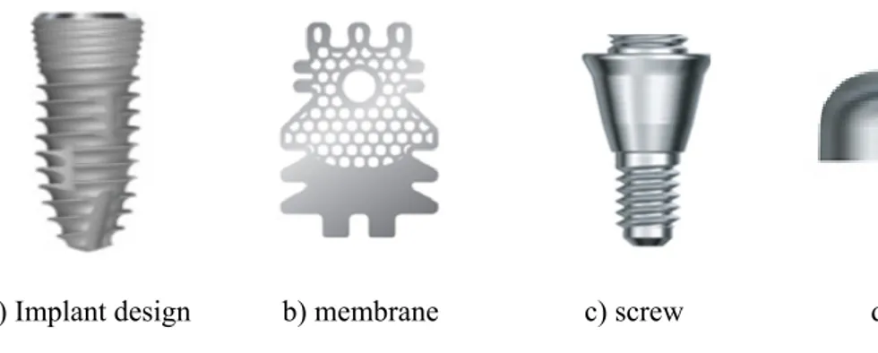
(1) Implant
A total of 30 Hydroxyapatite coated implants (3.5 X 10mm; Neo CMI Implant Neobiotech, Seoul, Korea) were used.
11
(2) Bone Graft Materials
In this experiment, OSTEONTM (Dentium. Seoul, Korea) which is biphasic calcium phosphate bone graft material was used as the control group. It is synthetic bone graft material with similar pore system to human cancellous bone, particle size of 0.5~1.0mm, hydroxyapatite 70% and β-TCP 30%. For the experimental group 1, OSTEONTM coated with ErhBMP-2 0.05mg/ml and for the experimental group 2 EGCG 5mg/ml (Sigma, purity 97.5~102.5%, MO, USA) and ErhBMP-2 0.05mg/ml (Cowellmedi®, Busan, Korea) was used together.
(3) Membrane
The defect site was grafted with bone substitution material and then used titanium mesh membrane (CTi-mem®, Neobiotech, Seoul, Korea).
a) Implant design b) membrane c) screw d) cap
12
Table 1. Experimental designBone Graft Materials Healing time N(=implant)
Group 1 BCP
6 Weeks 5
12 Weeks 5
Group 2 ErhBMP-2 -coated BCP
6 Weeks 5
12 Weeks 5
Group 3 EGCG /ErhBMP-2 – coated BCP
6 Weeks 5
12 Weeks 5
2.3 Surface coating and lyophilization
1) Surface coating process of bone graft material
Graft material was coated by combining ErhBMP-2 and EGCG with HA component in OSTEONTM (Dentium, Seoul, Korea). The method can be classified into 3 stages. First, to combine the OH- of HA with silane coupling agent, that is, 3-aminoproyltriethoxysilane (APTES) (Sigma, MO,USA). The Second was to combine N-succinimidyl-3-maleimidopropionate (SMP) (Sigma, MO,USA), which is a bifunctional cross-linker, with amino radicals. The third was to combine EGCG and ErhBMP-2 with SMP.
13
Fig. 2. Schematic diagram of surface coating process of BCP
2) Lyophilization Process
The graft material which had completed the coating procedure was lyophilized.
The ErhBMP-2 solution of 0.05 mg/ml (phosphate buffer solution) and The EGCG solution of 5 mg/ml in concentration was prepared in a vial containing 0.5 g of BCP, and was stored at -70°C of deep-freezer. The frozen solution was then placed in a lyophilizer (Ilshin Lab., Korea) for a day to sublimate the liquid phase in a vial without denaturalization of ErhBMP-2 and EGCG. The graft material was frozen on precooled shelves and then was lowered in temperature to -45 degrees. It was kept for 3 hours at that temperature. Then , it went through the primary drying and was kept for 2 hours inside the pressure chamber where the inside pressure was maintained at 7~10mTorr. Secondary drying was performed from -20°C to 20°C.
14
3) Sterilization of bone graft material
In order to minimize the degeneration of the substance coated on graft material, gas sterilization was performed at low concentration for a long time: At the temperature of 37 degrees, with ethylene oxide, for 4.5 hours.
2.4 Surgical procedures
All of the surgical treatments were carried out under anesthesia. Atropine (0.04mg/kg: Kwangmyung Pharmaceutical Ind. Co. Ltd., Seoul, Korea) was injected in their blood vessels and xylazine (2mg/Kg: Rompun, Bayer Korea Co., Seoul, Korea) and ketamine (10mg/Kg: Ketara, Yuhan Co., Seoul, Korea) were injected in their muscles. During the treatment, intubation was implemented and then inhalation anesthesia (Gerolan, Choongwae Pharmaceutical Co., Seoul, Korea) was carried out. Additional local anesthesia with 2% Lidocaine (1:80000 epinephrine, lidocaine HCl, Yuhan Co., Seoul, Korea) was added on surgical sites. Laboratory animals were monitored by electrocardiogram during operation.
6 weeks after 4 teeth (P2,P3,P4, and M1) of the left mandible were extracted, 4 teeth on the other side were extracted. 12 weeks after the extraction, crestal incision was conducted to elevate a full thickness flap. Three bone defects were formed where each size was 4 X 5mm for group 1, 2, and 3 respectively, at interval of 11mm around the buccal sites of alveolar bone. Enough intervals were positioned to prevent interaction among adjacent
15
implants. Those defects were equally formed 4mm width, 5mm high, irrigating them with enough saline. The defects were informed through probing. Each group was treated with bone graft materials and membranes after implantation. Membranes were fixed by fixing screws (IS CTi Spacer, Neobiotech, Seoul, Korea) and caps (CTi Cover Cap, Neobiotech, Seoul, Korea). Before closing the wounds, periosteal releasing incisions were conducted and vertical mattress suture was used for primary closing with 4-0 monosyn (Glyconate absorbable monofilament, B-Braun, Aesculap, PA, USA).
Ampicillin (Jong-geun Dang Pharmaceutical Company, Seoul, Korea: 500mg/day iv) had been intravenously injected to prevent wound infection for 3 days after the treatment. Then 0.2% Chlorhexidine (Chlorhexamed, Bu-kwang Pharmaceutical Company, Seoul, Korea) was used for oral cleansing twice a day for 2 weeks and the animals were fed liquid foods for the protection of the wounds. Suture materials were removed after 2 weeks. When it had been 6 and 12 weeks after implantation for the left and right side, respectively, thiopental was excessively injected to euthanize the animals.
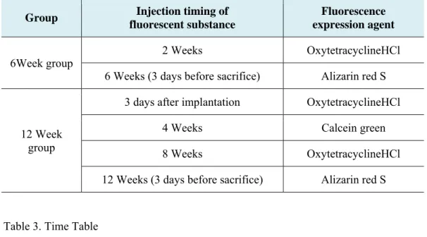
2.5 Fluorescence Analysis
Fluorescence expression agents were injected in specimen for observation under a fluorescent microscope. Oxy TC (oxytetracycline HCl; yellow; Pfizer, Seoul, Korea; 20mg/kg; iv) was injected at 3 days after implantation, Calcein green (calcein green, Sigma, St Louis, MO; 20mg/kg; iv) at 4 weeks later, oxytetracycline HCl at 8 weeks later and 3 days before sacrifice, Alizarin red S (Alizarin red S; Junsei Chemical, Tokyo,
16
Japan; 20mg/kg; iv) was injected. Topographic localization of the new bone formation and remodeling activity was analyzed in 2 weeks, 4 weeks, 8 weeks and 12 weeks. Their fluorescence microscopic image (DM LB, Leica, Germany) were taken at the wave range of 543nm ~ 617nm (red filter) and 515nm ~ 560nm (green filter) and Bone mineral apposition rate (BMAR) were measured.
Table 2. Fluorescence expression agent
Group Injection timing of
fluorescent substance
Fluorescence expression agent
6Week group 2 Weeks OxytetracyclineHCl
6 Weeks (3 days before sacrifice) Alizarin red S
12 Week group
3 days after implantation OxytetracyclineHCl
4 Weeks Calcein green
8 Weeks OxytetracyclineHCl
12 Weeks (3 days before sacrifice) Alizarin red S
Table 3. Time Table
Mineral apposition Total Bone Mass × 100 BMAR(%) =
17
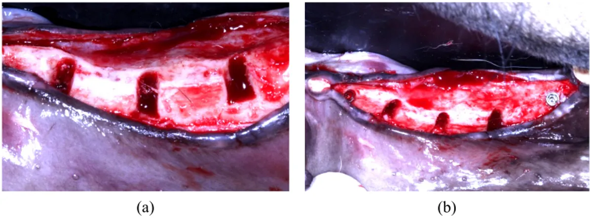
(a) (b)
Fig. 3. Formation of bone defects
(a) Dehiscence bony defect (buccal view) (b) Dehiscence bony defect (occlusal view)
(a) (b)
(c)
Fig. 4. Implantation and bone graft (a) Implant placement (buccal view) (b) Implant placement (occlusal view)
18
Fig. 5. Radiographic image after implant placement2.6 Specimen Preparation
A block including an implant was sectioned and was fixed with 10% neutral buffered formalin (pH7.0) for 2 weeks. After that, it was dehydrated by ethanol and embedded in methylmethacrylate (Technovit 720VLC, HeraeusKulzer, Dormagen, Germany). It was cut along the center axis of the implant and into bucco-lingual plane by a cutting system (Exakt 300, Kulzer, Norderstedt, Germany). The central section of each specimen was cut 15 ㎛ in thickness by a microgrinding system (Exak, Apparatebau, Norderstedt, Germanay). The sectioned specimens were dyed with Hematoxylin & Eosin (H&E). Dyed specimens were histologically analyzed under an optical microscope (Leica DM 2500, Leica Microsystems, Germany). And then, Image Pro Plus 4.5 program (Media Cybermetics, Silver Spring, USA) and SPOT (SPOT ver. 4.1, Diagnostic instrument Inc., Sterling heights, MI) were used to implement computer-assisted histometric measurement.
19
Measured site
1) Defect height
: a half of implant length 2) Bone regeneration height
: linear distance from the defect base to top of regenerate bone 3) Bone density
: percentage of mineralized bone at the base of defect in the same square (1mm X 1mm)
4) Bone-to-implant contact (BIC)
: percentage regenerated bone to implant contact 5) BMAR
: bone mineral apposition rate from the implant surface to 150 ㎛ region
2.7 Statistical Analysis
All data were expressed as mean±standard deviation. Data obtained from histological analysis were processed by SAS. Because the number of implants was not large, non-parametric analysis and non-parametric statistics analysis was implemented. The comparative statistics of groups were obtained through Kruskal Wallis test and ANOVA (A two-way analysis of variance) test. To know the significant differences by duration of treatment (6 week group versus 12 week group), Mann Whitney U test and independent two-sample t-test was used.
20
III. Results
3.1. Clinical observation
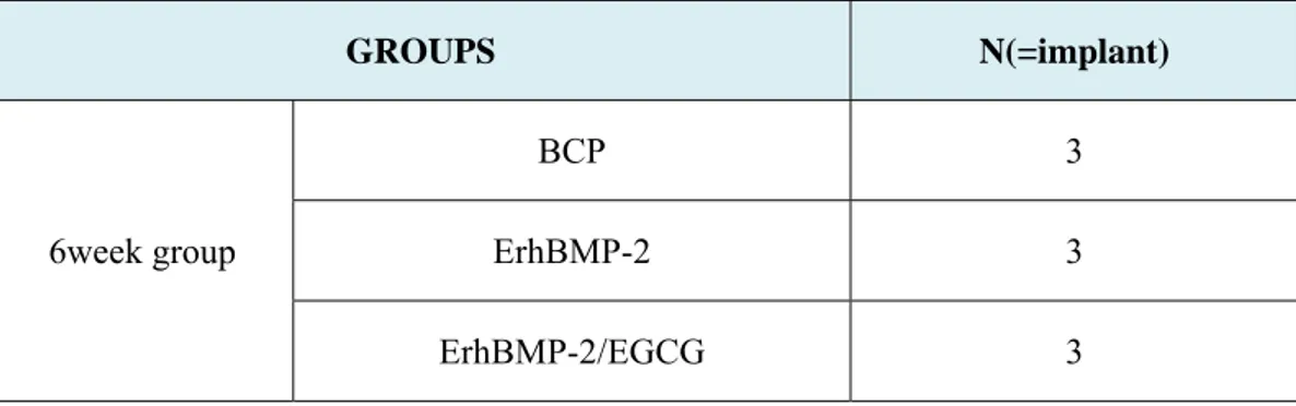
The five dogs were raised under the normal condition, and each showed unique shape of alveolar bone, gingival thickness, and intra-oral status. The clinical healing pattern in all samples was progressed without any singularity. There were no findings of inflammation or other complications on the operated site, and also a healthy tone and gingival shape was observed. However, membrane was exposed in some implants, in particular, in the entire 6 week group. 3 of 5 dogs appeared to have an intensive membrane exposure. No edema and acute inflammation response such as redness was not seen around the exposed membrane there for removal procedure of membrane was not carried out additionally.
Table 4. Number of implants where a membrane was exposed by group
GROUPS N(=implant)
6week group
BCP 3 ErhBMP-2 3 ErhBMP-2/EGCG 3
21
3.2. Histological observation
The shape of alveolar bone varied according to each dog, so different shape of new bone was formed on the buccal defect side. The shape of bone which formed in the buccal side was affected by membrane exposure. Thick connective tissue formed under the membrane and the upper side of new bone was collapsed where the membrane was exposed. In addition, many of the inflammatory cells infiltrated under the membrane and buccal grafting material was lost. It had an effect on the osseointegration of the lingual bone side.
(a) (b) (c)
(d) (e) (f) Fig. 6. The representative histological images (x 12.5)
(a) 12 week BCP group , (b) 12 week ErhBMP-2 group,( c) 12 week ErhBMP-2/EGCG group, (d) 6 week BCP group, (e) 6 week 2 group, (f) 6 week ErhBMP-2/EGCG group
22
Histological findings of the 6 week group
In the buccal side, new bone was forming actively at the periphery of the defect margin. Whereas in the 12 week group, newly formed bone had matured and the bone marrow had decreased compared with the 6 week group. The immature bone encircled the graft materials and new blood vessels formed actively in the bone marrow space.
x12.5
x50
x50
x100
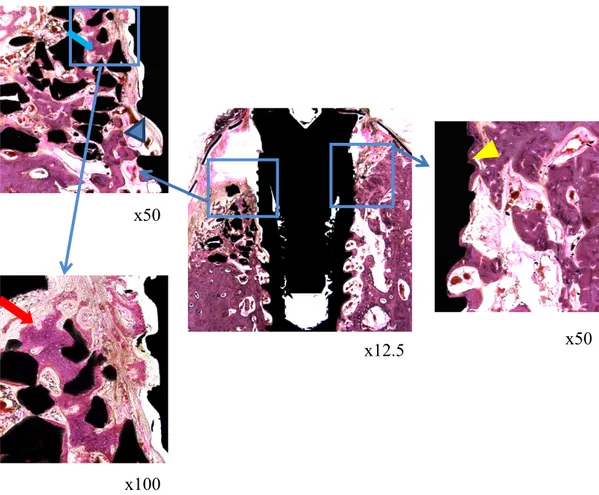
Fig. 7. Histological images of the 6 week BCP group
Appearance of new bone around the bone graft material (blue arrow). There are invasions of connective tissue between the implant and the boundary (blue arrow head). In the lingual side, direct bone contact was formed. (yellow arrow head). In the new bone, osteocyte in lacunae could be observed. (red arrow)
23
x50
x12.5
x50
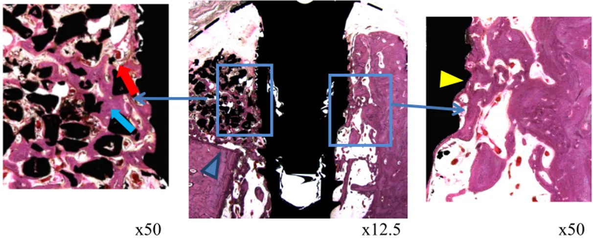
Fig. 8. Histological images of the 6 week ErhBMP-2 group
Compared to the BCP group, the buccal bone shows more bone formation around the implant (Blue arrow). Compared to other groups, higher blood vessel proliferation was shown. (red arrow) The clear boundary between new bone and existing bone.(Blue arrow head) In the lingual side, bone is contacting with implant uniformly
x50
x12.5
x50
Fig. 9. Histological images of the 6 week ErhBMP-2/EGCG group
Compared to the BCP group, the buccal bone shows more bone formation around the implant. (red arrow) Bone to implant contact was shown up to the top of implant in buccal side and active bone formation is underway in lingual side (blue arrow, yellow arrow head)
24
Histological findings of the 12 weeks group
New bones were maturated around grafting materials and the interface. The boundary of new bones and the existing bones was vague in the base of defects. Most of grafting materials came into direct contact with new bones. The particles of the margin were progressively absorbed and became smooth.
x50
x12.5
x50
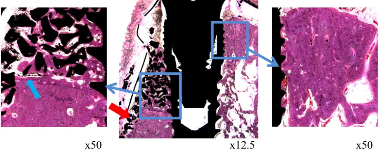
Fig. 10. Histological images of the 12 week BCP group
Characteristically, small particles being absorbed can be seen in the buccal side. (blue arrow) Compared to the 6 weeks, the margin of grafting materials became smooth (red arrow)
x50
x12.5
x50
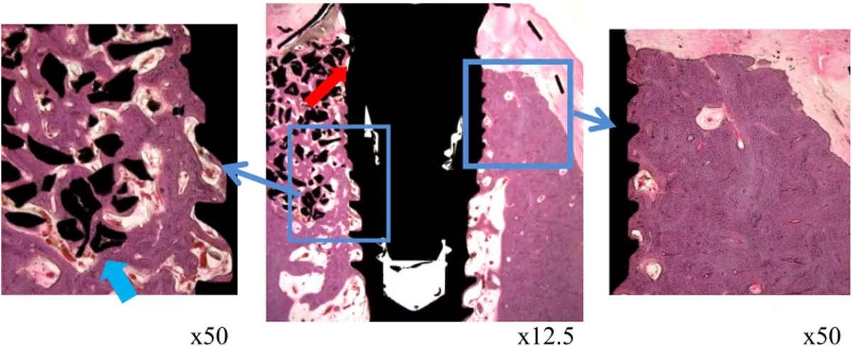
Fig. 11. Histological images of the 12 week ErhBMP-2 group
Although not originally intended, the graft material escapes from the membrane and forms new bone around it. It can be thought that the coated graft material has excellent bone formation ability. (red arrow) There is a clear boundary between the new bone and the defect.(Blue arrow)
25
x50
x12.5
x50
Fig. 12. Histological images of the 12 week ErhBMP-2 / EGCG group
The buccal bone shows superior bone formation and bone to implant contact from the base to the top of implant. (red arrow) It also shows the absorption of the graft material. Unlike the BMP group, vague boundary was shown between the defect and the new bone. (blue arrow)
26
3.3. Histometrical observation
In 7 specimens out of the 6 week group, the bone grafting materials did not remain. In 3 specimens out of the 12 week groups, the bone grafting materials did not remain.
After checking specimens, specimens were re-grouped and compared between groups in which the bone grafting materials still remained.
Bone to Implant Contact ( %)
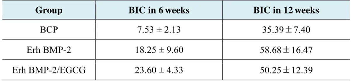
When comparing between groups having the bone grafting materials, BIC appeared to be higher in order of Erh BMP-2/EGCG Group, ErhBMP-2 group, and lastly the BCP group in 6 weeks.
When comparing groups having the bone grafting materials, the higher BIC was shown in order of the Erh BMP-2 group, the Erh BMP-2/EGCG group, and lastly the BCP group in 12 weeks.
Comparing by period, there were significant differences from the 6 weeks to the 12 weeks in BCP, Erh BMP-2, Erh BMP-2/EGCG group. Higher BIC value was showed in 12 week group. (p<0.05)
Table 5. Mean and standard deviation of BIC in the 6 and 12week group
Group BIC in 6 weeks BIC in 12weeks
BCP 7.53 ± 2.13 35.39±7.40
Erh BMP-2 18.25 ± 9.60 58.68±16.47
27
* : significantly different (p<0.05)Bone Density (%)
:
Percentage of mineralized bone at the base of defect in the same square (1mm X 1mm) In the 6 week group bone density increased in the order of the BCP group, the ErhBMP-2 group, and then the ErhBMP-2/EGCG group. In the 12 week group as well, the bone density increased in the order of the BCP group, the ErhBMP-2 group, and then the ErhBMP-2/EGCG group.Table 6. Mean and standard deviation of BD in the 6 and 12 week group
Group BD in 6 weeks BD in 12 weeks
BCP 9.48±8.99 (%) 25.37±14.31 (%) Erh BMP-2 37.60±20.33 (%) 36.63±10.9 (%) Erh BMP-2/EGCG 51.15±36.34 (%) 37.71±20.08 (%)
*
*
*
28
Bone regeneration height (mm)
: Linear distance between the defect base and the coronal level of regeneration bone
BRH appeared to be higher in order of Erh BMP-2/EGCG Group, Erh BMP-2 group, and lastly the BCP group in 6 weeks.
In 12 weeks, all three groups showed over 4mm of bone regeneration height.
Comparing by period, there were significant differences from the 6 weeks to the 12 weeks in BCP group. BCP group showed higher BRH value in 12 weeks with significant difference. (p<0.05)
29
Table 7. Mean and standard deviation of BRH in the 6 and 12 week group
Group BRH in 6 weeks BRH in 12 weeks
BCP 1.60±1.64(mm) 4.48±0.56(mm)
Erh BMP-2 3.60±1.86(mm) 4.23±0.64(mm)
Erh BMP-2/EGCG 4.63±0.54(mm) 4.86±0.10(mm)
* : significantly different (p<0.05)
Fluorescence analysis
Through fluorescence analysis, actively bone forming area was observed and the bone mineral apposition rate was measured.
Seen through the green filter (515nm~560nm wave length), Calcein green emitted green and under the red filter (450nm~490nm wavelength ) alizarin red S emitted red.
30
In the 12 week group the green emitting region showed the bone formation of the 4 weeks and the red emitting region showed the bone formation of the 12 weeks. In the 6 week group the red emitting region showed the bone formation of the 6 weeks.
In 4 weeks period bright fluorescence color appeared at the space between bone defect base and graft material; in 12 weeks period bright fluorescence color appeared at the boundary of bone grafting material and the interface of implant. Like the 4 weeks, in the 6weeks bright fluorescent color observed in the space between the base of the bone defect and graft material. Comparing the 4 weeks and the 12 weeks, there were more regions of bright fluorescent color in the 4 weeks group.
However, when comparing actively bone forming area around implant (from outer surface of implant to 150 ㎛ of it), ErhBMP-2/ EGCG group showed higher BMAR value in 4 weeks and the 12 weeks. There were no statistically significant differences.
(a) (b) (c)
Fig. 13. Fluorescent microscopic images in the 12 week group(a) Histological findings in the same region (b) Green emitting region (4 weeks) (c) Red emitting region (12weeks)
31
(a) (b) Fig. 14. Fluorescent microscopic images in the 6 week group
(a) Histological findings in the same region (b) Red emitting region(6 weeks)
Table 8. Mean and standard deviation of BMAR in the 4 and12 weeks
Group BMAR in 4 weeks BMAR in 12 weeks
BCP
5.39 ± 2.09 (%)
6.68 ± 2.82 (%)
Erh BMP-2
5.95 ± 2.76 (%)
6.38 ± 2.98 (%)
Erh BMP-2/EGCG9.03 ± 3.67 (%)
9.35 ± 5.09 (%)
32
IV. Discussion
Buccal bone loss occurs often due to periodontal disease or trauma. It can cause defects around the implant such as perforation or dehiscence, since the buccal bone is absorbed rapidly compared with the lingual bone. Insufficient bone volume threatens long-term prognosis of implant and increases implant placement incorrect area. Concerning about these problems, GBR procedure was tried and has become a successful procedure.
Among various bone grafting materials, biphasic calcium phosphate bone substitute (BCP) shows good results in cell experiments, animal experiments and clinical applications. BCP is composed with β-tricalcium phosphate (β-TCP) and Hydroxyapatite (HA). The OSTEONTM used in this experiment has HA : β- TCP ratio of 7 : 3 and pore size 300 to 500 ㎛, and it showed a good result in previous paper as a bone grafting material.14, 15 However, BCP has a fatal disadvantage in that it lacks osteogenetic potential. To complement the disadvantage of BCP, various surface treatments have been made to improve the bone formation.
In this experiment, BCP was coated with ErhBMP-2 and green tea extract (EGCG). Many studies were made about the influence of EGCG on bone formation. EGCG induces the apoptotic cell death of osteoclast and promotes the differentiation of multipotent mesenchyme stem cell to osteoblast which is increasing mineralized bone nodules and improving bone mineral density.29, 33 BMP, one of growth factors, promotes bone formation by inducing mesenchymal stem cell to differentiate into osteoblast. In previous studies, bone formation was enhanced when BMP was applied to bone defect.18,
33
34, 35 However, there were some anxieties about safety due to the adverse effect of BMP, when high concentration BMP was applied. Hence, restricted application of BMP at low concentration in clinics is recommend.
In this study, dehiscence defect was treated BCP coated with low concentration ErhBMP-2 and EGCG in combination and osteogenetic potential was evaluated in 6 and 12 weeks after implantation. The grafting material was used with the membrane which contributed to the formation and stabilization of blood clots and minimized the formation of empty space under the membrane. There are many reasons to form connective tissue under a membrane. Fine movement of 10~20 ㎛ makes mesenchymal cell differentiate into fibroblast.7, 36 In this study, the membrane was fixed with screws and caps to minimize fine movement. The fixing screw of the membrane not only prevents minute movement but also induces tight contact between the bone and the membrane. Therefor bone formation is enhanced by preventing adipose tissues and connective tissues from penetration into the space under the membrane. However, the structure of screws and caps, which project to top of the bone as much as 3 mm, makes soft issues hard to get primary closure.
To minimize the tension remaining inside soft tissues, releasing incision was given sufficiently. But there were membrane exposure and loss of bone grafting material in many specimens. In previous animal experiments wound dehiscence and membrane exposure were often reported in the GBR procedure.37, 38 In the study of Kim et al. the membrane condition of two groups – one is the group showing exposed membrane and the other is that showing unexposed membrane - were compared and they found that soft
34
tissues and organic film were inserted in the group where membrane was exposed. Hence, the exposed membrane influenced the volume and quality of newly formed bone underneath.39 In this study, the specimen with exposed membrane formed thick soft tissues under the membrane and most of bone grafting material was lost so that it was excluded from the histometrical analysis.
For the coating concentration of the materials used in the present study, the concentration that showed good results in existing literature was selected. In Park’s paper, bone formation was measured after adding EGCG of various concentrations 1μM, 5 μM, 25 μM and 50 μM. NIH3T3 cell line 3 X 10⁵ each were transplanted into a chalet of 50mm diameter and bone formation was induced by adding vitamin C and EGCG. At the concentration of 25 μM of EGCG, bone nodules formed the most. On the basis of this experiment, EGCG of 25 μM concentration was coated on BCP. For the ErhBMP-2 the concentration of 0.05mg/ml was used. When the high concentration BMP was used, several side effects of BMP-2 including formation of bone at unwanted site and swelling of soft tissue have been reported.40, 41 Wikesjö UM et al. compared regeneration ratio by applying rhBMP-2 of three different concentrations 0.05, 0.1 and 0.2 mg/ml to periodontal defect lesion. The result showed that there is no difference among the groups with the regeneration ratios 86, 96 and 88%.42 Hence, the lowest concentration 0.05mg/ml was selected to avoid the adverse effect from high concentration rhBMP-2, while it can produce optimum effect.
When osteoinductive protein was applied to defect areas with bone grafting material, various application methods were considered to make it continuously work at the local site. In previous studies, BCP was used often after being wetted in BMP solution.
35
Excellent results in bone formation were shown in this method.22, 43 However, it has disadvantages in handling the grafting material, applying accurate dose and controlling flow. To complement such disadvantages, coating method of ErhBMP-2 and EGCG on BCP was taken. After surface coating, it was progressively freeze-dried from -40℃ to 20℃(lyophilization protocol) that makes storage and operation easier.
There are several different opinions on sterilizing methods for the graft material. In order to minimize the degeneration of the substance coated on graft material, gas sterilization at low concentration for a long time was performed. It was reported that the sterilizing method influenced the osteoinductivity of BMP that was coated on graft material. Munting et al. reported that when ethylene oxide was used, it lowered osteoinductivity of BMP and gamma irradiation was less harmful.44Ijiri S et al. reported that when BMP was sterilized at low concentration for a long time with ethylene oxide, it influenced osteoinductivity of BMP less.45
In the previous study, various healing time was selected after defect model formation. Compared with human bone, bone remodeling duration is shorter in dogs. 6 weeks, 12 weeks and 17 weeks is needed to bone remodeling in rabbits, dogs and human respectively.46 In the paper of Kim et al., it was reported that most of the healing was made within 8 weeks when GBR was done at surface modified implant.15 Schwarz et al analyzed histological findings for each period after GBR procedure. At 4 weeks, new bone was actively formed from the defect base to the top of the implant. At 6 weeks, grafting material was stabilized and bone filling area increased very much.47 In this experiment 2 healing times were selected and bone formation processes for each group
36
were compared; one is 12 week group at that time sufficient healing was done, and the other is 6 week group at that time bone was forming actively.
In the 6 week group, BCP, ErhBMP-2 and ErhBMP-2/EGCG group showed higher value in order when the value of BIC, BV and BRH were compared. Effects of ErhBMP-2/EGCG were remarkable at the early bone healing process. Since the sample size was small, there was no significant difference in non-parametric statistical analysis among the three groups. Comparing by period, BIC, BRH and BMAR values increased by time in the three groups. BIC value in 12 week group was shown higher with significant difference.
It is known that EGCG improves osteoblastic activity and suppresses osteoclastic activity and that it influences bone metabolism. In the previous studies it was suggested that bone formation increased by increasing the number of osteoblast and by enhancing the activity of osteoblast at the early stage of bone formation. Choi EM et al. reported that suppressing the formation of IL-6 and TNF-αincreased the survival of osteoblast.48 Chen et al reported that bone mineralization was enhanced through Runx2-mediated mechanism,31 Mount et al reported that osteoblastic activity can be improved through Wnt signaling.49 And it was reported that EGCG activated osteoblastogensis through vascular endothelial growth factor-mediated mechanism.50, 51 Luo T et al reported that, when BMP-2 and VEGF(vascular endothelial growth factor) were applied around the implant, more bone formation was observed.52 Hence, it is thought that by enhancing VEGF mediate mechanism, EGCG can contribute to increase bone formation.
37
The osteogenesis effect of EGCG shown at in vitro experiments appeared also at in vivo experiments in a similar manner, and in this experiment ErhBMP-2/EGCG showed high measured value.
In the 6 week group, EGCG effect was remarkable, and it seems that it is related with the release kinetics of BMP and EGCG. Tazaki Jet al coated 125I-labeled BMP-2 on hydroxyapatite and measured the remaining BMP-2 in rats. One day after grafting BMP-2 remained 34%, and 3 weeks it remained 3% only.53 Xu X et al used hyaluronic acid-based hydrogel particles as a carrier at their in vitro study and found that most of BMP-2 was released within 13 days.54 Rodriguez R et al applied EGCG to aα-TCP and measured with UV-visible spectrophotometer. It was reported that initial 40% of EGCG was released within 24 hours and rest of EGCG was released progressively for next two weeks.55 Because most of rhBMP-2 and EGCG were released in the initial stage, it is thought that there was more osteoinductive effect in the 6 week group.
BIC of the 6 week group and that of the 12 week group was compared. The BIC in 12 week was higher than 6 week with significant differences. (p<0.05) It coincides with other studies that reported the increase of BIC according to healing time. Yan MN et al measured BIC around implant at 6 weeks and 12 weeks after bone defect formation and it increased from 10% to 50%.35 In the experiment of Sykaras N et al. the BIC in rhBMP-2 treated group was measured at 8 weeks and 12 weeks and it increased from 18.65% to 43.78%.56
In fluorescence analysis, the bone mineral apposition pattern was compared for various periods. In the 4 week group, bright fluorescent color appeared at the space between bone
38
defect base and bone grafting material; in the 12 week group, bright fluorescent color appeared at the boundary of bone grafting material and the interface of implant. When the 4 week group is compared with the 12 week group, the area showing fluorescent color appeared more in the 4 week group. It shows that the bone formation was more active in the 4 week group. In the 12 week group, the resorption of grafting material and were more active than new bone formation. Continuous bone remodeling around the implant showed still at 12 weeks, which is coincides with the result that the value of BIC in 12 week group is higher than that of 6 weeks group.
In this study, osteogenic potential was compared to confirm the effectiveness of the combination of low concentration ErhBMP-2 and EGCG by histological analysis, fluorescence analysis, histometric analysis (BIC, BV and BRH). The surface modification of BCP with low-concentrate BMP and EGCG showed better result than other group. Consequently, ErhBMP-2/EGCG group was more effective in enhancing the osteogenetic potential around dental implant.
As mentioned before, because there was much bone leakage due to the limit in experiment design, the sample size was reduced. The total sample size was too small. So the result changed quite a bit according to oral cavity condition or bone quality of the individual dog, it was hard to get statistically significant data between the groups of the experiment. However, results with a significant difference are expected to be obtained, when the number of specimen is increased sufficiently. Circumferential bone defect design is more affective to evaluate bone forming ability and the difference between the groups clearly, because this design is influenced less by other elements.
39
Until now the studies on EGCG have been made mostly in vitro. However, vivo studies are rare. Additional study is needed to evaluate of proper concentration, releasing kinetics and the synergy effect when it is used together with ErhBMP-2 and EGCG in vivo.
40
V. Conclusion
In this study, osteogenic potential was compared to confirm the effectiveness of the combination of low concentration ErhBMP-2 and EGCG by histological analysis, fluorescence analysis and histometric analysis (BIC, BD and BRH). Within the limitation of this study, the results were obtained as follows.
1. When comparing between the three groups, ErhBMP-2/EGCG group appeared to have higher value in bone to implant contact (BIC), bone density (BD), bone regeneration height (BRH) and bone mineral apposition rate (BMAR). But significant difference was not found.
2. BIC, BRH and BMAR values increased when comparing by time in the three groups.
- BCP, ErhBMP-2 and ErhBMP-2/EGCG groups showed higher BIC value in the 12 week group than the 6 week group with significant differences.(p<0.05) - BCP group showed higher BRH value in 12 weeks with significant
difference.(p<0.05)
3. In fluorescence analysis, bone remodeling around graft material and implant was more active in the ErhBMP-2/EGCG group.
Consequently, the surface modification of BCP with low-concentrate BMP and EGCG was effective in enhancing the osteogenetic potential.