The relationship between rheumatoid
arthritis and periodontal disease
Chuan-Xin Su
Department of Medical Science
The relationship between rheumatoid
arthritis and periodontal disease
Chuan-Xin Su
Department of Medical Science
The relationship between rheumatoid
arthritis and periodontal disease
Directed by Professor Soo-Kon Lee
The Master’s Thesis
submitted to the Department of Medical Science,
the Graduate School of Yonsei University
in partial fulfillment of the requirements for the
degree of Master of Medical Science
Chuan-Xin Su
This certifies that the Master’s Thesis of
Chuan-Xin Su is approved.
The Graduate School
Yonsei University
Acknowledgements
The time of my master course has elapsed shortly soon and it’s
time to complete my course. Although it’s not so long period of my
life to study about research, I have had much more knowledge and
experience about research as other people. On future reflection it
seems that I have not realized my various thought for my master
course, but I’m so happy to meet many nice persons. All of them
helped me to improve my studies and I’m so grateful to their help.
First of all, I thank my professor, Soo-Kon Lee who is my
adviser and aids about experimental studies. I always thank him and
give all of the best regards to him. And professor Yong-Beom Park,
he has supported for research in silence and created a pleasant
atmosphere, I’m also thankful for his kindness. When I faced with
obstacle about dental, professor Chang-Sung Kim and his team from
dental hospital rendered assistance to me pleasantly, I want to
express my gratitude to him.
I appreciate all of my laboratory colleagues who have got
together. First of all, I really thank for our oldest member, Ji-Hye
Kim, who helped me a lot as my really elder sister. I also would like
to thank for You-Eun Song, although she only stayed in our lab half
a year. She helped me so much about research and other things. Dr.
Sang-won Lee also gave me much help. Although he is a doctor in
our hospital, he gave me many good advices.
Finally, I would like to thank my family, for their
encouragement, understanding, support and love, and without them I
would not have this dissertation.
TABLE OF CONTENTS
ABSTRACT ... 1
Ⅰ. INTRODUCTION ... 5
Ⅱ. MATERIALS AND METHODS ... 8
1.Induction of collagen induced arthritis ... 8
2.Assessment of arthritis severity ... 9
3.Histopathological and immunohistochemical study ... 10
4.Micro-CT imaging ... 11
5.Statistical analysis ... 12
Ⅲ. RESULTS ... 13
1.The severity of arthritis in mice ... 13
2.The histopathological finding and the immunohistochemical staining
for TNF-
α, IL-1ß and iNOS in joints ... 15
3.Micro-CT findings ... 18
4.The histopathological finding and the immunohistochemical staining
for TNF-
α, IL-1ß and iNOS in jaws ... 20
Ⅳ. DISCUSSION ... 24
Ⅴ. CONCLUSION... 29
REFERENCES ... 30
LIST OF FIGURES
Figure 1. The severity of arthritis in mice. ... 14
Figure 2. Histopathological finding and immunohistochemical
stain-ing for TNF-
α, IL-1ß and iNOS in joint tissues ... 16
Figure 3. The scores of histopathological finding and
immunohisto-chemical staining ... 17
Figure 5. Micro-CT measurement results proved the severity of PD
in mice ... 19
Figure 6. Histopathological finding and immunohistochemical
stain-ing for TNF-
α, IL-1ß and iNOS in periodontal tissue ... 21
Figure 7. The scores of histopathological finding and
ABSTRACT
The relationship between rheumatoid arthritis and
periodontal disease
Chuan-Xin Su
Department of Medical Science
The Graduate School, Yonsei University
(Directed by Professor Soo-Kon Lee)
Background: Peridontitis has many similar characteristics of synovitis in
rheumatoid arthritis (RA). Recent reports have suggested a significant relationship
between RA and periodontal disease (PD) in RA patients. The aim of this study was to
in RA and PD in animal model.
Method: Twenty-five male DBA/1 mice 8 weeks old were divided into five
groups. Group 1 (CONTROL) and Group 2 (CONTROL+TNFi) were the control
groups without collagen injection. The other three groups which were experimental
groups were given a subcutaneous injection of bovine type II collagen (100μg/mice)
emulsified in complete Freund’s adjuvant (Arthrogen-CIA, Redmond, WA) (1:1, w/v)
into the base of tail. Three weeks later, the mice were given a booster subcutaneous
injection of bovine type II collagen (100μg/mice) in incomplete Freund’s adjuvant (1:1,
v/v) into the base of tail. After the development of arthritis (2 weeks after the booster
collagen injection), Group 3 (CIA+TNFi) was given the anti-TNF-α (5.5mg/kg), and
the Group 4 (CIA+MTX) was given the intraperitoneal injection of methotrexate
(MTX) (35mg/kg) twice a week for five weeks. At the same time the Group 5 (CIA)
injected with anti-TNF-α. Arthritis score and paw thickness were measured and
histopathological assessment of joint sections was performed. The expression of
proinflammatory cytokines and enzymes was evaluated by immunohistochemical
staining. Jaws were scanned and reconstructed into a 2 and 3-dimensional structure
with micro-CT. The average exposure areas of teeth were measured to calculate the
severity of PD.
Results: The anti-TNF-α therapy and MTX attenuated the severity of arthritis
and histopathological findings in CIA mice. Micro CT confirmed that the periodontal
disease existed in CIA mice. There were significant differences in the average area
measurements (CONTROL 0.0177 vs CONTROL+TNFi 0.0229 CIA 0.02205 p<0.05)
between Group 1 and Group 2 or Group 5. The average exposed area was significantly
lower in Group 5 than in Group 3 or Group 4 (CIA 0.02205 vs CIA+TNFi 0.02466,
Conclusions: Collagen induced arthritis was associated with the occurrence of
periodontitis. The MTX/TNFi drugs didn’t affect PD while they treated CIA. Further
The relationship between rheumatoid arthritis and
periodontal disease
Chuan-Xin Su
Department of Medical Science
The Graduate School, Yonsei University
(Directed by Professor Soo-Kon Lee)
Ⅰ. INTRODUCTION
Rheumatoid arthritis (RA) is a chronic, systemic inflammatory disorder that may
affect many tissues and organs, but principally attacks the joint architecture and
pericardium, pleura, and sclera, and also nodular lesions, most common in
subcutaneous tissue under the skin. Environmental factors such as age, gender,
smoking as well infections play a role in the pathogenesis of RA3.Although bacterial
infections and oral pathogens have been related to the pathogenesis of RA4-8, the
involved pathologic mechanisms are unknown; autoimmunity plays a pivotal role in its
chronicity and progression.
Periodontal disease (PD) refers to a number of inflammatory diseases affecting
the tissues that surround and support the teeth. PD is caused by the bacteria which
adhere to and grow on the tooth's surfaces. A diagnosis of periodontitis is established
by inspecting the soft gum tissues around the teeth with a probe and radiographs by
visual analysis, to determine the amount of bone loss around the teeth. Periodontal
ligament (PDL) resides between the cementum of the roots of the teeth and the alveolar
integrity of the PDL. PDL-derived cells produce various inflammatory mediators to
recognize the somatic components including lipopolysaccharide (LPS) 9.
The oral cavity harbours a large reservoir of pathogenic organisms that may
contribute to chronic bacteremia and potential damage of distant organs, including the
joints10. Also, there is evidence of a specific antibody response against anaerobic oral
bacteria as well as the presence of oral bacterial DNA in the synovial fluid of RA
patients 11.
The purpose of the study was to illustrate the relationship between RA and PD. I
hypothesized that CIA was a risk factor for the development of PD. The hypothesis
was that if we treat collagen induced arthritis with drugs such as methotrexate or
Ⅱ. MATERIALS AND METHODS
1. Induction of collagen induced arthritis
All animals were treated in accordance with the guidelines and regulations for
the use and care of animals of Yonsei University, Seoul, Korea. Firstly, twenty five
male DBA/1 mice at 8 weeks of age (SLC, Shizoka, Japan) were divided into five
groups. Group 1 (CONTROL) and Group 2 (CONTROL+TNFi) were the control
groups without collagen injection. The other three groups which were experimental
groups were given an subcutaneous injection of bovine type II collagen(100μg/mice)
emulsified in complete Freund’s adjuvant (Arthrogen-CIA, Redmond, WA) (1:1, w/v)
to the base of the tail. Three weeks later, the mice were given a booster subcutaneous
injection of bovine type II collagen (100μg/mice) in incomplete Freund’s adjuvant (1:1,
collagen injection), Group 3 (CIA+TNFi) was given the anti-TNF-α (5.5mg/kg twice
a week, Wyeth, Pfizer Inc). The Group 4 (CIA+MTX) was given the intraperitoneal
injection of methotrexate (MTX) (35mg/kg, Yuhan Co. Ltd) twice a week for five
weeks. At the same time the Group 5 (CIA) and the Group 1 (CONTROL) were
untreated while Group 2 (CONTROL+TNFi) which was normal was treated by anti-
TNF-α.
2. Assessment of arthritis severity
Mice were observed twice a week for 70 days after primary immunization.
Arthritis severity was evaluated by visual inspection. All 4 legs of the mice were
evaluated and scored from 0 to 4 according to the following scale: 0 = no signs of
arthritis, 1= swelling and/or redness of the paw or 1 digit, 2 = 2 joints involved, 3 =
Paw thickness was measured with a Vernier caliper. Arthritis scoring and paw
thickness measurement were performed by 2 independent observers.
3. Histopathological and immunohistochemical study
Mice were anesthetized and sacrificed on day 70, and joints and jaws were
removed for histopathological examination after routine fixation, decalcification, and
paraffin embedding of tissue. Tissue sections were prepared and stained with
hematoxylin and eosin (H&E). Sections were sequentially incubated with specific
antibodies directed against murine TNF-α (Hycult Biotechnology, Uden, The
Netherlands), IL-1β, and iNOS, (Santa Cruz Biotechnology, Santa Cruz, CA) followed
by the appropriate secondary antibodies (ISU Abxis, Seoul, Korea). All tissue samples
were counterstained with hematoxylin. After immunohistochemical staining,
expression of the different markers in the periodontal tissue of paw and knee joints was
pathologists, and the average of their scores were calculated. A score of 0 represented
minimal expression, 1 represented mild expression, and 2 represented moderate
expression whereas 3 represented abundant expression of a marker. Minor differences
between observers were resolved by mutual agreement.
4. Micro-CT imaging
Mice were observed twice a week for 70 days after primary collagen injection,
euthanized, and their jaws were excised and fixed in 4% formalin for 2 days. Left
mandibules were analyzed by Ultrasound Computerized Tomography (UCT) (skysan;
SCANCO USA, Inc.)12. Using 2 and 3-dimenional image reconstructions, the
cementoenamel junction (CEJ) and marginal bone crest (MBC) were manually drawn.
Alveolar bone resorption was determined as the distance and exposed root surface area
5. Statistical analysis
All statistical analyses were conducted using SPSS package for Windows,
version 17 (SPSS Inc., Chicago, IL). The representative values were the means of those
obtained from CIA mice, and all values in the experimental groups were compared to
controls. All results and measurements were expressed as the mean ± standard
deviation. Statistical comparisons between the 2 groups were evaluated by
Mann-Whitney U test and t-test, and correlations between parameters measured by micro-CT
were calculated using Spearman’s correlation coefficient. The level of significance was
Ⅲ. RESULTS
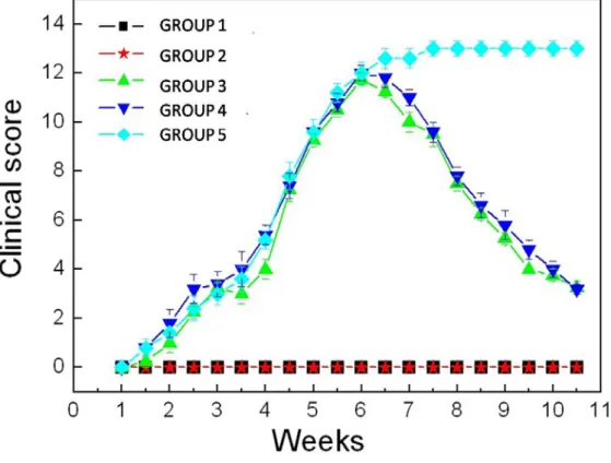
1. The severity of arthritis in mice
Group 3, 4, 5 showed definite evidence of arthritis such as redness and swelling
in the joints after collagen injected. After the treatment of TNFi and MTX, arthritis
score improved significantly in Group 3 and Group 4, although Group 5 showed
Figure 1. The severity of arthritis in mice. CIA mice model were treated by collagen at
the 1st week and the 4th week. As the developing of the disease, CONTROL+TNFi,
CIA+TNFi and CIA+MTX groups were treated by TNFi and MTX with others
untreated at 6th week. The improvement of arthritis was remarkable in the treated CIA
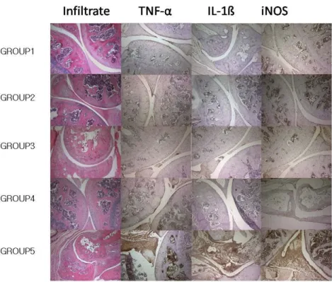
2. The histopathological finding and the immunohistochemical staining for TNF-α, IL-1ß and iNOS in joints
The histopahtological evaluation showed that the joint sections were not
inflamed in Group 1, Group 2, Group 3 and Group 4 which showed the intact bone
structure. However, the severe inflammations were found in Group 5.
Immnunohistochemical analysis of paws and knee joints tissues obtained from Group 5
exhibited markedly positive staining for TNF-α, IL-1ß and iNOS. However, TNF-α,
IL-1ß and iNOS were not expressed in treated groups even Group 2 and Group 1
Figure 2. Histopathological finding and immunohistochemical staining for TNF-α,
IL-1ß and iNOS in joint tissues. Histopathological evaluation revealed severe
inflammation in the joint sections of untreated CIA mice. In contrast, the extent of
arthritis and bone destruction was significantly reduced in the joints of mice be treated.
TNF-α, IL-1β and iNOS were showed markedly positive in CIA group. In
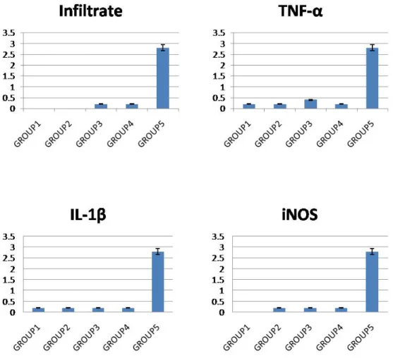
Figure 3. The scores of histopathological finding and immunohistochemical staining.
When we compared the values between the untreated and each treated group, cytokines
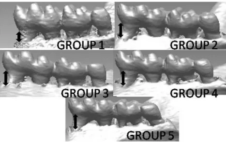
3. Micro-CT findings
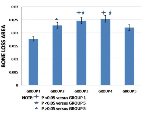
CEJ-bone crest distance was measured for the severity of periodontal disease
and the extent of the periodontal tissue breakdown was compared among the groups to
investigate the difference. The area of exposed root surface and the length of exposed
root (mesial and distal aspect) were measured by image program (Figure 4 and 5).
Figure 4. Micro-CT imaging pictures. The distance between the cementoenamel
Figure 5. Micro-CT measurement results proved the severity of PD in mice. It was easy
to see that the mice in Group 1 were normal than other groups. Even the mice in Group
2, Group 3 and Group 4 also got PD. And the Group 3 and Group 4 showed heavier PD
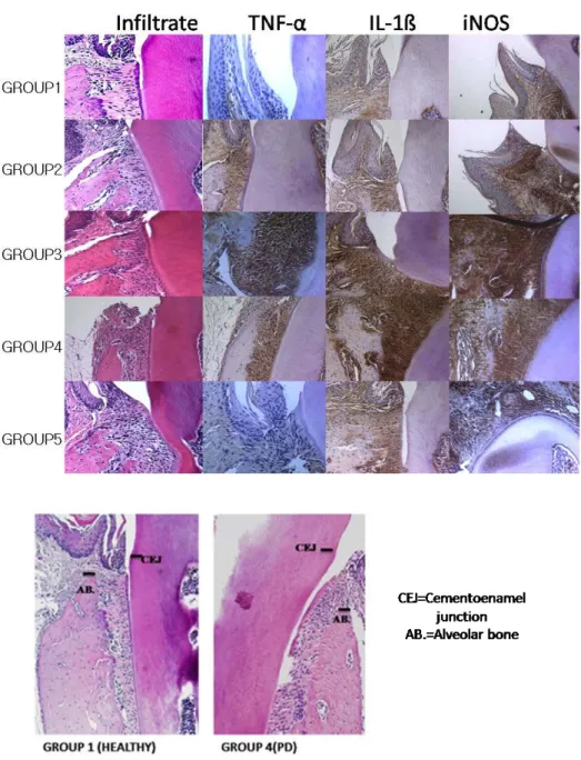
4. The histopathological finding and the immunohistochemical staining for TNF-α, IL-1ß and iNOS in jaws
All groups without CONTROL group showed the damage to the alveolar bone.
The epithelium became spongiotic and hyperplastic and the underlying stroma was
densely infiltrated with lymphocytes and plasma cells. Immnuno-histochemical
analysis of jaws obtained from CIA group exhibited markedly positive staining for
TNF-α, IL-1ß and iNOS. However, TNF-α, IL-1ß and iNOS were also expressed in
treated groups even CONTROL+TNFi group in periodontal tissues. There was no
IL-was thin cell layer with some rete ridge extending into the underlying gingival fibrous
tissue. However, in Group 5(CIA) and other groups, detachment of gingiva from tooth
surface and downward displace of epithelial attachment. The levels of TNF- α, IL- 1β
and iNOS in the periodontal tissues were significantly higher than that in Group 1. The
healthy tissue compared with the inflammated periodontal tissue showed that
detachment of gingival from tooth surface and downward displacement of epithelial
attachment. Gums separated from the teeth, forming pockets. Gum tissue and bone
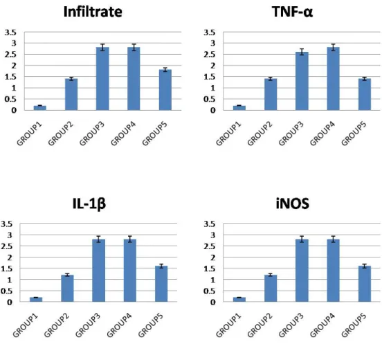
Figure 7. The scores of histopathological finding and immunohistochemical staining.
When we compared the values among the groups, cytokines were markedly positive in
CONTROL+TNFi group, CIA+TNFi group, CIA+MTX group and CIA group.
Ⅳ. DISCUSSION
In this study, I found that CIA was associated with the occurrence of periodontal
disease. The anti-inflammatory drugs didn’t affect on PD while they treated CIA. PD
was getting worse by the treatment of MTX and anti-TNF-αin CIA group while CIA
was improved by it. The clinical score and the histopathological and
immunohistochemical study for mice showed the arthritis was improved by the
treatment of TNFi/MTX while the CONTROL group, CONTROL+TNFi group and
CIA group stable. The results of histopathological and immunohistochemical study and
the Micro-CT image showed the PD existed in all groups except the CONTROL group.
There were significant differences in the average area measurements (CONTROL
0.0177 vs CIA 0.02205 CONTROL+TNFi 0.0229 p<0.05) between CONTROL group
CIA+TNFi 0.02466, CIA+MTX 0.0254, p<0.05).
TNF-α and IL-1β were all potent inducers of the production of acute phase
reactants by hepatocytes and thereby mediate the acute phase response seen in CIA and
PD. INOS, which was mainly associated with the periodontal inflammation13, enabled
its participation in anti-microbial and anti-tumor activities in order to take part in the
oxidative burst of macrophages. I found that the clinical score of mice models showed
that the mice in CONTROL group and CONTROL+TNFi group were normal while the
mice in CIA+TNFi group and CIA+MTX group got arthritis and be healing after
treated and the mice in CIA group were abnormal. All mice in CIA group got RA
while others didn’t. And the immuohistochemical results of joints tissue also proved
TNF-α, IL-1β and iNOS were significantly positive in CIA group. However, the
immuohistochemical results of jaws tissue proved that the TNF-α , IL-1β and iNOS
positive in CIA+TNFi group and CIA+MTX group.
MTX was introduced more than 50 years ago to treat cancer because of its
proliferative effects. Since the early 1980s, it has become the disease-modifying
anti-rheumatic drug (DMARD) of choice in the treatment of RA and was used in many
other rheumatic diseases as well. It was known that MTX has a side effect of bone
marrow suppression. There were also ulcerative mucositis, decreased hepatic and renal
function as side effects of MTX. However, according to the recent research, MTX
caused progressed periodontitis because of the chronic long-term neutropenia and the
change of microflora14,15. And the dose of MTX in our experiment was high. We were
not sure whether the low dose MTX also caused the serious periodontitis. That might
explain the progressed PD in CIA+MTX group but could not explain the severity of
periodontitis.
role on TNF-αof the inflammatory response in many organ systems. However, the
paper showed that anti-TNF-α make excessive bone resorption resulting in bone
loss16,17. In my research, it was clear that the TNF-α had significantly expression in
the groups which were being treated by TNFi not only the CIA+TNFi group but also
CONTROL+TNFi group in periodontal tissues while they were normal in the same
group of joint tissues. From the results of Micro-CT, it was easily to see that the
exposure area was the largest in CIA+TNFi group and CIA+MTX group. And it was
lager in CONTROL+TNFi group and CIA group than in CONTROL group.
There were many researchers hypothesized the relationship between RA and
PD18-20, and there were some researchers who test the association between them using
clinical patients 21-25. They demonstrate that RA may be associated with PD.
Despite I proved there was association between CIA and PD, I still need further
progress in CIA models. Mice might be observed at the regular intervals for the effect
of the drugs in periodontal tissues.
In conclusion, based on the findings of the present study, a role of RA in the
progression of periodontal disease was proposed. However, the drug’s affection was
not proposed. Further, studies were needed to confirm the findings of our study in the
Ⅴ. CONCLUSION
CIA associated with the occurrence of PD. The DMARD didn’t improved PD
while they treated CIA. Further studies will be necessary to investigate the cause of
REFERENCES
1. Fox DA, Etiology and pathogenesis of rheumatoid arthritis. In: Koopman WJ.
Arthritis and allied conditions. A textbook of rheumatology. 1st ed. Lippincott,
Williams & Williams, Baltimore; 2001. p. 1085-102.
2. Lee DM, Weinblatt ME. Rheumatoid arthritis. Lancet 2001; 15 358(9285): 903-11.
3. Alamanos Y, Drosos A A. Epidemiology of adult rheumatoid arthritis. Autoimmun
Rev 2005; 4: 130-6.
4. Cai XY, Yang C, Zhang ZY, Qiu WL, Ha Q, Zhu M. A murine model for septic
arthritis of the temporomandibular joint. J Oral Maxillofac Surg 2008; 66(5): 864-9.
5. Smith RL, Merchant TC, Schurman DJ. In vitro cartilage degradation by Escherichia
coli and Staphylococcus aureus. Arthritis Rheum 1982; 25(4): 441-6.
7. Rosenstein ED, Greenwald RA, Kushner LJ, Weissmann G. Hypothesis: The
humoral immune response to oral bacteria provides a stimulus for the development
of rheumatoid arthritis. Inflammation 2004; 28(6): 311-8.
8. Hara Y, Kaneko T, Yoshimura A, Kato I. Serum rheumatoid factor induced by
intraperitoneal administration of periodontopathic bacterial lipopolysaccharide in
mice. J Periodont Res 1996; 31(7): 502-7.
9. Kasamatsu A, Uzawa K, Shimada K, Shiiba M, Otsuka Y, Seki N, et al. Elevation of
galectin-9 as an inflammatory response in the periodontal ligament cells exposed to
Porphylomonas gingivalis lipopolysaccharide in vitro and in vivo. Int J Biochem
Cell Biol 2005; 37(2): 397-408.
10. LaPorte DM, Waldman BJ, Mont MA, Hungerford DS. Infections associated
with dental procedures in total hip arthroplasty. J Bone Joint Surg Br 1999; 81(4):
11. Trombone AP, Claudino M, Colavite P, de Assis GF, Avila-Campos MJ, Silva
JS, et al. Periodontitis and arthritis interaction in mice involves a shared
hyper-inflammatory genotype and functional immunological interferences. Genes Immun
2010; 29. [Epub ahead of print]
12. Waarsing JH, Day JS, Weinans H. An improved segmentation method for in
vivo microCT imaging. J Bone Miner Res 2004; 19(10): 1640-50.
13. Popkov VL, Fil'chukova IA, Lapina NV, Galenko-Yaroshevskii VP, Dukhanin
AS. Activity of nitric oxide synthase and concentration of nitric oxide end
metabolites in the gingiva under experimental pathological conditions. Bull Exp Biol
Med 2005; 140(4): 391-3.
14. Yoshinari N, Kameyama Y. Aoyama Y. Effect of long-term
methotrexate-induced neutropenia on experimental periodontal lesion in rats. J Periodontal Res
15. Wu JZ. Chemosensitivity testing of adenocystic tongue and gingival cancer
cell lines. Zhonghua Kou Qiang Yi Xue Za Zhi 1992; 27(2): 107-8, 28.
16. Lerner UH. Inflammation-induced bone remodeling in periodontal disease and
the influence of post-menopausal osteoporosis. J Dent Res 2006; 85(7): 596-607.
17. Pers JO, Saraux A, Pierre R, Youinou P. Anti-TNF-α Immunotherpy is
associated with increased gingival inflammation without clinical attachment loss in
subjects with rheumatoid arthritis. J Periodontal 2008; 79(9): 1645-51.
18. Liao F, Li Z, Wang Y, Shi B, Gong Z, Cheng X. Porphyromonas gingivalis
may play an important role in the pathogenesis of periodontitis-associated
rheumatoid arthritis. Med Hypotheses 2009; 72(6): 732-5.
19. Mercado F, Marshall RI, Klestov AC, Bartold PM. Is there a relationship
between rheumatoid arthritis and periodontal disease? J Clin Periodontol 2000;
20. Zhang DZ, Zhong DY, Deng J, Wang JB. Relationship between periodontal
disease and rheumatoid arthritis. Hua Xi Kou Qiang Yi Xue Za Zhi 2005; 23(6):
498-501.
21. De Pablo P, Dietrich T, McAlindon TE. Association of periodontal disease and
tooth loss with rheumatoid arthritis in the US population. J Rheumatol 2008; 35(1):
70-6.
22. Dissick A, Redman RS, Jones M, Rangan BV, Reimold A, Griffiths GR, ET
AL. Association of periodontitis with rheumatoid arthritis: a pilot study. J
Periodontol 2010; 81(2): 223-30.
23. Laurell L, Hugoson A, Hakansson J, Pettersson B, Sjostrom L, Berglof FE, et
al. General oral status in adults with rheumatoid arthritis. Community Dent Oral
Epidemiol 1989; 17(5): 230-3.
adults with rheumatoid arthritis. Community Dent Oral Epidemiol 1989; 17(5):
234-6.
25. Kobayashi T, Murasawa A, Komatsu Y, Yokoyama T, Ishida K, Abe A, et al.
Serum cytokine and periodontal profiles in relation to disease activity of rheumatoid
ABSTRACT (IN KOREAN)
류마티스 관절염과 치주질환의 관계
<지도교수: 이수곤>
연세대학교 대학원 의과학과
소전신
배경:치주질환
(PD)은 류마티스 관절염(RA)의 활막염과 유사한 특성을 가지고 있다. 최근 보고에서는 관절염환자에서 류마티스 관절염과 치주질환은 중요한 관계를 가지고 있다고 하지만 collagen-induced arthritis 모델에서의 치주질환에 대한 연구는 아직 없는것으로 알려져 있다. 이 연구의 목적은 RA 와 PD 의 관계를 밝히고 동물 모델에서 RA 와 PD 의치료 반응을 관찰 하는 것이다.
방법: 8 주된 수컷, 25 마리의 DBA/1 mice 를 5 개 그룹으로 나누었다.
그중 3 개 그룹(CIA+TNFi, CIA+MTX and CIA)은 type II 콜라겐을 주사하였고
3 주후 booster injection 을 실행하였다. type II 콜라겐을 주사한 그룹
(CIA+TNFi)중에서 한그룹은 anti-tumor necrosis factor (TNF) therapy 를
실행하였고 다른 한 그룹은 methotrexate (MTX)(CIA+MTX)를 주사하였다.
Collagen 을 injection 하지 않은 그룹(CONTROL)들 중에서 한 그룹은
anti-TNF-α therapy(CONTROL+TNFi)를 실행하였다. Arthritis score 와 paw
thickness 를 측정하였고 관절 절편의 조직병리학 평가도 진행하였다.
Proinflammatory cytokine 와 효소들의 발현은 면역조직화학적 방법으로
평가하였다. Jaw 를 micro-CT 스캔 하였고 2D 와 3D 구조를 복원하였다.
평균 노출면적과 치아의 길이를 측정하여 PD 의 중증도을 검증하였다.
조직병리학 결과를 호전시켰다. CIA mice 에 치근막염이 발생 하였다는
것은 micro CT 결과로 확인되였다. 치아의 평균면적의 측정결과는
CONTROL 그룹에서 CIA 그룹 혹은 CONTROL+TNFi 그룹에(0.0177 vs
0.02205 vs 0.0229 p<0.05) 비해 의미있게 낮았고, CIA 그룹이 CIA+MTX 그룹 혹은 CIA+TNFi 그룹에( 0.02205 vs 0.02466 vs 0.0254, p<0.05)비해 의미있게 낮았다. 결론: 류마티스 관절염과 치근막염의 발병은 연관이 있다. 그러나 DMARD 약물은 관절염를 호전시키나 PD 는 호전시키지 않을 뿐만 아니라 악화시켰다. 향후 이 차이들의 원인을 찾아내기 위해서는 더 많은 연구가 필요할것이다.