© 2014 Korean Breast Cancer Society. All rights reserved. http://ejbc.kr | pISSN 1738-6756
INTRODUCTION
Breast cancer has variable clinical, pathologic and molecu-lar features, resulting in diversity in behavior, response to therapy, and clinical outcome [1]. Breast cancer is divided into subtypes based on hormone receptor status and Her-2-neu (HER2) analysis [2]. These characteristics are commonly used to predict breast cancer prognosis and choose appropriate treatment options [3,4].
Heterogeneity of breast cancer is also associated with differ-ent types of genetic alterations such as mutations in oncogenes and tumor suppressor genes. One of the most frequent sites of gene mutation is the p53 gene [5]. The p53 protein has been
identified as a transcription factor with sequence-specific DNA-binding properties and the ability to regulate entry into the S phase of the cell cycle [6]. It plays a key role in many cel-lular pathways and influences the induction of apoptosis in malignant cells [7].
The p53 gene has been described as the most mutated gene in breast cancer, with approximately 30% of tumors having a p53 mutation [5]. A recent study of The Cancer Genome Atlas Network evaluated whole-exome sequencing data and identi-fied the frequency of p53 gene mutation as 37% in breast can-cer overall and as high as 80% in basal-like breast cancan-cer [8], which is characterized by expression of genes usually found in basal epithelial cells and it is placed within a cluster of estro-gen receptor negative and HER2 negative tumors being asso-ciated with poor prognosis [9,10].
The prognostic significance of p53 gene mutations has often been studied, but the impact of individual p53 gene mutations on outcomes in breast cancer remains controversial [11,12]. There are many types of p53 mutations, the most common type being missense mutation followed by frame shift, non-sense, and others [13]. According to several studies, the
influ-Patterns and Biologic Features of p53 Mutation Types in Korean Breast
Cancer Patients
Hyung Won Kim*, Hak Min Lee*, Seung Hyun Hwang, Sung Gwe Ahn, Kyung-A Lee1, Joon Jeong
Departments of Surgery and 1Laboratory Medicine, Gangnam Severance Hospital, Yonsei University College of Medicine, Seoul, Korea
ORIGINAL ARTICLE
Purpose: The p53 gene is one of the most frequently mutated genes in breast cancer. We investigated the patterns and biologic features of p53 gene mutation and evaluated their clinical signifi cance in Korean breast cancer patients. Methods: Patients who underwent p53 gene sequencing were included. Mutational analy sis of exon 5 to exon 9 of the p53 gene was carried out using polymerase chain reactiondenaturing high performance liquid chromatography and direct sequencing. Results: A total of 497 patients were eligible for the present study and p53 gene muta tions were detected in 71 cases (14.3%). Mutation of p53 was significantly associated with histologic grading (p<0.001), estro gen receptor and progesterone receptor status (p<0.001), HER2 status (p<0.001), Ki67 (p=0.028), and tumor size (p=0.004). The most frequent location of p53 mutations was exon 7 and
missense mutation was the most common type of mutation. Compared with patients without mutation, there was a statistical ly significant difference in relapsefree survival of patients with p53 gene mutation and missense mutation (p=0.020, p=0.006, respectively). Only p53 missense mutation was an independent prognostic factor for relapsefree survival in multivariate analysis, with an adjusted hazard ratio of 2.29 (95% confidence interval, 1.08–4.89, p=0.031). Conclusion: Mutation of the p53 gene was associated with more aggressive clinicopathologic characteris tics and p53 missense mutation was an independent negative prognostic factor in Korean breast cancer patients.
Key Words: Breast neoplasms, Mutation, p53 Genes
Correspondence to: Joon Jeong
Department of Surgery, Gangnam Severance Hospital, Yonsei University College of Medicine, 211 Eonju-ro, Gangnam-gu, Seoul 135-720, Korea Tel: +82-2-2019-3379, Fax: +82-2-3462-5994
E-mail: [email protected]
*These authors contributed equally to this work.
Received: October 24, 2013 Accepted: January 10, 2014
Cancer
ence of the type of p53 mutation on survival of breast cancer patients also varies [12,14]. There are few studies on the type and clinical significance of p53 gene mutations in Korean breast cancer patients. Therefore, we used DNA sequence-based analysis to analyze the patterns and biologic features of p53 gene mutations and evaluated the clinical significance of these mutations in Korean patients with breast cancer.
METHODS
Patient selection
This study initially included 606 patients who were surgical-ly treated for primary invasive breast cancer and who had un-dergone p53 gene sequencing between December 2002 and December 2009 at Gangnam Severance Hospital, Yonsei Uni-versity College of Medicine, Seoul, Korea. Patients who were treated with neoadjuvant chemotherapy and those who were diagnosed with recurrent or metachronous breast cancer were excluded from this study. Patients with ductal carcinoma in situ were also excluded. As a result, 497 patients were included in our analysis. This study was reviewed and approved by the Institutional Review Board (IRB) of Gangnam Severance Hos-pital (Local IRB approval number, 2011-0253).
The clinical data of each patient were reviewed and patho-logical findings including tumor size, tumor grade, presence of multifocal or multicentric disease, and the number of lymph node metastases were recorded. The results of estrogen recep-tor (ER) and progesterone receprecep-tor (PR) analysis were also re-corded. Her-2-neu status was assessed and HER2 positivity was defined by a score of 3+ on immunohistochemical (IHC) staining or HER2 gene amplification in fluorescence in situ hybridization. According to criteria suggested by the St. Gallen panelists [2], we classified breast cancer into four subtypes as follows: 1) luminal A: ER positive and/or PR positive, HER2 negative, Ki-67-positive <14%; 2) luminal B: ER positive and/ or PR positive, HER2 negative, Ki-67 ≥14% or HER2 positive, any Ki-67; 3) HER2: ER negative and PR negative, HER-2 positive, any Ki-67; 4) Triple negative: ER negative and PR negative, HER2 negative, any Ki-67. The pathologic T stage and lymph node (N) stage were classified according to the sev-enth edition of the American Joint Committee on Cancer clas-sification system [15]. The modified Scarf-Bloom-Richardson grading system was used for tumor grading. All eligible pa-tients were followed up for recurrence and survival. Relapse-free survival (RFS) was defined as the time between initial di-agnosis and locoregional recurrence, distant metastasis, or death from any cause related to breast cancer. Breast cancer-specific survival (BCSS) was defined as the time between initial diagnosis and death from any cause related to breast cancer.
p53 mutation analysis
Mutational analysis for exon 5 to exon 9 of the p53 gene was carried out using polymerase chain reaction (PCR)-denatur-ing high performance liquid chromatography (DHPLC) and direct sequencing. About 1 mg samples from freshly frozen surgical specimens and extraction of DNA was performed us-ing the Easy-DNATM kit (Invitrogen, Carlsbad, USA) and used as a template for PCR. Specific primers, as shown in Ta-ble 1, were used at 20 μmol. Each PCR was performed with an initial denaturation step of 5 minutes at 95°C, followed by 50 cycles of 94°C for 10 seconds, 62°C for 10 seconds, 72°C for 15 seconds; and 72°C for 5 minutes in a DNA thermal cycler (GeneAmp PCR System 2400; Perkin-Elmer, Branchburg, USA). Each PCR product was first screened for mutations by DHPLC (WAVE; Transgenomic, Omaha, USA), followed by sequence analysis if heteroduplex formation was detected [16,17]. The DHPLC device was operated according to the manufacturer’s instructions. For heteroduplex formation, PCR products were denatured at 95°C for 5 minutes and annealing was performed at 55°C for about 40 minutes. Afterwards, these products were automatically loaded into the DNASep Car-tridge (Transgenomic) column. The column was washed with buffer D (75% acetonitrile solution) at 0.9 mL/min at 60°C for 30 minutes, and stabilized using buffer A (0.1 M triethylam-monium acetate [TEAA] solution, pH 7.0) and buffer B (0.1 M TEAA with 25% acetonitrile, pH 7.0) at 0.9 mL/min for 60 minutes. The eluted DNA was detected using an ultra-violet light detector at a wavelength of 260 nm. Heteroduplexes and homoduplexes appear as separate forms in the chromatogram and the separation of these products was monitored on a computer screen. Sequencing analysis was performed using commercial reagents and an automated sequencer (ABI Prism BigDye Terminator v3.1 cycle sequencing kit and ABI 310 Genetic Analyzer; Applied Biosystems, Foster City, USA). Both forward and reverse strands were sequenced to confirm Table 1. Primer sequence used to amplify p53 gene
Sequence of primer Length (bp) Exon 5 Forward 5’-ATCTGTTCACTTGTGCCCTG 274
Reverse 5’-AACCAGCCCTGTCGTCTCTC
Exon 6 Forward 5’-AGGGTCCCCAGGCCTCTGAT 197 Reverse 5’-CACCCTTAACCCCTCCTCCC
Exon 7 Forward 5’-CCAAGGCGCACTGGCCTCATC 205 Reverse 5’-CAGAGGCTGGGGCACAGCAGG
Exon 8 Forward 5’-TTCCTTACTGCCTCTTGCTT 194 Reverse 5’-TGTCCTGCTTGCTTACCTCG
Exon 9 Forward 5’-CGCCGTGCAGTTATGCCTCAGATTC 279 Reverse 5’-CCCCCGCCCGGCCCCAATTGCAGGTAAAAC bp=base pair.
nucleotide alterations.
Data analyses and statistical methods
Univariate analysis was performed for survival analysis of types of p53 mutation and the Kaplan-Meier method was ap-plied to estimate disease-free survival and overall survival curves. The significance of differences between the estimated
survival curves was tested using log-rank statistics. All statisti-cal analyses were performed using the SPSS program version 18.0 (SPSS Inc., Chicago, USA). A p-value less than 0.05 was considered to indicate a statistically significant difference.
RESULTS
Among a total of 606 patients who underwent p53 gene se-quencing between December 2002 and December 2009, 497 patients were eligible for the present study. Among them, p53 gene mutations were detected in 71 cases (14.3%). The associ-ation of p53 mutassoci-ation with clinical and pathological charac-teristics was investigated (Table 2). Mutation of p53 was sig-nificantly associated with histologic grade (p<0.001), estro-gen receptor and progesterone receptor status (p<0.001), HER2 status (p<0.001), Ki-67 (p=0.028), and tumor size (p=0.004). p53 mutation was more prevalent in patients with high grade, hormone receptor negative, HER2 positive, and large tumors. Subgroup analysis showed that p53 mutation was observed more frequently in patients with HER2 or tri-ple-negative breast cancer subtypes (32.4%) (p<0.001).
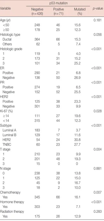
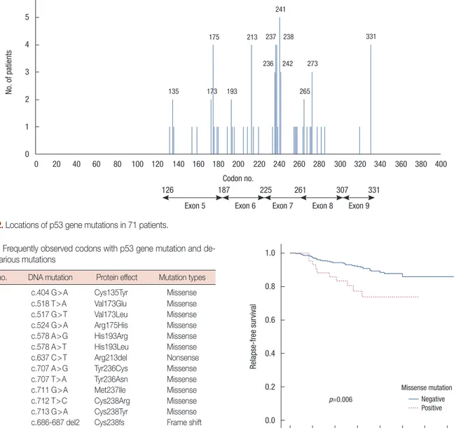
Among the 71 patients with p53 mutation, the types and lo-cations of the mutation were investigated. As shown in Figure 1, missense mutation was the most common type of p53 muta-tion (43 patients, 60.6%) and frame shift mutamuta-tion was the sec-ond most common (16 patients, 18.3%). Nonsense mutations were observed in 10 patients (11.3%), silent mutation in one patient (1.4%), and splicing mutation in six patients (8.5%). The most frequent location of p53 mutations was exon 7 (23 patients, 32.4%), followed by exon 5 (16 patients, 22.5%) (Fig-ure 2). Mutations in exons 6, 8, and 9 were observed in 10 (14.1%), 10 (14.1%), and five patients (7.0%), respectively. Mu-tation in an intron was detected in six patients (8.5%). The don numbers with p53 mutation are presented in Figure 2; co-Table 2. Clinical and pathologic characteristics of patients with p53
mutation Variable p53 mutation p-value Negative (n=426) Positive(n=71) Mutated(%) Age (yr) 0.181 <50 248 46 15.6 ≥50 178 25 12.3 Histologic type 0.056 Ductal 364 66 15.3 Others 62 5 7.4 Histologic grade <0.001 1 119 5 4.0 2 173 31 15.2 3 101 34 25.2 ER <0.001 Positive 290 21 6.8 Negative 136 50 26.9 PR <0.001 Positive 274 19 6.5 Negative 152 52 25.5 HER2 <0.001 Positive 125 38 23.3 Negative 301 33 9.9 Ki-67 (%) 0.028 ≥14 111 27 19.6 <14 315 44 12.3 Subtype <0.001 Luminal A 183 7 3.7 Luminal B 129 17 11.6 HER2 54 24 30.8 TNBC 60 23 27.7 T stage 0.004 1 210 23 9.9 2 201 48 19.3 3 15 0 0 N stage 0.881 0 238 38 13.8 1 125 22 15.0 2 45 9 16.7 3 18 2 10.0 Chemotherapy 0.007 Yes 345 66 16.1 Hormone therapy <0.001 Yes 303 23 7.1 Radiation therapy 0.283 Yes 175 26 12.9
ER=estrogen receptor; PR=progesterone receptor; HER2=human epider-mal growth factor receptor 2; TNBC=triple-negative breast cancer.
Missense Frame shift Nonsense Silent Splicing 43 8 13 6 1
dons 175, 213, 237, 238, 241, and 331 were hot spots for muta-tion in the present study. Frequently observed codons with p53 mutation and the detailed contents of various mutations were summarized in Table 3.
During the median follow-up period of 67 months (1–146 months), 51 patients had recurrence and 17 died from breast cancer. One patient already had distant metastasis at the time of diagnosis and she was excluded from survival analysis. In
univariate analysis, we compared RFS and BCSS of the patients according to presence of p53 mutation. The 5-year RFS rates for patients with and without p53 mutation was 81.3% and 92.2%, respectively, and there was a statistically significant dif-ference (p=0.020). The 5-year BCSS rates for patients with and without p53 mutation was 97.0% and 97.5%, respectively, and this was not statistically significant (p=0.401). We further in-vestigated survival according to presence of p53 missense mu-tation. The 5-year RFS rates for patients with and without mis-sense mutation was 77.3% and 97.0%, respectively, and there was a statistically significant difference in RFS between patients Table 3. Frequently observed codons with p53 gene mutation and
de-tails of various mutations
Codon no. DNA mutation Protein effect Mutation types
135 c.404 G>A Cys135Tyr Missense
173 c.518 T>A Val173Glu Missense
c.517 G>T Val173Leu Missense
175 c.524 G>A Arg175His Missense
193 c.578 A>G His193Arg Missense
c.578 A>T His193Leu Missense
213 c.637 C>T Arg213del Nonsense
236 c.707 A>G Tyr236Cys Missense
c.707 T>A Tyr236Asn Missense
237 c.711 G>A Met237Ile Missense
238 c.712 T>C Cys238Arg Missense
c.713 G>A Cys238Tyr Missense c.686-687 del2 Cys238fs Frame shift
241 c.722 C>A Ser241Tyr Missense
c.722 C>T Ser241Phe Missense
242 c.724 T>G Cys242Gly Missense
c.723delC Cys242Alafsx5 Frame shift
265 c.794 T>C Leu265Pro Missense
273 c.817 C>T Arg273Cys Missense
c.818 G>A Arg273His Missense 331 c.991 C>T Gln331stop Nonsense
c.991delC Gln331fs Frame shift
Rela
pse-free sur
vival
0 20 40 60 80 100 120 140 Follow-up period (mo)
1.0 0.8 0.6 0.4 0.2 0.0 Missense mutation p=0.006 Negative Positive
Figure 3. Kaplan-Meier relapse-free survival curve according to p53 missense mutation. No. of pa tients 0 20 40 60 80 100 120 140 160 180 200 220 240 260 280 300 320 340 360 380 400 Codon no. 6 5 4 3 2 1 0 241 238 237 273 242 236 331 265 193 175 213 135 Exon 5 126 187 225 261 307 331
Exon 6 Exon 7 Exon 8 Exon 9 173
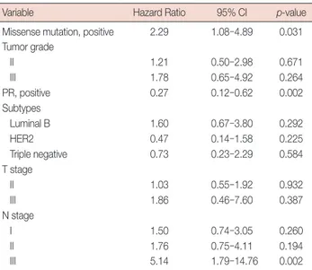
with missense mutation and those without missense mutation (p=0.006) (Figure 3). Missense mutation also showed mar-ginal significance in BCSS (p=0.060).
Multivariate analysis included tumor grade, PR, subtypes, T stage, N stage, p53 mutation, and missense mutation as co-variates that showed marginally significant association with relapse-free survival in univariate analysis (Table 4). After ad-justment for tumor grade, PR, subtypes, T stage, and N stage, presence of p53 mutation was not an independent prognostic factor of RFS (hazard ratio (HR), 1.70; 95% confidence inter-val (CI), 0.86–3.36; p=0.125). However, after adjustment for the same variables, p53 missense mutation was an indepen-dent prognostic factor of RFS with a relative hazard ratio of 2.29 (95% CI, 1.08–4.89; p=0.031) (Table 5).
DISCUSSION
In the present study we investigated patterns of mutation within exon 5 and exon 9 of the p53 gene in Korean patients with breast cancer. Gene sequencing revealed the presence of p53 mutation in 14.3% of patients, with missense mutations the most common type of mutation. Mutations occurred most commonly in exon 7, and codons 175, 213, 237, 238, 241, and 331 were hot spots of p53 mutation.
A number of previous studies have investigated the types and sites of mutations in the p53 gene in breast cancer. The p53 gene encodes three main protein domains: the transactivation domain, the DNA binding domain, and the oligomerization domain [18]. The DNA binding domain, encoded by exon 5 and exon 8, is the most common site of p53 gene mutation, ac-counting for approximately 90% of the p53 mutations reported in breast cancer [19,20]. Moreover, according to the Interna-tional Agency of Research on Cancer (IARC) TP53 mutation
database, the types of mutations observed indicate a high prev-alence of missense mutations (http://www.irac.Fr/p53). These missense mutations are scattered throughout the coding se-quence, but 97% of them cluster in exons encoding the DNA-binding domain and within this domain mutation “hotspots” have been identified at codons 175, 245, 248, 249, 273, and 282. As a result, the majority of studies have focused on mutations occurring within exon 5 and exon 8. Our results showed that the “hotspot” codons of p53 mutation in our patients were dif-ferent from those in the IARC database or previous Western studies and, with the exception of codon 175, the usual hotspots were not observed. Analysis of the type of p53 mutation showed that missense mutations were the most common type of mutations in this study but the frequency of missense muta-tion was relatively low compared with that in the IRAC data-base or other studies.
We found that p53 mutation was more prevalent in high-grade, large, hormone receptor-negative, HER2-positive tu-mors. These results were similar to those of previous studies [12,20]. Olivier et al. [12] reported that, in addition to tumor grade and hormone receptor status, p53 mutation was more frequent in large tumors and node-negative tumors. Consider-ing the division of breast tumors accordConsider-ing to the subtype classification, previous studies showed that p53 mutations were more frequently found in patients with triple-negative or HER2-positive breast cancer subtypes [8,21,22]. The study of Curtis et al. [22] involving a detailed, genome-scale analysis of nearly 2,000 breast cancer cases reported that p53 mutations were found in 34% of basal-like, 22% of HER2, 13% of lumi-Table 4. Univariate analysis of clinicopathologic variables for
relapse-free survival in breast cancer patients
Variable p-value (log-rank test)
Age, over 50 0.299 Histology, ductal 0.484 Tumor grade 0.074 ER, positive 0.115 PR, positive 0.002 HER2, positive 0.782 Ki-67, over 14% 0.689 Subtype 0.084 p53 mutation, positive 0.020
Missense mutation, positive 0.006
T stage 0.009
N stage <0.001
ER=estrogen receptor; PR=progesterone receptor; HER2=human epider-mal growth factor receptor 2.
Table 5. Multivariate analysis of factors associated with relapse-free survival (Cox proportional hazards model)
Variable Hazard Ratio 95% CI p-value
Missense mutation, positive 2.29 1.08–4.89 0.031 Tumor grade II 1.21 0.50–2.98 0.671 III 1.78 0.65–4.92 0.264 PR, positive 0.27 0.12–0.62 0.002 Subtypes Luminal B 1.60 0.67–3.80 0.292 HER2 0.47 0.14–1.58 0.225 Triple negative 0.73 0.23–2.29 0.584 T stage II 1.03 0.55–1.92 0.932 III 1.86 0.46–7.60 0.387 N stage I 1.50 0.74–3.05 0.260 II 1.76 0.75–4.11 0.194 III 5.14 1.79–14.76 0.002
PR=progesterone receptor; HER2=human epidermal growth factor receptor 2; CI=confidence interval.
nal B, and 5% of luminal A molecular subtypes. The Cancer Genome Atlas Network [8] reported rates of p53 mutation of up to 80% in the basal-like subtype, 72% in the HER2 sub-type, 29% in the luminal B subsub-type, and 12% in the luminal A subtype. Although mutation rates were quite different from those reported in these studies, the present study also showed that p53 mutation was detected most frequently in tumors with triple-negative or HER2 subtypes. The discrepancies in the incidence and location of p53 mutations between previous studies and the present study may be due to ethnic differences in the study populations and/or heterogeneous samples and analytical methods (e.g., whole-exome sequencing). Further investigations are needed to explain these discrepancies.
There are several studies on the prognostic value of p53 mutation types. Børresen-Dale [5] suggested that the presence of mutations in the DNA binding domain of p53 is associated with aggressive tumors and poor prognosis. Végran et al. [14] also suggested that only missense mutations occurring in the DNA binding domain were significantly associated with worse disease-free survival and overall survival. Alsner et al. [23] showed that patients with missense mutations affecting the DNA binding or zinc binding domains displayed a very aggressive phenotype with short survival. These findings are supported by our data showing that missense mutation of the p53 gene is an independent prognostic factor and is associated with poor clinical outcome, although overall p53 mutation was not an independent prognostic factor. Olivier et al. [12] also showed that missense mutations, especially in the DNA-binding domain encoded by exon 5 and exon 8, and specific missense mutations (i.e., codon 179 and R248W) are associat-ed with worse prognosis. However, they found that nonmis-sense mutations in DNA-binding domains had a similar poor prognostic value. Moreover, additional analysis of the whole coding sequence in 651 cases revealed that mutations located outside exon 5 and exon 8 were detected in 4% of patients and were associated with worse prognosis compared with wild type. Thus, they recommended conducting mutation analysis on all coding exons and splicing junctions. The present study did not investigate all coding exons (2 to 11) and could not as-sess the prognostic significance of p53 mutations occurring outside of the DNA-binding domain. A whole exon sequenc-ing study with a large population seems to be warranted.
In summary, the present study analyzed the patterns and biologic features of p53 mutation types and evaluated the clin-ical significance of these mutations in Korean patients with breast cancer. Our data showed that exon 7 was the most fre-quent site of p53 mutation, and missense mutation was the most common type of mutation. Mutation of p53 was more prevalent in high-grade, large, triple-negative tumors, and
HER2-positive tumors. Missense p53 mutation was an inde-pendent predictive factor associated with poor prognosis. We need additional studies with a large number of patients for clinical implication of results and for explanation of the ob-served differences in “hotspot” codons and the incidence of p53 gene mutation between this and other studies.
ACKNOWLEDGMENTS
We acknowledge the generous help of Dong Su Jang for graphic design.
CONFLICT OF INTEREST
The authors declare that they have no competing interests.
REFERENCES
1. Rakha EA, El-Sayed ME, Green AR, Lee AH, Robertson JF, Ellis IO. Prognostic markers in triple-negative breast cancer. Cancer 2007;109: 25-32.
2. Goldhirsch A, Wood WC, Coates AS, Gelber RD, Thürlimann B, Senn HJ, et al. Strategies for subtypes: dealing with the diversity of breast can-cer: highlights of the St. Gallen International Expert Consensus on the Primary Therapy of Early Breast Cancer 2011. Ann Oncol 2011;22: 1736-47.
3. Kaplan HG, Malmgren JA. Impact of triple negative phenotype on breast cancer prognosis. Breast J 2008;14:456-63.
4. Grann VR, Troxel AB, Zojwalla NJ, Jacobson JS, Hershman D, Neugut AI. Hormone receptor status and survival in a population-based cohort of patients with breast carcinoma. Cancer 2005;103:2241-51. 5. Børresen-Dale AL. TP53 and breast cancer. Hum Mutat
2003;21:292-300.
6. Levine AJ, Momand J, Finlay CA. The p53 tumour suppressor gene. Nature 1991;351:453-6.
7. Yonish-Rouach E, Resnitzky D, Lotem J, Sachs L, Kimchi A, Oren M. Wild-type p53 induces apoptosis of myeloid leukaemic cells that is in-hibited by interleukin-6. Nature 1991;352:345-7.
8. Cancer Genome Atlas Network. Comprehensive molecular portraits of human breast tumours. Nature 2012;490:61-70.
9. Perou CM, Sørlie T, Eisen MB, van de Rijn M, Jeffrey SS, Rees CA, et al. Molecular portraits of human breast tumours. Nature 2000;406:747-52. 10. Sotiriou C, Neo SY, McShane LM, Korn EL, Long PM, Jazaeri A, et al. Breast cancer classification and prognosis based on gene expression profiles from a population-based study. Proc Natl Acad Sci U S A 2003; 100:10393-8.
11. Berns EM, van Staveren IL, Look MP, Smid M, Klijn JG, Foekens JA. Mutations in residues of TP53 that directly contact DNA predict poor outcome in human primary breast cancer. Br J Cancer 1998;77:1130-6. 12. Olivier M, Langerød A, Carrieri P, Bergh J, Klaar S, Eyfjord J, et al. The
clinical value of somatic TP53 gene mutations in 1,794 patients with breast cancer. Clin Cancer Res 2006;12:1157-67.
prognosis in breast cancer: a meta-analysis. Br J Cancer 1999;80:1968-73.
14. Végran F, Rebucci M, Chevrier S, Cadouot M, Boidot R, Lizard-Nacol S. Only missense mutations affecting the DNA binding domain of p53 influence outcomes in patients with breast carcinoma. PLoS One 2013; 8:e55103.
15. Edge S, Byrd DR, Compton CC, Fritz AG, Greene FL, Trotti A. AJCC Cancer Staging Manual. 7th ed. New York: Springer; 2010.
16. Xiao W, Oefner PJ. Denaturing high-performance liquid chromatogra-phy: a review. Hum Mutat 2001;17:439-74.
17. Keller G, Hartmann A, Mueller J, Höfler H. Denaturing high pressure liquid chromatography (DHPLC) for the analysis of somatic p53 muta-tions. Lab Invest 2001;81:1735-7.
18. Levrero M, De Laurenzi V, Costanzo A, Gong J, Wang JY, Melino G. The p53/p63/p73 family of transcription factors: overlapping and dis-tinct functions. J Cell Sci 2000;113(Pt 10):1661-70.
19. Olivier M, Hainaut P. TP53 mutation patterns in breast cancers: search-ing for clues of environmental carcinogenesis. Semin Cancer Biol 2001; 11:353-60.
20. Berns EM, Foekens JA, Vossen R, Look MP, Devilee P, Henzen-Logmans SC, et al. Complete sequencing of TP53 predicts poor response to sys-temic therapy of advanced breast cancer. Cancer Res 2000;60:2155-62. 21. Bull SB, Ozcelik H, Pinnaduwage D, Blackstein ME, Sutherland DA,
Pritchard KI, et al. The combination of p53 mutation and neu/erbB-2 amplification is associated with poor survival in node-negative breast cancer. J Clin Oncol 2004;22:86-96.
22. Curtis C, Shah SP, Chin SF, Turashvili G, Rueda OM, Dunning MJ, et al. The genomic and transcriptomic architecture of 2,000 breast tumours reveals novel subgroups. Nature 2012;486:346-52.
23. Alsner J, Yilmaz M, Guldberg P, Hansen LL, Overgaard J. Heterogeneity in the clinical phenotype of TP53 mutations in breast cancer patients. Clin Cancer Res 2000;6:3923-31.