Disease and Collateral Revascularization
Young Seok Park1, Young Joo Jeon2, Hyun Seok Kim2, Kyu Young Chae3, Seung-Hun Oh4, In Bo Han1, Hyun Sook Kim4, Won-Chan Kim4, Ok-Joon Kim4, Tae Gon Kim1, Joong-Uhn Choi1, Dong-Seok Kim5*, Nam Keun Kim3*
1 Department of Neurosurgery, CHA Bundang Medical Center, CHA University School of Medicine, Seongnam, South Korea, 2 Institute for Clinical Research, CHA Bundang Medical Center, CHA University School of Medicine, Seongnam, South Korea,3 Department of Pediatrics, CHA Bundang Medical Center, CHA University School of Medicine, Seongnam, South Korea,4 Department of Neurology, CHA Bundang Medical Center, CHA University School of Medicine, Seongnam, South Korea, 5 Department of Pediatric Neurosurgery, Severance Hospital, Brain Korea 21 Project for Medical Science, Brain Research Institute, Yonsei University College of Medicine, Seoul, Korea
Abstract
We conducted a case-control study to investigate whether vascular endothelial growth factor (VEGF 22578, 21154, 2634, and 936) and kinase insert domain containing receptor (KDR 2604, 1192, and 1719) polymorphisms are associated with moyamoya disease. Korean patients with moyamoya disease (n = 107, mean age, 20.9615.9 years; 66.4% female) and 243 healthy control subjects (mean age, 23.0616.1 years; 56.8% female) were included. The subjects were divided into pediatric and adult groups. Among the 64 surgical patients, we evaluated collateral vessel formation after 2 years and divided patients into good (collateral grade A) or poor (collateral grade B and C) groups. The frequencies and distributions of four VEGF (22578, 21154, 2634, and 936) and KDR (2604, 1192, and 1719) polymorphisms were assessed from patients with moyamoya disease and compared to the control group. No differences were observed in VEGF 22578, 21154, 2634, and 936 or KDR 2604, 1192, and 1719 polymorphisms between the control group and moyamoya disease group. However, we found the 2634CC genotype occurred less frequently in the pediatric moyamoya group (p = 0.040) whereas the KDR 2604C/1192A/1719T haplotype increased the risk of pediatric moyamoya (p = 0.024). Patients with the CC genotype of VEGF 2634 had better collateral vessel formation after surgery. Our results suggest that the VEGF 2634G allele is associated with pediatric moyamoya disease and poor collateral vessel formation.
Citation: Park YS, Jeon YJ, Kim HS, Chae KY, Oh S-H, et al. (2012) The Role of VEGF and KDR Polymorphisms in Moyamoya Disease and Collateral Revascularization. PLoS ONE 7(10): e47158. doi:10.1371/journal.pone.0047158
Editor: Thiruma V. Arumugam, University of Queensland, Australia
Received May 21, 2012; Accepted September 10, 2012; Published October 12, 2012
Copyright: ß 2012 Park et al. This is an open-access article distributed under the terms of the Creative Commons Attribution License, which permits unrestricted use, distribution, and reproduction in any medium, provided the original author and source are credited.
Funding: This work was supported by the National Research Foundation of Korea (2010-0023851). The funders had no role in the study design or decision to publish.
Competing Interests: The authors have declared that no competing interests exist. * E-mail: [email protected] (NKK); [email protected] (DSK)
Introduction
Although many studies have investigated the etiology of moyamoya disease, unsatisfactory progress has been made. Moyamoya disease etiology may be idiopathic, environmental, or genetic. The search for genetic loci linked to moyamoya disease has uncovered associations with chromosomes 3, 6, 7, 8, and 17 and the HLA haplotype [1–8], but relevant genes have not been identified [9]. Recent linkage analyses from East Asian families with moyamoya disease demonstrated a linkage of 17q25.3 with the disease at a locus that is 21480 bp from the transcription site of the Raptor gene. However, an allele of this gene was not detected in samples from Caucasian patients with this disease [10]. Vascular endothelial growth factor (VEGF) is involved in vasculogenesis and vascular permeability in various intracranial lesions [11]. In ischemic disease, cerebral angiogenesis is caused by the release of VEGF [12,13]. VEGF affects vasculogenesis, endothelial cell proliferation and migration, vascular permeability, and stromal degradation through the activation of proteolytic enzymes that are involved in angiogenesis [14,15]. VEGF binds its receptor tyrosine kinases, VEGF receptor-1 and VEGF receptor-2 (also known as kinase insert domain containing receptor, or KDR)
but KDR is the key receptor mediating angiogenesis [16] and is essential for endothelial cell survival and integrity [17].
Although excess VEGF in moyamoya disease has been demonstrated [11,18] and the association is convincing, the specific role for VEGF remains unclear. Therefore, we studied the relationship of VEGF and KDR polymorphisms and moyamoya disease. VEGF is a major angiogenic factor and a prime regulator of endothelial cell proliferation [19]. The gene that encodes VEGF is located on chromosome 6 and is comprised of a 14-kb coding region with eight exons and seven introns [20]. VEGF is activated transcriptionally and posttranscriptionally by hypoxia in tumor necrosis and in various models of ischemia [21,22]. Ischemia stimulates VEGF expression in the brain suggesting that it may be important for the vascular response to cerebral ischemia [23,24,25]. Several single nucleotide polymorphisms (SNPs) have been described in the VEGF gene (National Center for Biotech-nology Information, Gene association no: NT 007592). The VEGF gene includes at least 4 relatively common polymorphisms that may influence VEGF expression: 22578C.A (rs699947), 21154G.A (rs1570360), 2634G.C (rs2010963), and 936C.T (rs3025039) [26,27,28].
Three of these polymorphisms are located in the promoter region at 22578, 21154, and 2634 relative to the translation start site. The 22578A, 21154A, and 2634G alleles are all associated with decreased VEGF expression [26,27]. In addition to promoter region polymorphisms, the T allele of the common 936C.T polymorphism in the 39-untranslated region is also associated with significantly decreased serum VEGF levels [28]. Recently, several SNPs of the VEGF gene have been associated with cancer risk and prognosis [29], as well as coronary arterial disease [30]. Moreover, KDR 2604C, 1192A, and 1719A alleles of chromosome 4 were associated with decreased VEGF binding
activity and coronary artery disease [31]. These results indicate the importance of VEGF-KDR signaling in human disease.
To our knowledge, no previous study has evaluated both KDR and VEGF polymorphisms with moyamoya disease and collateral vessel formation after surgery. The aim of present study was to evaluate the frequencies of the VEGF 22578C.A, 21154G.A, 2634G.C, and 936C.T and KDR 2604T.C (rs2071559), 1192G.A (rs2305948), and 1719T.A (rs1870377) polymor-phisms in Korean patients with moyamoya disease in an effort to determine the relationship of these polymorphisms with moyamoya disease.
Methods Subjects
One hundred seven consecutive Korean patients with moya-moya disease [mean age, 20.9615.9 years; 71 females (66.4%), 36 males (33.6%)] were recruited for this study. Moyamoya disease was defined as the presence of clinical ischemic or hemorrhagic symptoms in combination with vascular lesions in magnetic resonance imaging (MRI) or magnetic resonance angiography (MRA).
The control group was comprised of 243 healthy subjects [mean age, 23.0616.1 years; 138 female (56.8%); 105 male (43.2%)] from the same geographic region as the moyamoya patients. These age- and sex-matched subjects were recruited from outpatient clinics at Severance Hospital (Seoul, Korea) and CHA Bundang Medical Center (Seongnam, Korea).
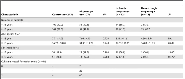
Moyamoya disease has a bimodal pattern of incidence so we divided the patients into pediatric (,18 years) and adult ($18 years) groups. We further divided the moyamoya patients into ischemic or hemorrhagic groups based on clinical and MRI findings. We performed indirect bypass surgery in 64 patients and direct superficial temporal artery to middle cerebral artery bypass plus encephalo-duro-arterio-myo-synangiosis (STA-MCA plus EDAMS) in one patient. We graded newly developed collateral vessels according to the method of Matsushima et al. [32]. Briefly, Grade A represented synangiosis-induced filling of greater than Table 1. Demographic characteristics between controls and moyamoya patients.
Characteristic Control (n = 243) Moyamoya (n = 107) P* Ischemic moyamoya (n = 92) Hemorrhagic moyamoya (n = 15) P* Number of subjects ,18 years 102 (42.0) 56 (52.3) 54 (58.7) 2 (13.3) $18 years 141 (58.0) 51 (47.7) 38 (41.3) 13 (86.7) Age (means6SD) ,18 years 7.7164.05 7.9864.13 0.920 8.1164.12 4.5063.54 NA $18 years 36.72610.05 34.98611.29 0.248 34.63611.45 36.00611.21 0.689 Sex [male, n(%)] ,18 years 54 (52.9) 22 (39.3) 0.100 21 (38.9) 1 (50.0) 1.000{ $18 years 51 (21.0) 14 (27.5) 0.260 12 (31.6) 2 (15.4) 0.472{ Collateral vessel formation score (n = 64)
0 – 2
1 – 22
2 – 40
*P values were calculated using the Mann-Whitney test for continuous data and x2
-test for categorical data. {
Fisher’s exact test. NA; not applicable. doi:10.1371/journal.pone.0047158.t001
Figure 1. Linkage disequilibrium(LD) of VEGF and KDR polymorphisms. There was strong LD between 21154G.A (rs 1570460) and 2634G.C (rs2010963) (D9 = 0.92), 22578C.A (rs 699947)and 1154G.A (rs1570360) (D9 = 0.89). There was strong LD between 1719T.A(rs1870377) and 1192G.A (rs2305948) (D9 = 0.79). doi:10.1371/journal.pone.0047158.g001
two-thirds of MCA circulation, Grade B represented between one-third and two-one-thirds, and Grade C represented less than one-one-third. We further divided the 64 indirect bypass surgical patients by collateral vessel formation after 2 years into good (collateral grade A) and poor (collateral grade B, C) using MRA. Table 1 shows the demographic characteristics of the moyamoya patients and control subjects.
All participants gave informed written consent prior to enrollment in the study. The institutional review boards of Severance Hospital (4-2008-0308) and CHA Bundang Medical Center (PBC09-103) approved this study.
VEGF Genotyping
We investigated four relevant single nucleotide polymorphism (SNP) candidates in the VEGF gene. We used the G-DEX blood extraction kit (iNtRON Biotechnology, Inc., Seongnam, South Korea) according to the manufacturer’s instructions for DNA extraction. We obtained all SNP sequences from the HapMap database (www.hapmap.org) [33]. The VEGF 2578C.A, -1154G.A, -634G.C, and 936C.T polymorphisms were analyzed by polymerase chain reaction–restriction fragment length polymorphism (PCR-RFLP) method.
We used following primers: VEGF 22578C.A polymorphism, forward 59-GGA TGG GGC TGA CTA GGT AAG-39 and reverse 59-AGC CCC CTT TTC CTC CAA C-39, that amplifies a 308 bp (C allele) or 326 bp (A allele) product; VEGF
21154G.A polymorphism, forward 59-CGC GTG TCT CTG GAC AGA GTT TCC-39 and reverse 59-CGG GGA CAG GCG AGC TTC AG-39, that amplifies a 173 bp product; VEGF 2634G.C polymorphism, forward 59-CAG GTC ACT CAC TTT GCC CCG GTC-39 and reverse 59-GCT TGC CAT TCC CCA CTT GAA TCG-39, that amplifies a 204 bp product; and VEGF 936C.T polymorphism, forward 59-AAG GAA GAG GAG ACT CTG CGC AGA GC-39 and reverse 59-TAA ATG TAT GTA TGT GGG TGG GTG TGT CTA CAG G-39, that amplifies a 208 bp fragment.
The VEGF 22578C.A and 2634G.C polymorphisms were identified by digesting the PCR product with the restriction endonuclease AvaII (New England Biolabs, Beverly, MA, USA). The VEGF 21154G.A polymorphism was identified by digesting the PCR product with the restriction endonuclease MnlI (New England Biolabs). The VEGF 936C.T polymorphism was identified by digesting the PCR product with the restriction endonuclease NlaIII (New England Biolabs). All restriction digests were performed at 37uC for 16 h.
KDR Genotyping
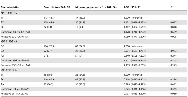
We identified 3 well-known SNPs in the KDR gene, including one in the promoter region (2604) and two in the coding region (1192 and 1719). All SNP sequences were obtained from the HapMap database (http://www.hapmap.org) [33]. We used previously described primers and PCR-RFLP conditions for Table 2. The frequencies of the VEGF 22578C.A, 21154G.A, 2634G.C, and 936C.T polymorphisms between control subjects and patients with moyamoya disease.
Characteristics Controls (n = 243, %) Moyamoya patients (n = 107, %) AOR (95% CI) P* VEGF 22578C.A
CC 128 (52.7) 62 (57.9) 1.000 (reference)
CA 99 (40.7) 36 (33.6) 0.769 (0.470–1.258) 0.296 AA 16 (6.6) 9 (8.4) 1.188 (0.491–2.873) 0.702 Dominant (CC vs. CA+AA) 0.827 (0.520–1.315) 0.422 Recessive (CC+CA vs. AA) 1.296 (0.547–3.072) 0.556 VEGF–1154G.A
GG 173 (71.2) 80 (74.8) 1.000 (reference)
GA 62 (25.5) 25 (23.4) 0.873 (0.510–1.496) 0.622 AA 8 (3.3) 2 (1.9) 0.517 (0.106–2.519) 0.414 Dominant (GG vs. GA+AA) 0.834 (0.495–1.405) 0.495 Recessive (GG+GA vs. AA) 0.546 (0.113–2.653) 0.454 VEGF 2634G.C GG 90 (37.0) 37 (34.6) 1.000 (reference) GC 103(42.4) 48 (44.9) 1.127 (0.666–1.906) 0.655 CC 50 (20.6) 22 (20.6) 1.083 (0.575–2.042) 0.804 Dominant (GG vs. GC+CC) 1.124 (0.695–1.817) 0.634 Recessive (GG+GC vs. CC) 1.024 (0.579–1.811) 0.934 VEGF 936C.T CC 172 (70.8) 67 (62.6) 1.000 (reference) CT 64 (26.3) 37 (34.6) 1.530 (0.926–2.529) 0.097 TT 7 (2.9) 3 (2.8) 1.230 (0.301–5.027) 0.773 Dominant (CC vs. CT+TT) 1.484 (0.912–2.416) 0.112 Recessive (CC+CT vs. TT) 1.079 (0.270–4.313) 0.915 *Adjusted by age and gender. AOR, adjusted odds ratio. CI, confidence interval.
KDR polymorphism analyses [31]. For SNP 2604, two DNA fragments (174 bp and 116 bp) were observed for the C allele and one band (290 bp) was produced for the T allele. For the 1192 polymorphism, two DNA fragments (30 bp and 232 bp) indicated the A allele and one band (252 bp) was produced for the G allele. For SNP 1719, two DNA fragments (191 bp and 213 bp) were observed for the A allele and one band (404 bp) was produced for the T allele. The genotyping reproducibility was confirmed by bi-directional sequencing of 400 randomly selected samples. Measurement of Plasma Total Homocysteine (tHcy), Folic Acid (FA), and Vitamin B12 (VB12)
Blood was collected from moyamoya patients into a tube containing anticoagulant 12 hours after a meal. The tube was centrifuged for 15 min at 1000 6g, and the plasma was separated. The concentration of tHcy (n = 37, 10.1862.81mmol/L) in the plasma was measured by fluorescent polarizing immunoassay (FPIA) with IMx (Abbott Laboratories, Chicago, IL, USA). The plasma concentration of FA (n = 30, 10.2563.50 ng/ml) and VB12 (n = 35, 848.066319.22 pg/ml) was determined using a radioassay kit (ACS 180; Bayer, Tarrytown, NY, USA).
Measurement of Whole Blood Nitric Oxide (NO)
Blood was collected from moyamoya patients into a tube containing anticoagulant 12 hours after a meal. NO production was evaluated by determining the circulating levels of nitrosyl-hemoglobin complexes. In the present study, the paramagnetic properties of nitrosyl-heme adducts were used to detect whole blood NO-hemoglobin derivatives (n = 33, 5.3066.93 arbitrary unit [AU]) by electron paramagnetic resonance (EPR) spectros-copy.
Statistical Analyses
To analyze the demographic characteristics of moyamoya disease, we used the Mann–Whitney and chi-square (x2) tests for continuous and categorical data, respectively. The associations among pediatric and adult patients were estimated by computing the odds ratios (ORs) and 95% confidence intervals (CIs) using Fisher’s exact test. The adjusted odds ratios (AORs) for VEGF and KDR polymorphisms were calculated using multiple logistic regression analyses using gender and age. The genotype distribu-tion of each polymorphism was expected under Hardy-Weinberg equilibrium. Statistical analyses were performed using GraphPad Prism 4.0 (GraphPad Software, Inc., San Diego, CA, USA) and StatsDirect software (version 2.4.4; StatsDirect Ltd., Altrincham, UK). Haplotype analyses were performed using HAPSTAT (version 3.0; University of North Carolina, Chapel Hill, NC, USA) and Haploview 4.2 (Broad Institute, Cambridge, MA, USA). StatsDirect Statistical Software (Version 2.4.4; StatsDirect Ltd, Altrincham, UK) was used to calculate the adjusted OR and 95% CI. The linkage disequilibrium between loci was measured using the absolute value of Lewontin’s D [34].
Results
Figure 1 shows the linkage disequilibrium of VEGF and KDR polymorphisms from the present study. A comparison of genotype frequencies between moyamoya patients and control subjects of the VEGF 22578C.A, 21154G.A, 2634G.C, and 936C.T polymorphisms and the KDR 2604T.C, 1192G.A, and 1719T.A polymorphisms is shown in Table 2. There were no statistically significant differences between moyamoya patients and controls in any of the polymorphisms evaluated (Tables 2 and 3). In subgroup analyses (Tables 4 and 5), the CC genotype of the VEGF 2634 was less frequent in pediatric moyamoya disease (p = 0.040; CC vs. GG) and comparison with the GG+GC Table 3. The frequencies of the KDR 2604T.C, 1192G.A, and 1719T.A polymorphisms between control subjects and patients with moyamoya disease.
Characteristics Controls (n = 243, %) Moyamoya patients (n = 107, %) AOR (95% CI) P* KDR 2604T.C
TT 113 (46.5) 47 (43.9) 1.000 (reference)
TC 108 (44.4) 50 (46.7) 1.131 (0.698–1.833) 0.617 CC 22 (9.1) 10 (9.3) 1.102 (0.482–2.517) 0.818 Dominant (CC vs. CA+AA) 1.128 (0.710–1.792) 0.609 Recessive (CC+CA vs. AA) 1.039 (0.470–2.296) 0.925 KDR 1192G.A
GG 186 (76.5) 80 (74.8) 1.000 (reference)
GA 52 (21.4) 22 (20.6) 0.994 (0.563–1.754) 0.983 AA 5 (2.1) 5 (4.7) 2.140 (0.596–7.693) 0.244 Dominant (GG vs. GA+AA) 1.101 (0.646–1.875) 0.725 Recessive (GG+GA vs. AA) 2.139 (0.597–7.665) 0.243 KDR 1719T.A
TT 46 (18.9) 26 (24.3) 1.000 (reference)
TA 114 (46.9) 45 (42.1) 0.764 (0.417–1.401) 0.384 AA 83 (34.2) 36 (33.6) 0.799 (0.427–1.495) 0.482 Dominant (TT vs. TA+AA) 0.775 (0.446–1.346) 0.365 Recessive (TT+TA vs. AA) 0.997 (0.613–1.620) 0.989 *Adjusted by age and gender. AOR, adjusted odds ratio. CI, confidence interval.
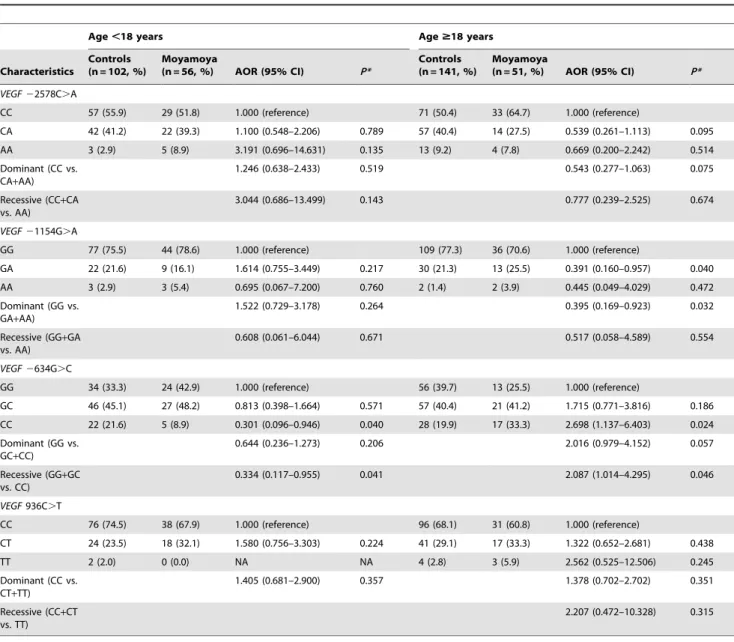
genotype was also significantly different in pediatric moyamoya patients. In the adult subgroup, the VEGF 2634CC genotype was more frequent in moyamoya disease (p = 0.024; CC vs. GG). The frequencies of the KDR polymorphisms in both the pediatric and adult subgroups were not significantly different.
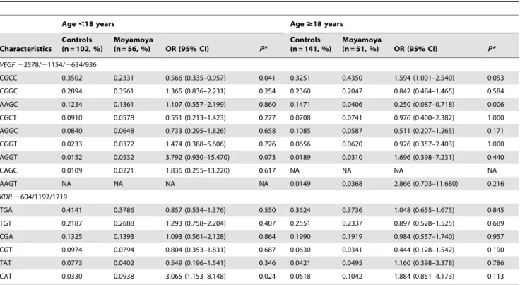
We conducted haplotype analyses of the VEGF and KDR polymorphisms (Table 6). The C-G-C-C haplotype (VEGF 22578/21154/2634/936) in pediatric moyamoya patients was significantly different. In addition, the C-A-T haplotype (KDR 2604/1192/1719) increased the risk of pediatric moyamoya. In adult moyamoya, the A-A-G-C haplotype (VEGF 22578/21154/ 2634) was significantly different whereas other haplotypes were not. We have estimated and provided several haplotype frequen-cies in Table 6.
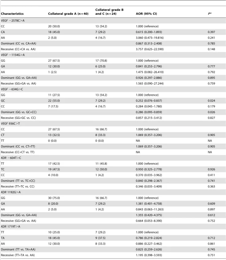
We performed indirect bypass surgery in 64 patients. The genotypes containing the VEGF 2634C allele had better collateral vessel formation after surgery whereas 2634GG was associated with poor collateral grade (Table 7). We also investigated the haplotype frequency differences related to collateral grades.
However, there were no statistical differences (Table S1). Our results potentially implicate the VEGF 2634G.C polymorphism in the development of collateral vessel formation in moyamoya disease. The other VEGF and KDR polymorphisms we studied did not exhibit statistically significant differences in collateral vessel formation.
To assess the clinical significance of the VEGF and KDR polymorphisms, we surveyed the association between the studied polymorphisms and various vascular risk factors (tHcy, FA, VB12, and NO). Blood tHcy, FA, VB12, and NO are essential for vascular homeostasis regulation [35]. There were no significant distinctions according to VEGF polymorphisms but the KDR 2604C (p = 0.017) and 1192A (p = 0.032) alleles were linked to decreased NO levels (Table S2). The association of the KDR 2604 and 1192 polymorphisms with NO levels suggests that KDR haplotypes containing 2604C or 1192A may adversely affect vascular homeostasis.
Table 4. The frequency of the VEGF polymorphisms according to age.
Age ,18 years Age $18 years Characteristics Controls (n = 102, %) Moyamoya (n = 56, %) AOR (95% CI) P* Controls (n = 141, %) Moyamoya (n = 51, %) AOR (95% CI) P* VEGF 22578C.A CC 57 (55.9) 29 (51.8) 1.000 (reference) 71 (50.4) 33 (64.7) 1.000 (reference) CA 42 (41.2) 22 (39.3) 1.100 (0.548–2.206) 0.789 57 (40.4) 14 (27.5) 0.539 (0.261–1.113) 0.095 AA 3 (2.9) 5 (8.9) 3.191 (0.696–14.631) 0.135 13 (9.2) 4 (7.8) 0.669 (0.200–2.242) 0.514 Dominant (CC vs. CA+AA) 1.246 (0.638–2.433) 0.519 0.543 (0.277–1.063) 0.075 Recessive (CC+CA vs. AA) 3.044 (0.686–13.499) 0.143 0.777 (0.239–2.525) 0.674 VEGF 21154G.A GG 77 (75.5) 44 (78.6) 1.000 (reference) 109 (77.3) 36 (70.6) 1.000 (reference) GA 22 (21.6) 9 (16.1) 1.614 (0.755–3.449) 0.217 30 (21.3) 13 (25.5) 0.391 (0.160–0.957) 0.040 AA 3 (2.9) 3 (5.4) 0.695 (0.067–7.200) 0.760 2 (1.4) 2 (3.9) 0.445 (0.049–4.029) 0.472 Dominant (GG vs. GA+AA) 1.522 (0.729–3.178) 0.264 0.395 (0.169–0.923) 0.032 Recessive (GG+GA vs. AA) 0.608 (0.061–6.044) 0.671 0.517 (0.058–4.589) 0.554 VEGF 2634G.C GG 34 (33.3) 24 (42.9) 1.000 (reference) 56 (39.7) 13 (25.5) 1.000 (reference) GC 46 (45.1) 27 (48.2) 0.813 (0.398–1.664) 0.571 57 (40.4) 21 (41.2) 1.715 (0.771–3.816) 0.186 CC 22 (21.6) 5 (8.9) 0.301 (0.096–0.946) 0.040 28 (19.9) 17 (33.3) 2.698 (1.137–6.403) 0.024 Dominant (GG vs. GC+CC) 0.644 (0.236–1.273) 0.206 2.016 (0.979–4.152) 0.057 Recessive (GG+GC vs. CC) 0.334 (0.117–0.955) 0.041 2.087 (1.014–4.295) 0.046 VEGF 936C.T CC 76 (74.5) 38 (67.9) 1.000 (reference) 96 (68.1) 31 (60.8) 1.000 (reference) CT 24 (23.5) 18 (32.1) 1.580 (0.756–3.303) 0.224 41 (29.1) 17 (33.3) 1.322 (0.652–2.681) 0.438 TT 2 (2.0) 0 (0.0) NA NA 4 (2.8) 3 (5.9) 2.562 (0.525–12.506) 0.245 Dominant (CC vs. CT+TT) 1.405 (0.681–2.900) 0.357 1.378 (0.702–2.702) 0.351 Recessive (CC+CT vs. TT) 2.207 (0.472–10.328) 0.315 *Adjusted by age and gender. NA; not applicable. AOR, adjusted odds ratio. CI, confidence interval.
Table 5. The frequency of the KDR polymorphisms according to age.
Age ,18 years Age $18 years Characteristics Controls (n = 102, %) Moyamoya (n = 56, %) AOR (95% CI) P* Controls (n = 141, %) Moyamoya (n = 51, %) AOR (95% CI) P* KDR 2604T.C CC 54 (52.9) 25 (44.6) 1.000 (reference) 59 (41.8) 22 (43.1) 1.000 (reference) CA 40 (39.2) 27 (48.2) 1.413(0.711–2.808) 0.324 68 (48.2) 23 (45.1) 0.929 (0.467–1.847) 0.833 AA 8 (7.8) 4 (7.1) 1.096(0.298–4.031) 0.890 14 (9.9) 6 (11.8) 1.141 (0.388–3.356) 0.811 Dominant (CC vs. CA+AA) 1.350(0.697–2.616) 0.374 0.951 (0.495–1.826) 0.879 Recessive (CC+CA vs. AA) 0.889(0.251–3.143) 0.855 1.198 (0.433–3.320) 0.728 KDR 1192G.A GG 20 (19.6) 16 (28.6) 1.000 (reference) 26 (18.4) 10 (19.6) 1.000 (reference) GA 47 (46.1) 22 (39.3) 0.572 (0.246–1.323) 0.193 67 (47.5) 23 (45.1) 1.082 (0.440–2.664) 0.864 AA 35 (34.2) 18 (32.1) 0.654 (0.232–1.572) 0.342 48 (34.0) 18 (35.3) 0.965 (0.382–2.435) 0.940 Dominant (GG vs. GA+AA) 0.611 (0.284–1.313) 0.207 1.003 (0.440–2.286) 0.994 Recessive (GG+GA vs. AA) 0.927 (0.460–1.869) 0.833 1.069 (0.543–2.103) 0.848 KDR 1719T.A TT 77 (75.5) 44 (78.6) 1.000 (reference) 109 (77.3) 36 (70.6) 1.000 (reference) TA 22 (21.6) 9 (16.1) 0.739 (0.310–1.762) 0.495 30 (21.3) 13 (25.5) 1.283 (0.602–2.734) 0.518 AA 3 (2.9) 3 (5.4) 1.848 (0.349–9.773) 0.470 2 (1.4) 2 (3.9) 3.003 (0.403–22.390) 0.283 Dominant (TT vs. TA+AA) 0.877 (0.398–1.934) 0.746 1.378 (0.667–2.846) 0.386 Recessive (TT+TA vs. AA) 2.026 (0.382–10.736) 0.407 2.675 (0.362–19.768) 0.335 *Adjusted by age and gender. NA; not applicable. AOR, adjusted odds ratio. CI, confidence interval.
doi:10.1371/journal.pone.0047158.t005
Table 6. Haplotype frequencies of the VEGF and KDR polymorphisms according to age.
Age ,18 years Age $18 years Characteristics Controls (n = 102, %) Moyamoya (n = 56, %) OR (95% CI) P* Controls (n = 141, %) Moyamoya (n = 51, %) OR (95% CI) P* VEGF 22578/21154/2634/936 CGCC 0.3502 0.2331 0.566 (0.335–0.957) 0.041 0.3251 0.4350 1.594 (1.001–2.540) 0.053 CGGC 0.2894 0.3561 1.365 (0.836–2.231) 0.254 0.2360 0.2047 0.842 (0.484–1.465) 0.584 AAGC 0.1234 0.1361 1.107 (0.557–2.199) 0.860 0.1471 0.0406 0.250 (0.087–0.718) 0.006 CGCT 0.0910 0.0578 0.551 (0.213–1.423) 0.277 0.0708 0.0741 0.976 (0.400–2.382) 1.000 AGGC 0.0840 0.0648 0.733 (0.295–1.826) 0.658 0.1085 0.0587 0.511 (0.207–1.265) 0.171 CGGT 0.0233 0.0372 1.474 (0.388–5.606) 0.726 0.0656 0.0620 0.926 (0.357–2.403) 1.000 AGGT 0.0152 0.0532 3.792 (0.930–15.470) 0.073 0.0189 0.0310 1.696 (0.398–7.231) 0.440 CAGC 0.0109 0.0221 1.836 (0.255–13.220) 0.617 NA NA NA NA AAGT NA NA NA NA 0.0149 0.0368 2.866 (0.703–11.680) 0.216 KDR 2604/1192/1719 TGA 0.4141 0.3786 0.857 (0.534–1.376) 0.550 0.3624 0.3736 1.048 (0.655–1.675) 0.845 TGT 0.2187 0.2688 1.293 (0.758–2.204) 0.407 0.2551 0.2337 0.897 (0.528–1.525) 0.689 CGA 0.1325 0.1393 1.093 (0.561–2.128) 0.864 0.1990 0.1919 0.984 (0.557–1.740) 0.957 CGT 0.0974 0.0794 0.804 (0.353–1.831) 0.687 0.0630 0.0341 0.444 (0.128–1.542) 0.190 TAT 0.0773 0.0402 0.549 (0.196–1.541) 0.346 0.0421 0.0495 1.160 (0.398–3.378) 0.786 CAT 0.0330 0.0938 3.065 (1.153–8.148) 0.024 0.0618 0.1042 1.884 (0.851–4.173) 0.113 *P-values after 10,000 permutation test. NA; not applicable. OR, odds ratio. CI, confidence interval.
Discussion
In this study, we found VEGF or KDR polymorphisms influence moyamoya disease in subgroup analyses as well as the formation of revascularization after bypass surgery. VEGF is involved in
vasculogenesis in different intracranial lesions [11], is an endo-thelial cell mitogen that induces transient vascular leakage, and a potent angiogenic factor [36]. VEGF promotes angiogenesis in cerebral ischemia [11,37] and causes pathologic vessel formation [12].
Table 7. Comparison of the VEGF and KDR genotype frequencies according to collateral vessel formation score.
Characteristics Collateral grade A (n = 40)
Collateral grade B
and C (n = 24) AOR (95% CI) P* VEGF 22578C.A
CC 20 (50.0) 13 (54.2) 1.000 (reference)
CA 18 (45.0) 7 (29.2) 0.615 (0.200–1.893) 0.397 AA 2 (5.0) 4 (16.7) 3.060 (0.473–19.816) 0.241 Dominant (CC vs. CA+AA) 0.867 (0.313–2.408) 0.785 Recessive (CC+CA vs. AA) 3.757 (0.625–22.590) 0.148 VEGF 21154G.A
GG 27 (67.5) 17 (70.8) 1.000 (reference)
GA 12 (30.0) 6 (25.0) 0.841 (0.253–2.796) 0.777 AA 1 (2.5) 1 (4.2) 1.475 (0.082–26.410) 0.792 Dominant (GG vs. GA+AA) 0.926 (0.297–2.886) 0.895 Recessive (GG+GA vs. AA) 1.565 (0.090–27.244) 0.759 VEGF 2634G.C GG 11 (27.5) 13 (54.2) 1.000 (reference) GC 22 (55.0) 7 (29.2) 0.252 (0.076–0.837) 0.024 CC 7 (17.5) 4 (16.7) 0.284 (0.045–1.780) 0.179 Dominant (GG vs. GC+CC) 0.286 (0.095–0.859) 0.026 Recessive (GG+GC vs. CC) 0.857 (0.215–3.412) 0.827 VEGF 936C.T CC 27 (67.5) 16 (66.7) 1.000 (reference) CT 13 (32.5) 8 (33.3) 1.069 (0.357–3.206) 0.905 TT 0 (0.0) 0 (0.0) NA NA Dominant (CC vs. CT+TT) 1.069 (0.357–3.206) 0.905 Recessive (CC+CT vs. TT) NA NA KDR 2604T.C TT 17 (42.5) 11 (45.8) 1.000 (reference) TC 19 (47.5) 12 (50.0) 0.950 (0.325–2.778) 0.926 CC 4 (10.0) 1 (4.2) 0.370 (0.035–3.962) 0.411 Dominant (TT vs. TC+CC) 0.840 (0.298–2.367) 0.741 Recessive (TT+TC vs. CC) 0.346 (0.035–3.409) 0.363 KDR 1192G.A GG 30 (75.0) 16 (66.7) 1.000 (reference) GA 8 (20.0) 7 (29.2) 1.381 (0.401–4.758) 0.609 AA 2 (5.0) 1 (4.2) 0.843 (0.063–11.263) 0.897 Dominant (GG vs. GA+AA) 1.355 (0.420–4.375) 0.612 Recessive (GG+GA vs. AA) 0.664 (0.053–8.390) 0.752 KDR 1719T.A
TT 10 (25.0) 7 (29.2) 1.000 (reference)
TA 18 (45.0) 9 (37.5) 0.786 (0.219–2.824) 0.712 AA 12 (30.0) 8 (33.3) 0.886 (0.227–3.462) 0.861 Dominant (TT vs. TA+AA) 0.825 (0.259–2.626) 0.745 Recessive (TT+TA vs. AA) 1.195 (0.398–3.593) 0.751 *Adjusted by age and gender. NA; not applicable. AOR, adjusted odds ratio. CI, confidence interval.
Moyamoya disease is characterized by the angiographic findings of arterial stenosis and occlusion of the circle of Willis [38]. It can lead to transient ischemic attacks or a cerebral infarction pattern in juveniles [39] and a hemorrhagic stroke pattern in adults [40,41]. Takekawa et al. [18] reported increased VEGF expression in autopsy specimens from adults with moyamoya disease and Sakamoto et al. reported that the total meningeal cellularity and VEGF expression in the moyamoya dura was significantly higher in moyamoya patients compared to controls [11]. Although increased VEGF concentrations have been demonstrated in moyamoya disease [11,18], the specific role of VEGF in moyamoya genetics remains unclear. We therefore reasoned that mutations and genetic polymorphisms of the VEGF gene may cause cerebral ischemia in moyamoya disease.
Vascular endothelial growth factor A (VEGF-A) is a disulfide-bonded dimeric glycoprotein that is a member of a protein family that includes VEGF-B, VEGF-C, VEGF-D, and placental growth factor (PGF). The gene that encodes VEGF-A is located on human chromosome 6 and comprises a 14-kb coding region with eight exons. VEGF cellular signaling activity depends on specific membrane receptors. The receptors include fms like tyrosine kinase-1 (Flt-1 or VEGFR-1), KDR (also known as VEGFR-2) [42,43], and Flt-4 (also known as VEGFR-3) [44,45]. VEGF acts on endothelial cells particularly through Flt-1 and KDR [46,47]. Epidermal growth factor (EGF) arising from hypoxia stimulates Flt-1 expression and inhibits KDR expression [48].
VEGF binding to KDR activates multiple signaling cascades that affect angiogenesis as well as endothelial cell survival, proliferation, and migration. Recently, several SNPs in the VEGF gene have been linked to cancer risk and prognosis [29] and coronary arterial disease [30], indicating the importance of the VEGF-KDR signaling pathway in human disease. Oh et al. reported the SNP 1719T allele conferred ischemic stroke risk in a dose dependent manner [49].
Revascularization after bypass surgery and the formation of new pial vessels may play a role in moyamoya disease. Several studies have suggested that endogenous VEGF production mediates compensatory revascularization during various physiological and pathological processes [37,50,51,52].
In addition, as suggested by the data in our study, VEGF 2634 G.C may be a possible prognostic biomarker after bypass surgery. Taken together, we can speculate that VEGF polymor-phisms influence moyamoya disease as well as the formation of synangiosis-induced collateral vessel after bypass surgery. Al-though revascularization with pial synangiosis helps ameliorate ischemic symptoms, some patients progress to cerebral infarction or hemorrhage, even after surgery. This likely reflects the degree of synangiosis and is dependent on the genetic characteristics of the patient. Therefore, VEGF or KDR polymorphisms can be used as prognostic factors after revascularization surgery.
There are some limitations in correlating VEGF or KDR polymorphisms with our clinical findings. Our data should be interpreted with caution because of the relatively small sample size. Patients require long-term follow up to assess clinical outcomes and variation in the clinical characteristics of moyamoya disease makes it difficult to identify specific genes that are associated with the disease. In addition, moyamoya disease is characterized by genetic heterogeneity and complex interactions between genes and other factors. Although there are limitations with sample size and long-term follow-up clinical findings, it is important to determine the relationship of VEGF or KDR polymorphisms with moyamoya disease and collateral vessel formation after surgery.
In summary, no differences in VEGF 22578, 21154, 2634, and 936 or KDR 2604, 1192, and 1719 polymorphisms were observed between total moyamoya disease patients and control subjects. However, in subgroup analyses, we found that the CC genotype in VEGF 2634 occurred less frequently in pediatric patients (p = 0.040) and occurred more often in adult moyamoya patients (p = 0.024). The genotypes including the VEGF 2634C allele had better collateral vessel formation after surgery. In addition, the C-G-C-C (VEGF 22578/21154/2634/936) hap-lotype and the C-A-T (KDR -604/1192/1719) haphap-lotype in pediatric patients, as well as the A-A-G-C (VEGF 22578/ 21154/2634/936) in adult moyamoya patients, had significant differences. Therefore, these results suggest that VEGF or KDR polymorphisms influence moyamoya disease as well as the formation of synangiosis-induced collateral vessel after bypass surgery.
Supporting Information
Table S1 Haplotype analyses of VEGF and KDR
poly-morphisms according to collateral score. *P-values after 10,000 permutation test.
(DOC)
Table S2 Association ofVEGF and KDR polymorphisms
with vascular risk factors. *P-values between major and minor alleles of each polymorphism analyzed by Mann-Whitney test. AU; arbitrary units.
(DOC)
Acknowledgments
We thank all the people who give the help for this study. Author Contributions
Conceived and designed the experiments: YSP DSK NKK. Performed the experiments: YSP YJJ HSK. Analyzed the data: YSP YJJ. Contributed reagents/materials/analysis tools: YSP SHO HSK WCK OJK TGK IBH JUC DSK. Wrote the paper: YSP YJJ DSK NKK.
References
1. Kang HS, Kim SK, Cho BK, Kim YY, Hwang YS, et al. (2006) Single nucleotide polymorphisms of tissue inhibitor of metalloproteinase genes in familial moyamoya disease. Neurosurgery 58: 1074–1080.
2. Ikeda H, Sasaki T, Yoshimoto T, Fukui M, Arinami T (1999) Mapping of a familial moyamoya disease gene to chromosome 3p24.2–p26. Am J Hum Genet 64: 533–537.
3. Inoue TK, Ikezaki K, Sasazuki T, Matsushima T, Fukui M (2000) J Child Neurol 15: 179–182.
4. Sakurai K, Horiuchi Y, Ikeda H, Ikezaki K, Yoshimoto T, et al. (2004) J Hum Genet 49: 278–281.
5. Yamauchi T, Tada M, Houkin K, Tanaka T, Nakamura Y, et al. (2000) Linkage of familial moyamoya disease (spontaneous occlusion of the circle of Willis) to chromosome 17q25. Stroke 31: 930–935.
6. Han H, Pyo CW, Yoo DS, Huh PW, Cho KS, et al. (2003) Associations of Moyamoya patients with HLA class I and class II alleles in the Korean population. J Korean Med Sci 18: 876–880.
7. Mineharu Y, Liu W, Inoue K, Matsuura N, Inoue S, et al. (2008) Autosomal dominant moyamoya disease maps to chromosome 17q25.3. Neurology 70: 2357–2363.
8. Park YS, Min KT, Kim TG, Lee YH, Cheong HJ, et al. (2011) Age-specific eNOS polymorphisms in moyamoya disease. Childs Nerv Syst 27: 1919–1926. 9. Nanba R, Tada M, Kuroda S, Houkin K, Iwasaki Y (2005) Sequence analysis and bioinformatics analysis of chromosome 17q25 in familial moyamoya disease. Childs Nerv Syst 21: 62–68.
10. Liu W, Hashikata H, Inoue K, Matsuura N, Mineharu Y, et al. (2010) A rare Asian founder polymorphism of Raptor may explain the high prevalence of
Moyamoya disease among East Asians and its low prevalence among Caucasians. Environ Health Prev Med 15: 94–104.
11. Sakamoto S, Kiura Y, Yamasaki F, Shibukawa M, Ohba S, et al. (2008) Expression of vascular endothelial growth factor in dura mater of patients with moyamoya disease. Neurosurg Rev 31: 77–81; discussion 81.
12. Cao Y, Hong A, Schulten H, Post MJ (2005) Update on therapeutic neovascularization. Cardiovasc Res 65: 639–648.
13. Jin KL, Mao XO, Nagayama T, Goldsmith PC, Greenberg DA (2000) Induction of vascular endothelial growth factor and hypoxia-inducible factor-1alpha by global ischemia in rat brain. Neuroscience 99: 577–585.
14. Mukhopadhyay D, Tsiokas L, Zhou XM, Foster D, Brugge JS, et al. (1995) Hypoxic induction of human vascular endothelial growth factor expression through c-Src activation. Nature 375: 577–581.
15. Ikeda E, Achen MG, Breier G, Risau W (1995) Hypoxia-induced transcriptional activation and increased mRNA stability of vascular endothelial growth factor in C6 glioma cells. J Biol Chem 270: 19761–19766.
16. Waltenberger J, Claesson-Welsh L, Siegbahn A, Shibuya M, Heldin CH (1994) Different signal transduction properties of KDR and Flt1, two receptors for vascular endothelial growth factor. J Biol Chem 269: 26988–26995. 17. Gerber HP, McMurtrey A, Kowalski J, Yan M, Keyt BA, et al. (1998) Vascular
endothelial growth factor regulates endothelial cell survival through the phosphatidylinositol 3’-kinase/Akt signal transduction pathway. Requirement for Flk-1/KDR activation. J Biol Chem 273: 30336–30343.
18. Takekawa Y, Umezawa T, Ueno Y, Sawada T, Kobayashi M (2004) Pathological and immunohistochemical findings of an autopsy case of adult moyamoya disease. Neuropathology 24: 236–242.
19. La Rosa S, Uccella S, Finzi G, Albarello L, Sessa F, et al. (2003) Localization of vascular endothelial growth factor and its receptors in digestive endocrine tumors: correlation with microvessel density and clinicopathologic features. Hum Pathol 34: 18–27.
20. Vincenti V, Cassano C, Rocchi M, Persico G (1996) Assignment of the vascular endothelial growth factor gene to human chromosome 6p21.3. Circulation 93: 1493–1495.
21. Leung DW, Cachianes G, Kuang WJ, Goeddel DV, Ferrara N (1989) Vascular endothelial growth factor is a secreted angiogenic mitogen. Science 246: 1306– 1309.
22. Shweiki D, Itin A, Soffer D, Keshet E (1992) Vascular endothelial growth factor induced by hypoxia may mediate hypoxia-initiated angiogenesis. Nature 359: 843–845.
23. Kovacs Z, Ikezaki K, Samoto K, Inamura T, Fukui M (1996) VEGF and flt. Expression time kinetics in rat brain infarct. Stroke 27: 1865–1872; discussion 1872–1863.
24. Hayashi T, Abe K, Suzuki H, Itoyama Y (1997) Rapid induction of vascular endothelial growth factor gene expression after transient middle cerebral artery occlusion in rats. Stroke 28: 2039–2044.
25. Lennmyr F, Ata KA, Funa K, Olsson Y, Terent A (1998) Expression of vascular endothelial growth factor (VEGF) and its receptors (Flt-1 and Flk-1) following permanent and transient occlusion of the middle cerebral artery in the rat. J Neuropathol Exp Neurol 57: 874–882.
26. Brogan IJ, Khan N, Isaac K, Hutchinson JA, Pravica V, et al. (1999) Novel polymorphisms in the promoter and 5’ UTR regions of the human vascular endothelial growth factor gene. Hum Immunol 60: 1245–1249.
27. Awata T, Inoue K, Kurihara S, Ohkubo T, Watanabe M, et al. (2002) A common polymorphism in the 5’-untranslated region of the VEGF gene is associated with diabetic retinopathy in type 2 diabetes. Diabetes 51: 1635–1639. 28. Renner W, Kotschan S, Hoffmann C, Obermayer-Pietsch B, Pilger E (2000) A common 936 C/T mutation in the gene for vascular endothelial growth factor is associated with vascular endothelial growth factor plasma levels. J Vasc Res 37: 443–448.
29. Schneider BP, Wang M, Radovich M, Sledge GW, Badve S, et al. (2008) Association of vascular endothelial growth factor and vascular endothelial growth factor receptor-2 genetic polymorphisms with outcome in a trial of paclitaxel compared with paclitaxel plus bevacizumab in advanced breast cancer: ECOG 2100. J Clin Oncol 26: 4672–4678.
30. Petrovic D, Verhovec R, Globocnik Petrovic M, Osredkar J, et al. (2007) Association of vascular endothelial growth factor gene polymorphism with myocardial infarction in patients with type 2 diabetes. Cardiology 107: 291–295. 31. Wang Y, Zheng Y, Zhang W, Yu H, Lou K, et al. (2007) Polymorphisms of KDR gene are associated with coronary heart disease. J Am Coll Cardiol 50: 760–767.
32. Matsushima T, Inoue T, Suzuki SO, Fujii K, Fukui M, et al. (1992) Surgical treatment of moyamoya disease in pediatric patients–comparison between the results of indirect and direct revascularization procedures. Neurosurgery 31: 401–405.
33. Consortium IH (2003) The international HapMap Project. Nature 426: 789– 796.
34. Hedrick PW (1987) Gametic disequilibrium measures: proceed with caution. Genetics 117: 331–341.
35. Fabian E, Kickinger A, Wagner KH, Elmadfa I (2011) Homocysteine and asymmetric dimethylarginine in relation to B vitamins in elderly people. Wien Klin Wochenschr 123: 496–501.
36. Thomas KA (1996) Vascular endothelial growth factor, a potent and selective angiogenic agent. J Biol Chem 271: 603–606.
37. Issa R, Krupinski J, Bujny T, Kumar S, Kaluza J, et al. (1999) Vascular endothelial growth factor and its receptor, KDR, in human brain tissue after ischemic stroke. Lab Invest 79: 417–425.
38. Suzuki J, Takaku A (1969) Cerebrovascular "Moyamoya" disease. Disease showing abnormal net-like vessels in base of brain. Arch Neurol 20: 288–299. 39. Fukui M (1997) Current state of study on moyamoya disease in Japan. Surg
Neurol 47: 138–143.
40. Kuroda S, Hashimoto N, Yoshimoto T, Iwasaki Y (2007) Radiological findings, clinical course, and outcome in asymptomatic moyamoya disease: results of multicenter survey in Japan. Stroke 38: 1430–1435.
41. Mineharu Y, Liu W, Inoue K, Matsuura N, Inoue S, et al. (2008) Autosomal dominant moyamoya disease maps to chromosome 17q25.3. Neurology 70: 2357–2363.
42. Takahashi T, Shibuya M (1997) The 230 kDa mature form of KDR/Flk-1 (VEGF receptor-2) activates the PLC-gamma pathway and partially induces mitotic signals in NIH3T3 fibroblasts. Oncogene 14: 2079–2089.
43. Soker S, Takashima S, Miao HQ, Neufeld G, Klagsbrun M (1998) Neuropilin-1 is expressed by endothelial and tumor cells as an isoform-specific receptor for vascular endothelial growth factor. Cell 92: 735–745.
44. Harry LE, Paleolog EM (2003) From the cradle to the clinic: VEGF in developmental, physiological, and pathological angiogenesis. Birth Defects Res C Embryo Today 69: 363–374.
45. Ferrara N, Gerber HP, LeCouter J (2003) The biology of VEGF and its receptors. Nat Med 9: 669–676.
46. Rahimi N, Dayanir V, Lashkari K (2000) Receptor chimeras indicate that the vascular endothelial growth factor receptor-1 (VEGFR-1) modulates mitogenic activity of VEGFR-2 in endothelial cells. J Biol Chem 275: 16986–16992. 47. Marchand GS, Noiseux N, Tanguay JF, Sirois MG (2002) Blockade of in vivo
VEGF-mediated angiogenesis by antisense gene therapy: role of Flk-1 and Flt-1 receptors. Am J Physiol Heart Circ Physiol 282: H194–204.
48. Okuda Y, Tsurumaru K, Suzuki S, Miyauchi T, Asano M, et al. (1998) Hypoxia and endothelin-1 induce VEGF production in human vascular smooth muscle cells. Life Sci 63: 477–484.
49. Oh SH, Min KT, Jeon YJ, Kim MH, Moon JS, et al. (2011) Association between kinase insert domain-containing receptor gene polymorphism and haplotypes and ischemic stroke. J Neurol Sci 308: 62–66.
50. Marti HJ, Bernaudin M, Bellail A, Schoch H, Euler M, et al. (2000) Hypoxia-induced vascular endothelial growth factor expression precedes neovasculariza-tion after cerebral ischemia. Am J Pathol 156: 965–976.
51. Kuo NT, Benhayon D, Przybylski RJ, Martin RJ, LaManna JC (1999) Prolonged hypoxia increases vascular endothelial growth factor mRNA and protein in adult mouse brain. J Appl Physiol 86: 260–264.
52. Dvorak HF, Brown LF, Detmar M, Dvorak AM (1995) Vascular permeability factor/vascular endothelial growth factor, microvascular hyperpermeability, and angiogenesis. Am J Pathol 146: 1029–1039.