저작자표시-비영리-변경금지 2.0 대한민국 이용자는 아래의 조건을 따르는 경우에 한하여 자유롭게 l 이 저작물을 복제, 배포, 전송, 전시, 공연 및 방송할 수 있습니다. 다음과 같은 조건을 따라야 합니다: l 귀하는, 이 저작물의 재이용이나 배포의 경우, 이 저작물에 적용된 이용허락조건 을 명확하게 나타내어야 합니다. l 저작권자로부터 별도의 허가를 받으면 이러한 조건들은 적용되지 않습니다. 저작권법에 따른 이용자의 권리는 위의 내용에 의하여 영향을 받지 않습니다. 이것은 이용허락규약(Legal Code)을 이해하기 쉽게 요약한 것입니다. Disclaimer 저작자표시. 귀하는 원저작자를 표시하여야 합니다. 비영리. 귀하는 이 저작물을 영리 목적으로 이용할 수 없습니다. 변경금지. 귀하는 이 저작물을 개작, 변형 또는 가공할 수 없습니다.
A THESIS FOR THE DEGREE OF DOCTOR OF
PHILOSOPHY
New Molecular and Pharmacological
Mechanisms of Camptothecin
in Cell Death and Cell Division
RAJAPAKSHA GEDARA
PRASAD THARANGA
JAYASOORIYA
Department of Marine Life Sciences
SCHOOL OF BIOMEDICAL SCIENCE
JEJU NATIONAL UNIVERSITY
REPUBLIC OF KOREA
i
Acknowledgments
Though the following dissertation is an individual work, I could never have researched the heights or explored the depths without the help, support, guidance and efforts lot of people. First I would like express my deepest appreciation to Professor Kim Gi-Young, who has the attitude and the substance of a genius: he continually and convincingly conveyed a spirit of adventure in regard to this research and an excitement in regard to teaching. Without his guidance and persistent help this dissertation would not have been possible.
I would like to thank to all of the staff members in department of Marine Life Sciences in Jeju National University, Prof. Choon-bok song, Prof. Moon-Soo Heo, Prof. Jehee Lee, Prof. Kyeong-Jun Lee, Prof. Kwang-Sik Choi, Prof. You-Jin Jeon, Prof, In-Kyu Yeo, Prof. Joon-Bom Lee, Prof. Young Dong Lee, Prof. Jung Suk-Geun, Prof. Seungheon Lee, Prof. Sang-Rul Park who gave me the possibility to complete this thesis. In addition, specially thank to Professor Yung Hyun Choi, Professor Moon-Soo, Professor Seungheon Lee and Professor Sang-Rul Park for being the members of supervising committee in my thesis.
Then I would like to thank to all of my lab members Chang-Hee Kang, Sang-Hyuck Kang, Hee-Ju Kim, those who always encourage and help me to be successful of my research. I also express my gratitude to the Prof. Mahanama De Zoysa and other Srilankan and all other friends who support me to success my work. Finally, I dedicated to my wonderful parents, R.G. Jayasooriya and R.A. Seela, sister, K.K. Keshila and my wife M.G. Dilshara for supporting my studies and urging me on. All of your continued support and urging after moving to JNU is deeply appreciated. Thanks for making me finish this thing. It’s about time! Remember, it’s ok to stress just not to stress out. To all of you, thanks for always being there for me.
ii Table of Contents Acknowledgments... i Table of Contents ... ii 한글요약... vi Summary ... x
List of figures ... xiv
Chapter 1 An introduction to anti-cancer mechanisms invitro ... 1
1.1 Camptothecin as anticancer drug ... 2
1.2 Cell cycle and check point control as anticancer target ... 4
1.3 Autophagy as a cell death and tumor suppressor mechanism ... 5
1.4 Regulation of the telomerase hTERT gene as a target for cellular oncogenic mechanisms ... 7
1.5 Anti-invasive mechanism of cancer cells ... 9
1.6 TRAIL-induced apoptosis: a relevant tool for anticancer therapy ... 10
1.7 Aims of this study ... 13
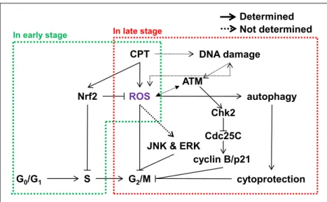
Chapter 2 Camptothecin induces G2/M phase cell cycle arrest resulting from autophagy-mediated cytoprotection: Implication of reactive oxygen species ... 14
Abstract ... 15
2.1 Introduction ... 16
2.2 Materials and methods ... 18
2.3 Results ... 22
2.3.1 CPT irreversibly induces G2/M phase arrest in various cancer cell lines ... 22
2.3.2 ROS are potential initiators of CPT-induced G2/M phase arrest ... 24
2.3.3 CPT-induced Nrf2 in the early stage delays cell cycle at the S phase ... 25
2.3.4 ATM-mediated Chk2 is a key checkpoint in CPT-induced G2/M phase arrest ... 28
2.3.5 CPT-induced Cdc25C degradation requires the proteasome pathway in G2/M phase arrest ... 31
iii
2.3.6 ERK and JNK regulate Cdc25C-mediated cyclin B and p21 expression in
CPT-induced G2/M phase arrest ... 35
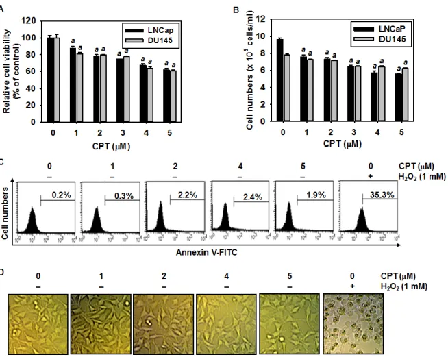
2.3.7 CPT decreases cell viability, but not induces cell death ... 37
2.3.8 CPT-induced autophagy blocks cell death and leads to G2/M phase arrest ... 39
2.4 Discussion ... 42
2.5 Conclusion ... 46
Chapter 3 Camptothecin induces mitotic arrest in LNCaP cells, resulting from Mad2-mediated cyclin B1 and Cdk1 expression: Implication of tubulin polymerization ... 47
Abstract ... 48
3.1 Introduction ... 49
3.2 Materials and method ... 51
3.3 Results ... 57
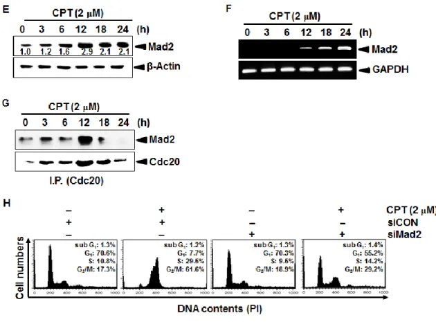
3.3.1 Mad2 is a key M phase check point in CPT-induced cell cycle arrest ... 57
3.3.2 CPT-induced Mad2 regulates cyclin B1 and Cdk1 ... 60
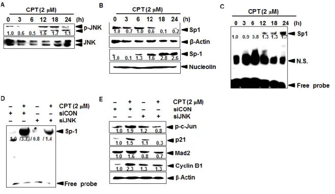
3.3.3 CPT increases Mad2 expression by inducing JNK-mediated Sp1 activation ... 62
3.3.4 CPT-induced phosphorylation of p21 promotes Mad2 expression ... 64
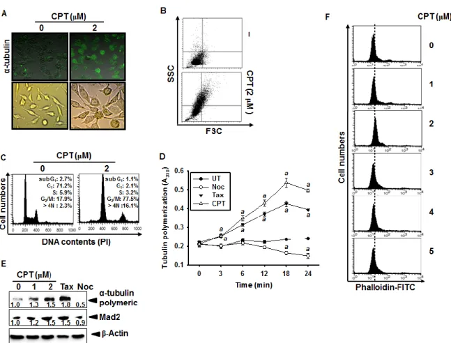
3.3.5 CPT stimulates Mad2 expression resulting from tubulin polymerization ... 66
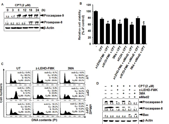
3.3.6 Accumulation of procaspase-9 and autophagy regulates CPT-induced M phase arrest ... 69
3.4 Discussion ... 72
3.5 Conclusion ... 76
Chapter 4 Camptothecin enhances c-Myc-mediated endoplasmic reticulum stress, leading to autophagy ... 77
Abstract ... 78
4.1 Introduction ... 79
4.2 Materials and methods ... 82
4.3 Results ... 86
4.3.1 CPT induces c-Myc-mediated ROS generation, accompanied by CHOP expression ... 86
iv
4.3.2 c-Myc regulates CPT-induced ER stress by inducing ROS generation ... 88
4.3.3 CPT promotes autophagy formation, resulting from ER stress ... 91
4.3.4 CPT promotes autophagy by increasing intracellular Ca2+ release ... 93
4.3.5 CPT induces JNK-dependent autophagy by enhancing AP-1 activity ... 96
4.4 Discussion ... 99
4.5 Conclusion ... 101
Chapter 5 CPT induces c-Myc- and Sp1-mediated hTERT expression in LNCaP cells: involvement of reactive oxygen species and PI3K/Akt ... 102
Abstract ... 103
5.1 Introduction ... 104
5.2 Materials and method ... 107
5.3 Results ... 112
5.3.1 CPT increases hTERT expression and activity, which is not associated with G2/M phase arrest ... 112
5.3.2 ROS regulate CPT-induced hTERT expression ... 115
5.3.3 CPT regulates c-Myc- and Sp1-dependent hTERT expression ... 117
5.3.4 CPT induces PI3K/Akt signaling involved in hTERT expression ... 120
5.3.5 CPT induces apoptosis in human leukemia cells... 122
5.4 Discussion ... 124
5.5 Conclusion ... 127
Chapter 6 Camptothecin suppresses matrix metalloproteinase-9 and vascular endothelial growth factor in DU145 cells through Nrf2-dependent HO-1 induction ... 128
Abatract ... 129
6.1 Introduction ... 130
6.2 Materials and methods ... 132
6.3 Results ... 137
6.3.1 CPT has no influence on cell viability ... 137
v
6.3.3 CPT inhibits VEGF expression and production ... 141
6.3.4 CPTdownregulates NF-κB activity ... 143
6.3.5 HO-1 induces CPT-induced MMP9 and VEGF inhibition ... 145
6.3.6 Nrf2 regulates CPT-induced MMP-9 and VEGF expression by inducing HO-1 expression ... 147
6.4 Discussion ... 150
6.5 Conclusion ... 152
Chapter 7 Camptothecin sensitizes human hepatoma Hep3B cells to TRAIL-mediated apoptosis via ROS-dependent death receptor 5 upregulation with the involvement of MAPKs... 153
Abastract... 154
7.1 Introduction ... 155
7.2 Materials and methods ... 157
7.3 Results ... 160
7.3.1 CPT sensitizes various types of cancer cells to TRAIL-mediated cell death ... 160
7.3.2 CPT/TRAIL activates apoptotic signals via the extrinsic and intrinsic pathways 161 7.3.3 DR5 upregulation is required for CPT/TRAIL-induced apoptosis ... 164
7.3.4 Reactive oxygen species (ROS) mediate CPT/TRAIL-induced upregulation of DR5 ... 166
7.3.5 ERK and p38 potentiate CPT/TRAIL-mediated DR5 expression ... 168
7.4 Discussion ... 170
7.5 Conclusion ... 172
vi
한글요약
Camptothecin(CPT)은 항암, 항균 효능을 갖는 단일화합물로써, 중국과 티베트의 원 주민들이 정통적으로 널리 약초로 쓰이던 희수나무에서 유래하였다. CPT는 1966년 처 음으로 보고 되었으며, pentacyclic alkaloid적 성질을 가지고 있다. 현재까지 CPT와 이의 유도체들은 전임상 단계까지의 연구가 성공적으로 진행되어있다. 초기 세포를 대상으 로 한 CPT의 type I topoisomerase (topo) 활성 변화 연구는 의약품 개발 부분에 새로운 흥미를 이끌어 내었다. 의약품로서 CPT는 췌장암과 난소암 치료에 사용되는 topotecan 과 irinotecan의 반합성 물질로서 재개발되었다. 거기에 더하여 기초연구에 있어서 CPT 는 방사선치료와 병행하여 치료하는 것으로 연구가 진행되었다. 이 학위논문에서는 CPT가 갖는 서로 다른 암세포주에서의 항암효과에 대한 in vitro 연구를 진행하였다. 이 학위논문에는 CPT의 활성에 대해서 6가지의 다른 챕터로 나눠 항암효과에 대한 신호전달 과정을 진행하였다. 첫 번째 단계는, CPT가 전립선암세포주인 LNCaP세포에서 cyclin B1, Cdk1, 및 다양한 단백질의 조절을 통해 G2/M arrest
유도한다는 내용이다. 더 나아가 CPT에 의한 G2/M arrest 는 활성산소와 Cdc25c의 인산 화 활성의 변화로 인한 것임을 확인하였다. 그리고, 우리는 Nrf2가 ROS를 조절함으로 써 G2/M 세포의 비율을 조절하는 것을 찾아내었다. 이번 연구에서 Nrf2를 억제하자 산 화스트레스에 의하여 DNA가 손상을 입고 G2/M arrest가 유도 되는 것을 확실하게 보여 주었다. 추가적으로, 이러한 활성은 MAPK인 ERK와 JNK의 활성화와 공동으로 일어나 는 것이 확인되었다. 마지막으로 우리는 CPT가 autophagy의 활성을 통하여 G2/M arrest 로 인한 apoptosis를 억제함으로써 세포 보호효과를 갖는 것을 보여주었다.
vii
두 번째 챕터에서는 LNCaP 세포를 이용하여 CPT의 세포주기 조절 과정을 분자세
포학적으로 이해할 수 있도록 연구를 진행하였다. 우리는 CPT가 유도하는 G2/M phase
arrest에서 Mad2의 역할을 조사하였다. CPT가 JNK의존적인 Sp1의 활성을 통하여 Mad2의 발현증가를 확인하였으며, mitotic arrest가 발생하는 과정에서 Mad2에 의해 cyclin B1과 Cdk1의 발현이 증가함으로 Mad2의 결핍이 prometaphase arrest를 유도하는
것을 확인하였다. 추가적으로 siRNA를 이용한 p21결핍은 CPT가 유도한 G2/M단계의
세포 비율은 감소하였지만, p21이 소모된 조건에서 sub-G1의 증가는 유도하지 못하였
다. 하지만, caspase-9억제제와 autophagy억제제를 전처리 한 조건에서는 apoptosis가 일 어나는 것을 방해하였다. 이러한 결과들은 CPT가 항암제 개발에 있어서 희귀하게도 세 포주기와 microtubule 조절을 하는 좋은 잠재력을 가진 것이다.
세 번째로 우리 연구의 목적은 서로 다른 암세포주에서 CPT가 autophagy를 통해 세포의 생존에 관여하는지 죽음에 관여하는지를 연구하는 것이다. c-Myc의 결핍은 LNCaP 세포에서 PERK, eIF2α와 ATF의 인산화를 제거하였는데, 이것은 c-Myc이 유도 한 UPR의 활성은 CPT에 의한 활성산소의존적임을 규명하였다. 우리는 CPT가 LNCaP 세포에서 c-Myc의 발현을 유도하는 것을 추가적으로 확인하였고, 이것은 autophagy와 JNK의 활성을 통해 생성된 활성산소에 매개되는 UPR 시그널을 유도한다. 추가적으로, 3MA를 통해 CPT가 유도한 autophagy를 억제하면, LNCaP의 세포죽음이 증가한다. 우 리의 연구결과는 AMPK의 인산화가 다른 조절인자들을 억제하는 것을 보여 준다. 칼슘 의 세포질 내 축적은 APMK의 인산화와 autophagy를 유도하는데 필수적이다. 그럼으로 이 결과들은 CPT가 유도한 autophagy가 LNCaP세포에서 보호 작용을 하는 것을 증명한 다.
viii
네 번째로 우리는 hTERT의 promoter가 포함된 luciferase 유전자 발현 실험 방법을 사용하여 CPT가 hTERT의 전사인자들을 조절하는 기전에 대하여 규명하였다. CPT는 농도 의존적으로 hTERT의 promoter 활성을 강하게 증가시켰다. 또한, CPT는 활성산소 의 생산을 유도하여 항산화 활성을 저해 하여 LNCaP세포에서 활성산소의 발현을 증가 시켰다. TRAP-ELISA kit을 사용하여 CPT가 유도한 telomerase의 활성이 항산화제의 전 처리를 하자 감소하는 것을 확인하였다. CPT의 처리는 LNCaP세포에서 c-Myc과 Sp1가 DNA와의 결합 활성을 증가시킴으로써 hTERT의 발현을 크게 증가 시켰다. 이 결과들 은 CPT가 hTERT의 인산화를 증가시키고 Akt의 인산화를 통해 핵 안으로의 이동을 방 해한다는 것으로 추정된다. 다섯 번째로, CPT가 MMP-9과 VEGF의 발현과 활성에 대한 연구를 진행하였다. CPT는 PMA와 TNF-α에 의해 유도된 MMP-9의 mRNA와 단백질발현을 강하게 억제하 였다. 추가적으로, CPT는 유방암 세포주인 MDA-MB-231과 방광암 세포주인 T24세포 에서 PMA 자극에 의해 발현된 MMP-9의 mRNA발현을 강하게 감소 시켰는데, 이것은 CPT가 서로 다른 암 세포주에서도 MMP-9의 발현을 조절 할 수 있다는 것을 의미한다. 더 나아가, CPT의 전처리는 유방암세포주인 MDA-MB-231과 방광암 세포주인 T24세 포에서 PMA와 TNF-α에 의해 유도된 VEGF의 mRNA 과발현을 강하게 억제하였다. 우 리는 CPT가 DU145 세포에서 NF-κB의 억제를 통하여 MMP-9과 VEGF의 발현 억제를 통하여 세포의 침윤작용을 방해하는 것을 추가적으로 확인하였다. 이것은 CPT가 암세 포의 전이와 침윤을 억제할 수 있는 항암제로서 좋은 후보가 될 수 있음을 의미한다. 이 번 연구에서 우리는 HO-1의 유도체인 CoPP가 PMA가 유도한 MMP-9과 VEGF의 발현 을 억제 하는 것을 확인 하였는데, 이것은 CPT에 의해 발현된 HO-1가 MMP-9과 VEGF
ix
의 발현을 억제하는 것을 의미한다. 우리는 CPT에 의해 활성화된 Nrf2 의존적으로 발 현된 HO-1이 PMA가 유도한 MMP-9과 VEGF의 발현을 억제한다는 것을 증명하였다. 이것은 Nrf2를 억제한다면 HO-1에 의해 억제되는 MMP-9과 VEGF의 발현을 증가 시킬 수 있다는 것을 의미한다.
마지막으로, 우리는 CPT와 TRAIL의 병행 처리가 간암세포주인 Hep3B 세포에서 세포죽음을 유도하는 것을 연구하였다. CPT와 TRAIL은 TRAIL에 저항성을 획득한 세 포에서도 세포의 증식을 억제하였다. Caspase-8의 억제제인 z-IETD-fmk는 CPT와
TRAIL에 의해 강하게 증가된 sub-G1 phase, annexin-V 염색된 세포비율, DNA 절편화를
강하게 억제하였고, 이것은 CPT와 TRAIL의 병행 처리가 caspase 의존적인 apoptosis를 유도하는 것을 의미한다. 그리고 우리는 CPT와 TRAIL의 병행 처리에 의해 증가된 세 포 죽음이 DR5의 발현 증가에 의한 것임을 찾아 내었고, DR5는 ROS와 MAPK인 ERK 와 p38의 활성에 의한 것임을 증명하였다.
x
Summary
Camptothecin (CPT) is a potent antitumor antibiotic isolated from extracts of Camptotheca acuminata, a tree native to China and Tibet which has been extensively used in traditional Chinese medicine. The structure was determined to be that of a pentacyclic alkaloid and was first reported in 1966. The success of CPT in preclinical studies led to clinical investigations. Due to the negligible water solubility of CPT, these trials were initiated using the water-soluble sodium salt. The discovery that the primary cellular target of CPT is type I DNA topoisomerase (topo) created renewed interest in the drug. Advances in the medicinal chemistry of CPT resulted in the semi-synthetic, more water-soluble analogues topotecan and irinotecan which are used clinically for the treatment of colon and ovarian
cancers, respectively. Additional CPT analogues are under investigation, and are also of
interest in combination regimens as radiation sensitizers.
This dissertation is mainly focused on anti-cancer effect of CPT in different cancer cell lines in vitro studies. This dissertation is divided in to six chapters based on the different phases of signaling cascade in which the CPT activity is involved. In the first phase, we
investigated that CPT-induced an irreversible G2/M phase cell cycle arrest in LNCaP cells
which is associated with a marked decrease in the expression of G2/M regulating proteins
such as cyclin B, cell division cycle 25C (Cdc25C) at Ser216. Further we reported that cell
cycle arrest caused by the generation of reactive oxygen species and ataxia telangiectasia mutated check point kinase 2-mediated phosphorylation of Cdc25C. Our finding
demonstrates that depletion of siNrf2 further induce the CPT induce S phase to G2/M phase
cell population mean that Nrf2 regulate the G2/M arrest at S phase. Thus present study opens
xi
damage and prominent G2/M arrest. These events are coordinated with the activation of
mitogen-activated protein kinases specially, extracellular-signal regulated kinase and c-jun-N-terminal kinase. Finally, we showed that CPT protects cells from apoptosis through
autophagy, direct to the G2/M phase cell cycle arrest.
Second chapter illustrated that the molecular mechanisms underline the cell cycle regulation activities of CPT in LNCaP cells. We analyze the role of Mad2 in CPT induce M phase arrest. We report that CPT induces Mad2 expression through JNK dependent Sp1 activity. Also our data show the Mad2 knockdown produced clear suppression of prometaphase arrest, the important of Mad2 in up-regulation of cyclin B1 and Cdk1 protein levels and the development of mitotic arrest. In addition to that transient transfection of
LNCaP cells with sip21 decreased the CPT-induced G2/M phase cell population but depletion
of p21 didn’t induce the sub G1 population. However, pretreatment of caspase 9 inhibitor and
autophagy inhibitor increased the apoptosis cell percentage combine treatment with CPT. Importantly CPT has good potential as a novel class of cell cycle and microtubule pathway for cancer therapy.
Next phase our aim to investigate the effect of CPT on autophagy for the cell survival or death mechanism in different cancer cell lines. Depletion of c-Myc abrogated the level of phosphorylation eIF2α, PERK and also ATF4 in LNCaP cells suggest that cMyc-induced UPR activation is ROS dependent in CPT-treated LNCaP cells. We found that CPT induced c-Myc expression in LNCaP cells, which was induced UPR signaling mediated by ROS leading to activation of JNK cascade as well as autophagy. Further CPT-induced autophagy inhibited by the 3MA, a autophagy inhibitor increased the cell death in LNCaP cells. Our data also showed phosphorylation of AMPK downregulated with the presence of those regulators.
xii
of autophagy. Therefore, these data confirmed that CPT-induced autophagy as cytoprotection mechanism in LNCaP cells.
Fourth phase we revealed the underlying mechanisms involved in CPT-induced transcriptional control of hTERT using the luciferase gene expression system containing hTERT gene promoter region. CPT significantly increased the promoter activities of hTERT dose-dependent manner. CPT significantly induces the ROS formation in LNCaP cells whereas pretreatment of antioxidant abolished the CPT-induced ROS production. Using TRAP-ELISA kit, we found that CPT-induced telomerase activity was decreased in pre-treatment of antioxidant. CPT pre-treatment resulted in a significant increase of c-Myc and Sp1 DNA binding activity in LNCaP cells and also CPT increases hTERT gene expression through enhance of c-Myc- and Sp1-binding on the regulatory regions of hTERT. These results suggest that CPT increases phosphorylation of hTERT and thereby possibly inhibit its translocation to the nucleus through the phosphorylation of Akt.
We, in the fifth phase, evaluated the effects of CPT on MMP-9 and VEGF expression and activity. CPT significantly downregulates PMA- and TNF-α-induced MMP-9 mRNA and protein expression. Additionally, CPT substantially downregulated the expression of PMA-stimulated MMP-9 mRNA in human breast carcinoma MDA-MB-231 cells and bladder carcinoma T24 cells, indicating that CPT suppresses MMP-9 expression in many different types of cancer cells. Moreover, pretreatment with CPT significantly inhibited PMA- and TNF-α-induced VEGF upregulation at the mRNA level in breast carcinoma MDA-MB-231 cells and bladder carcinoma T24 cells. We found that CPT reduces invasion of DU145 cells accompanying with downregulation of MMP-9 and VEGF via NF-κB inhibition, which indicates that CPT may be a good candidate to regulate cancer invasion. In this study, we found that CPT increases the expression of HO-1 and CoPP, an HO-1 inducer inhibits
PMA-xiii
induced MMP9 and VEGF expression, while the suppressive effect of those gene expression due to CPT is significantly reversed by potent HO-1 inhibitor ZnPP, which indicates that CPT-induced HO-1 expression is intimately associated with the downregulation of PMA-induced MMP-9 and VEGF. We demonstrated that CPT leads induction of Nrf2 by a mechanism dependent on HO-1 expression assuming that CPT reverses PMA-induced MMP-9 and VEGF expression via Nrf2 dependent HO-1 expression. It is confirmed by silencing of Nrf2 increases the MMP9 and VEGF expression accompanying with induction of HO-1.
Final chapter, we examined whether combined treatment with a sublethal dose of CPT and TRAIL (CPT/TRAIL) induces cell death in human hepatocarcinoma Hep3B cells. CPT/TRAIL effectively inhibits cell proliferation in TRAIL-resistant cells in a cell-type-nonspecific manner, although the combined treatment induces different antiproliferation rates. pretreatment with a caspase-8 inhibitor z-IETD-fmk significantly blocked apoptotic
characteristics, such as DNA fragmentation, the presence of an annexin-V+ population, and
the sub-G1 phase induced by CPT/TRAIL, indicating that CPT sensitizes Hep3B cells to
TRAIL-induced apoptosis in a caspase-dependent manner. We found that CPT/TRAIL increases cell death via upregulation of DR5 expression through the generation of reactive oxygen species (ROS) and the activation of extracellular signal-regulated protein kinase (ERK) and of p38 mitogen-activated protein kinases (MAPKs).
xiv
List of figures
Fig. 1. Chemical structure of Camptothecin ... 2
Fig. 2. Camptothecin (CPT)-induced G2/M phase arrest. ... 23
Fig. 3. Camptothecin (CPT)-induced ROS mediates G2/M phase cell cycle arrest. ... 25
Fig. 4. Camptothecin (CPT)-induced the expression of Nrf2 level. ... 27
Fig. 5. Effect of camptothecin (CPT) on ATM and Chks activation in LNCaP cells. ... 30
Fig. 6. Camptothecin (CPT)-induced Cdc25C degradation is mediated by a ubiquitin-proteasome pathway... 33
Fig. 7. Camptothecin (CPT)-induced G2/M phase arrest through JNK and ERK activity. ... 36
Fig. 8. Camptothecin (CPT)-decreased the cell proliferation in prostate cancer cells. ... 38
Fig. 9. Camptothecin (CPT)-induced autophagy as cytoprotection mechanism. ... 40
Fig. 10. Scheme of Camptothecin (CPT)-induced G2/M phase cell cycle arrest. ... 41
Fig. 11. Camptothecin (CPT)-induced Mad2 expression in LNCaP cells. ... 59
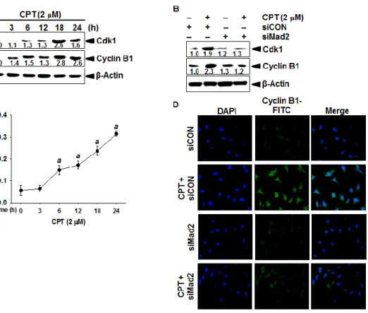
Fig. 12. Effect of camptothecin (CPT) on cyclin B1 and Cdk1. ... 61
Fig. 13. Effect of camptothecin (CPT) on JNK dependent-Sp1 activation. ... 63
Fig. 14. Effect of camptothecin (CPT) on phosphorylation of p21 in LNCaP cells. ... 65
Fig. 15. Effect of camptothecin (CPT) on tubulin polymerization. ... 68
Fig. 16. Effect of camptothecin (CPT) on accumulation of caspase-9 and autophagy. ... 70
Fig. 17. Schematic explanation of camptothecin (CPT)-induced mitotic arrest. ... 71
Fig. 18. Effect of camptothecin (CPT) on c-Myc dependent ROS production. ... 87
Fig. 19. Effect of camptothecin (CPT) on endoplasmic reticulum (ER) stress. ... 90
Fig. 20. Effect of camptothecin (CPT) on autophagy formation. ... 92
Fig. 21. Effect of camptothecin (CPT) for intracellular Ca2+ release. ... 95
Fig. 22. Effect of camptothecin (CPT) on JNK-dependent AP1 activity. ... 97
xv
Fig. 24. Camptothecin (CPT)-induced telomerase activity and hTERT expression. ... 114
Fig. 25. Nrf2 is essential for camptothecin-induced hTERT expression. ... 116
Fig. 26. Upregulation of c-Myc and Sp1 by camptothecin (CPT). ... 119
Fig. 27. Camptothecin (CPT)-induced phosphorylation of Akt in LNCaP cell. ... 121
Fig. 28. Camptothecin (CPT) decreases cell viability and proliferation ... 123
Fig. 29. Schematic explanation of camptothecin (CPT) in controlling hTERT. ... 124
Fig. 30 Effect of CPT on the viability of prostate cancer DU145 and LNCaP cells. ... 138
Fig. 31. Effect of CPT on MMP-9 activity and expression. ... 140
Fig. 32. Effects of CPT on VEGF expression. ... 142
Fig. 33. Effect of CPT on NF-κB DNA binding activity. ... 144
Fig. 34. Effect of CPT on the expression of HO-1 in DU145 cells. ... 146
Fig. 35. Effect of CPT on the expression of Nrf-2 in DU145 cells. ... 148
Fig. 36. A schematic model of CPT-induced downregulation of MMP-9 and VEGF expression in PMA/TNF-α-stimulated DU145 cells. ... 149
Fig. 37. CPT sensitizes TRAIL-induced cell death regardless of cell-type specificity. ... 161
Fig. 38. Effect of CPT and/or TRAIL on the expression of caspases and various intracellular regulators of apoptosis. ... 163
Fig. 39. Effect of CPT and/or TRAIL on DR5 expression. ... 165
Fig. 40. CPT and/or TRAIL induce ROS-mediated DR5 expression. ... 167
1
Chapter 1
2
1.1 Camptothecin as anticancer drug
Camptothecin (CPT), a naturally occurring cytotoxic alkaloid and novel effective anticancer drug, was originally isolated from Camptotheca acuminate [Zhou et al., 2010] as a strong inhibitor of the DNA-replicating enzyme topoisomerase I [Stewart and Schütz1987]. CPT has a planar pentacyclic ring structure, that includes a pyrrolo [3,4-β]-quinoline moiety, conjugated pyridone moiety and one chiral center at position 20 within the alpha-hydroxy lactone ring with (S) configuration (Fig. 1). Initially, it was discovered that CPT potentially targets topoisomerase I activity by inhibiting the rejoining step during the cleavage and relegation of DNA, resulting in topoisomerase I-induced DNA single-strand break repair pathways, ultimately leading to cell death [Pan et al., 2013; Strumberq et al., 2000]. Research also suggests that CPT possesses promising antitumor activities against a broad spectrum of cancer cell lines, such as those for melanoma, breast, colon, lung, and ovarian cancers, because many cancer cells are defective in the downregulation of topoisomerase I activity [Liu et al., 2010; Wang et al., 2008].
Fig. 1. Chemical structure of Camptothecin
Zeng et al., showed CPT induced apoptosis in cancer cells through targeting to the 3’UTR regions of Bak1, p53 and Mcl1 by miR-125b mediated mitochondrial pathways [Zeng et al., 2012]. Park et al., report indicated the CPT induces Cdk1 and cyclin E-associated kinase activities in response to DNA-damaging [Park et al., 1997]. Nevertheless, no reports
3
emphasize the CPT induce G2/M phase cell cycle arrest corresponding to the ROS/Nrf-2
signaling and MAPK cascades. Xiao reported that CPT induced s phase arrest which activate Chk1 and cause the rapid proteolysis of Cdc25A whereas elimination of Chk1 expression through knockdown abrogate s phase and protect Cdc25A degradation [Xiao et al., 2003]. It is also documented that CDK-602, a synthetic water-soluble CPT derivative and topoisomerase I inhibitor that has been shown to exert clinical anticancer effect on various types of tumor [Kim et al., 2015].
Kalpper reported that treatment of topoisomerase II inhibitor increases telomerase activity in HL60 cells and telosome reacts after DNA damage by upregulating telomerase activity and TRF2 expression [Kalpper et al., 2003]. It is further mention that activation of
telomere increased the accumulation in the G2/M phase cells. Despite Murofushi discussed
the highest levels of hTERT expression and telomerase activity, seen in the G1- and S-phases
in HepG2 cells which were 2-3-fold higher than the lowest levels in G0-phase and during
asynchronization [Murofushi et al., 2006]. According to Zeng data Camptothecin potentially inhibited DNA synthesis, followed the same kinetics as DNA-PK activation and RPA2 phosphorylation [Zeng et al., 2012].
CPT exposure in LoVo cells resulted c-Myc overexpression and elevation of p53 protein levels, suggesting a role of p53 in the c-Myc-imposed sensitization to the apoptotic effects of CPT [Babcock et al., 2013]. Camptothecin also inhibited DNA synthesis, followed the same kinetics as DNA-PK activation and RPA2 phosphorylation [Arango et al., 2003]. However, there is no report that CPT-induced c-Myc increase the UPR, resulted in induction of autophagy.
4
1.2 Cell cycle and check point control as anticancer target
The mitosis phase in normal human cells is initiated by the Cdk1/cyclin B1 complex through regulation of Cdc25C, however in cancer cells, these proteins are deregulated, so that normal growth control and checkpoints are evaded. Increased Cdc25C phosphatase activity could lead to the activation of Cdk1/cyclin B, which results from the dephosphorylation of Cdk1 by Cdc25C [Stanford et al., 2005]. In contrast, phosphorylation of Cdc25C on Ser-216 decreases its phosphatase activity through the cytoplasmic translocation of Cdc25C with 14-3-3 from the nucleus [Stanford et al., 2005]. This is known to be an important regulatory protein inducing delay or blocking mitotic entry.
The function of microtubules, other cytoskeletal components, has been frequently studied
in the G2/M cell cycle phase. For example, treatment with microtubulin-interfering agents,
such as nocodazole and taxol, induces tetraploidy arrest because sister chromatid segregation is interrupted [Kim et al., 2008]. This mitotic failure state could result in an excessive endoreduplication by prolonged exposure to microtubule-interfering agents. Additionally, Topo II inhibitors such as etoposide, amsacrine, and merbarone can induce endoreduplication by preventing the decatenation of replicated chromosomes, which subsequently fail to complete normal segregation at mitosis [Cortes et al., 2003].
All aerobic organisms experience physiological oxidant stress as a consequence of aerobic metabolism. In generating ATP, aerobic respiration releases the superoxide anion radical as a byproduct. Once produced, superoxide anion can form other ROS, such as
hydrogen peroxide (H2O2) or the highly reactive hydroxyl radical (OH−) [Uttara et al., 2009].
In normal physiological states, these are produced through electron leakage and uncoupling when molecular oxygen is consumed in the electron transport chain, although ROS can also be generated in response to various exogenous stimuli, such as radiation, surgery, or
5
chemicals. It is well established that ROS are mediators of intracellular signaling cascades. Excessive ROS generation can induce redox-signaling pathways, including those involved in oxidative stress, loss of cell function, cell cycle arrest, and apoptosis [Liou et al., 2010]. An accumulation of ROS can severely damage cellular macromolecules, especially DNA, and
induce G2/M phase arrest through ataxia telangiectasia mutant (ATM) activation [Nowsheen
et al., 2012]. Cytotoxic ROS signaling also induces mitochondrial-dependent apoptosis; this occurs through the activation of apoptosis signal-regulating kinase 1 (ASK1)/mitogen-activated protein kinase (MAPK) pathways, and of the proapoptotic Bcl-2 proteins Bax or Bak, with the subsequent effects of mitochondrial membrane permeabilization and cell death [Kim et al., 2012].
Many cytotoxic agents and/or DNA-damaging agents arrest cell cycling at G1, S or G2/M
phase. Progression from G2 to M phase is regulated by a number of the cyclin family
members, in particular cyclin B1 and Cdk1. cyclin B1, together with cyclin A, promotes the
G2/M transition.Meanwhile, Cdk1 is essential for the G1/S and G2/M phase transitions of the
eukaryotic cell cycle. The phosphorylation and dephosphorylation of Cdk1 have important regulatory roles in the control of the cell cycle [Asghar et al., 2015]. Moreover, the phosphatase activity of Cdc25C is also implicated in the regulation of the progression of
G2/M phase [Tyagi et al., 2005]. Nevertheless, no reports emphasize the CPT induce G2/M
phase cell cycle arrest corresponding to the ROS/Nrf-2 signaling and MAPK cascades.
1.3 Autophagy as a cell death and tumor suppressor mechanism
Autophagy is a homeostatic and evolutionarily conserved process that degrades cellular organelles and proteins, and maintains cellular biosynthesis during nutrient deprivation or metabolic stress [Leone et al., 2013]. Autophagy begins with the formation of
double-6
membrane vesicles, known as autophagosomes, that engulf cytoplasmic constituents. The autophagosomes then fuse with lysosomes, where the sequestered contents undergo degradation and recycling [Leone, et al., 2013]. Autophagy is important in all cells for the removal of damaged or long-lived proteins and organelles. Autophagy defects are associated with susceptibility to metabolic stress, genomic damage, and tumorigenesis in mice, indicating a role for autophagy in tumor suppression [Yang, et al., 2011]. Monoallelic loss of the essential autophagy gene, Beclin 1, has been found in 40 to 75% of human breast, prostate, and ovarian cancers [Laddha et al., 2014], suggesting that autophagy may play a role in preventing these tumors. Although autophagy is a mechanism of tumor suppression, it also confers stress tolerance that enables tumor cells to survive under adverse conditions. Stress in tumor cells is compounded by the high metabolic demand associated with rapid cell proliferation [Phan et al., 2014]. Autophagy is induced in tumor cells within hypoxic tumor regions. Stress-induced autophagy in tumor cells can lead to treatment resistance and tumor dormancy, with eventual tumor regrowth and progression [Yang et al., 2011]. In preclinical models, inhibition of prosurvival autophagy by genetic or pharmacological means was shown to kill tumor cells and trigger apoptotic cell death [Sui et al., 2013]. However, autophagy has been referred to as a double-edged sword because in certain cellular contexts, excessive or sustained tumor cell autophagy may be prodeath, particularly in apoptosis-defective cells [Kimmelman et al., 2011]. Understanding the role of autophagy in cancer treatment is critical, because many anticancer therapies have been shown to activate autophagy, although the consequences of autophagy activation in this context are unclear. The complex role of autophagy in cancer continues to emerge, and it is important to elucidate the mechanisms by which autophagy influences tumorigenesis as well as treatment response [Sui et al., 2013]. Analysis of autophagic signaling may identify novel therapeutic targets for modulation and
7 therapeutic advantage.
Consistent with previous reports, c-Myc activated the expression of genes involved in multiple physiological functions, including control of cell cycle progression, apoptosis, nucleotide biosynthesis and basal metabolism. c-Myc promote DNA damage correlated with induction of reactive oxygen species without induction of apoptosis in human fibroblasts [Lu et al., 2007]. In peculiar, deregulation of c-Myc controlled the p53-mediated DNA damage response, enabling cells with damaged genomes to enter the cycle, resulting in poor colonogenic survival [Cui et al., 2015]. Despite, c-Myc induces DNA double strand breaks independent of ROS production of murine lymphocytes in vivo as well as normal foreskin fibroblasts in vitro [Vafa et al., 2002]. Rapamycin treatment decreased bortezomib-induced expression of c-Myc protein in Elt3 cells where exogenous expression of c-Myc overcome the suppressive effect on ATF4/CHOP expression by c-Myc bound to ATF4 promoter [Ray et al., 2006]. Camptothecin exposure in LoVo cells resulted c-Myc overexpression and elevation of p53 protein levels, suggesting a role of p53 in the c-Myc-imposed sensitization to the apoptotic effects of CPT [Babcock et al., 2013]. Camptothecin also inhibited DNA synthesis, followed the same kinetics as DNA-PK activation and RPA2 phosphorylation [Arango et al., 2003]. However, there is no report that CPT-induced c-Myc increase the UPR, resulted in induction of autophagy.
1.4 Regulation of the telomerase hTERT gene as a target for cellular oncogenic mechanisms
Telomeres are localized in the physical ends of eukaryotic chromosomes in a nucleic acid-protein complex. Disruption of the telomere structure, by telomeric DNA cleavage or loss of telomere binding protein functions, is associated with senescence and cell death, whereas telomere dysfunction also induces a state of constant cell proliferation [Oulton et al.,
8
2000]. Telomerase is a ribonucleoprotein complex with specialized reverse transcriptase activity which plays a crucial role in sustainment of telomere length and inducing cell immortalization, as found in many cancers [Hahn et al., 2001]. Therefore, telomerase activity regulation has been considered as a strategy for control of senescence and cell death. Telomerase is structurally composed of an RNA subunit known as human telomerase RNA (hTR) and a protein subunit known as human telomerase reverse transcriptase (hTERT), which plays an important enzymatic role in telomerase activity. Previous studies revealed that knockdown of hTERT completely suppress cancer cell growth by telomerase inactivation, while overexpression of hTERT results in a significant decrease of flavonoid-mediated sensitivity in cancer cells [Janknecht et al., 2004]. There have been numerous studies focused on hTERT regulation in inducing cancer cell apoptosis [Moon et al., 2010], because cancer cells specifically express hTERT at much higher concentrations than normal cells.
Most cancer cells exhibit pronounced activation of telomerase which prevents telomere shortening and subsequently leads to immortal cell characteristics and tumorigenesis [Hahn et al., 2001]. This finding suggests that telomerase activity inhibition can act as a cancer treatment strategy. Due to their biological activity, it has been demonstrated that natural compounds possess chemotherapeutic functions including cell cycle arrest and induction of apoptosis [Ramos et al., 2007]. In addition, published studies indicated that many flavonoids target downregulation of telomerase in the apoptosis mechanism of cancer cells, without concurrent cytotoxicity in normal cells [Kale et al., 2008]. These results demonstrate that natural agents are good candidates for diminishing the occurrence of tumorigenesis by telomerase activity suppression.
9
1.5 Anti-invasive mechanism of cancer cells
Tumor metastasis is a multistep process by which a subset of individual cancer cells disseminates from a primary tumor initiation to distant secondary organs or tissues [Spano et al., 2012]. Previous studies have shown that most of cancer cells have high metastatic ability because of the constitutive expression of several angiogenic genes including vascular endothelial growth factor (VEGF) and matrix metalloproteinase-9 (MMP-9) [Sharma et al., 2011]. In especial, VEGF is an interesting inducer of metastasis and invasion both in vivo and in vitro, because it is a highly specific mitogen from endothelial cells to induce vascularization and angiogenesis in a malignant tissue [Chen et al., 2012]. MMP-9 is also a key effector molecule that promotes tumor cell invasion through type-IV collagen degradation-dependent extracellular matrix remodeling [Deryugina and Quigley, 2006]. MMP-9 expression has been observed in tumors of various organs, including the prostate, bladder, breast, brain, liver, and pancreatic carcinoma. Growing evidence shows that cancer cells secrete high levels of growth factors and matrix-degrading proteases and thus attributed to metastasis to distant organs including the liver, lungs, spine, bladder, bone, and lymph node [Dasgupta et al., 2012]. Therefore, it is good strategy to reduce cancer angiogenesis by inhibiting expression of VEGF and to suppress invasion by downregulate MMP-9 for treatment of several cancers.
NF-B is maintained in the cytoplasm as an inactive heterodimer form between p50 and p65 bound to its inhibitory factor B (IB) [DiDonato et al., 2012]. Upon stimulation, IB results in a rapid phosphorylation by IB kinase α/β, and subsequent ubiquitination as well as degradation by proteasome complex [Magnani et al., 2000]. In consequence, free NF-B subunits move into the nucleus and bind their specific promoter regions to regulate gene expression such as MMP-9 and VEGF. Pahl et al., (1999) showed obvious evidence that the
10
NF-κB pathway is closely involved in the expression of MMP-9 and VEGF in several types of cancer cells. A recent study also showed that the inhibition of NF-κB activity in human cancer cells decreases their tumorigenic and metastatic abilities by suppressing angiogenesis and invasion by suppression of MMP-9 and VEGF. These studies suggest that NF-κB-mediated VEGF and MMP-9 play important roles in the regulation of cell proliferation, invasion, and angiogenesis. Therefore; NF-κB is considered a good target to suppress NF-κB-dependent VEGF and MMP-9 expression to inhibit the invasion and metastasis.
1.6 TRAIL-induced apoptosis: a relevant tool for anticancer therapy
Apoptosis plays critical roles in a wide variety of physiological processes during fetal development and in adult tissues, and the characteristics of the apoptotic cells represent chromatin condensation and nuclear fragmentation, plasma membrane blebbing, and cell shrinkage [Reed ET AL., 2000]. The molecular machinery responsible for apoptosis has been elucidated, revealing a family of intracellular proteases and effector caspases, which are directly or indirectly responsible for the morphological and biochemical changes that characterize the phenomenon of apoptosis. TRAIL initiates apoptosis, upon binding to its death ligand DR4 or/and DR5 with a homotrimeric structures by inducing the recruitment of specific cytoplasmic proteins such as FADD to the intracellular DD of the receptors, which form DISC [Srivastava et al., 2001]. In addition to their carboxy-terminal DD, FADD contains an amino terminal death effect or domain (DED), which recruits DED-containing apoptosis initiating proteases, caspase-8, and caspase-10 to the receptors for the complete formation of DISC [Hellwig et al., 2012]. Both FADD and procaspase-8 contain these DED, and it is likely that aggregation of procaspase-8 molecules results in enzymatic conversion to active caspase-8 and further activation of the caspase cascade of enzymes. The recruitment of
11
caspase-8 can be inhibited by another DED containing protein called FADD-like IL-1β-converting enzyme (FLICE) inhibitory protein (c-FLIP) [Srivastava et al., 2001]. The cellular homologue c-FLIP can also inhibit apoptosis by preventing the recruitment and activation of proximal caspases. In the DISC, caspase-8 and -10 are cleaved through autoproteolysis and thus become enzymatically active and therefore initiate apoptotic cell death through cleavage of downstream effectorcaspase-3 and -7 [Festjens et al., 2006]. In contrary, DcRs could prevent apoptosis signaling via complex formation with TRAIL-R1 and/orTRAIL-R2, resulting in an ineffective DISC [Munoz-Pinedo et al., 2006]. Nevertheless, activation of caspase-8 is sufficient for subsequent activation of the effector caspase-3 to execute cellular apoptosis in some cell type (known as the intrinsic pathway or the type I apoptotic pathway). Additionally, cells move to the intrinsic pathway (mitochondria-dependent pathway) by causing changes in the inner mitochondrial membrane that results in a loss of the mitochondrial transmembrane potential and release of cytochrome c, Smac/DIABLO, and the serine protease HtrA2/Omi pro-apoptotic proteins from the intermembrane space into the cytosol [Shiozaki et al., 2002]. It is initiated by cleavage of Bcl-2 inhibitory BH3-domain-containing protein Bid by caspase-8, is required for cellular apoptosis [Bansal H., et al., 2012]. The truncated Bid (tBid) in turn interacts with Bax and Bak, and induces the oligomerization of Bax and Bak in mitochondrial membrane, which leads to the change of the mitochondrial membrane potential and release above molecules to the cytoplasm [Munoz-Pinedo et al., 2006]. Cytochrome c facilitates the interaction of apoptotic protease activating factor 1 (Apaf-1) with caspase-9 as an initiator caspase resulting in the formation of apoptosome in whichcaspase-9 is activated [Shiozaki et al., 2002]. In a parallel, Smac interacts with X-linked inhibitor of apoptosis protein (XIAP), releasescaspase-3 cleavage and thereby allows encountering the TRAILinduced extrinsic pathway. Activation of caspase9,
-12
3, and -7 can be blocked by inhibitor of apoptosis proteins (IAP), an evolutionarily conserved family of caspase inhibitory protein such as XIAP, c-IAP1, c-IAP2, and survivin. Proapoptotic members of the Bcl-2 family such as Bax, or its homologue Bak, which contain threeBcl-2 homology domains (BH3), are counteracted by the anti-apoptotic family members Bcl-2 or Bcl-xL, which contain an additional BH4 domain [Munoz-Pinedo, et al., 2006]. In previous, ectopic Bcl-xL overexpression conferred to acquire resistance against TRAIL in pancreatic cancer cell lines [Shiozaki, et al., 2002]. Hepatocytes also provide an example of cells that become resistant to Fas-induced apoptosis in vivo when Bid, or both Bax and Bak, are deleted [Chen et al., 2007]. Thus, the intrinsic pathway was required for TRAIL-mediated apoptosis through activation of proapoptotic Bcl-2 family such as Bid, Bax, and Bak which being essential for induction of the mitochondrial events.
Natural products have played a highly significant role as sources new drugs in recent decades. Therefore, natural products with strong synergistic activity with TRAIL, but minimal toxicity in normal cells are thought to be sources for new chemotherapeutic tools for TRAIL-based cancer therapy [Roberts et al., 2011]. Initially, it was discovered that CPT potentially targets topoisomerase I activity by inhibiting the rejoining step during the cleavage and relegation of DNA, resulting in topoisomerase I-induced DNA single-strand break repair pathways, ultimately leading to cell death [Strumberq et al., 2000; Pan et al., 2013]. To date, most studies have been focused on topoisomerase I activity in CPT-induced cancer cell death. However, the anticancer mechanisms of CPT in TRAIL-mediated cell death remain unclear.
13
1.7 Aims of this study
The principal goal of this thesis was to establish molecular evidence for the anti cancer mechanism and signaling pathway in human cancer cells upon the CPT treatment. The consecutive signaling molecules involved in anticancer mechanism are investigated from the transcription and translation levels. The main objectives of the study were
To characterized the molecules involved in cell cycle pathways to response CPT in prostate cancer cells
To identify the autophagy as cytoproctive mechanism under genomic stress condition To investigate the role of telomerase activity in CPT-induced cancer cells
To identify the CPT as anti-invasive agent in prostate cancer cells
To identify the CPT as sensitizer for various cancer cells to TRAIL-mediated cell death
14
Chapter 2
Camptothecin induces G
2/M phase cell cycle arrest resulting from
autophagy-mediated cytoprotection: Implication of reactive oxygen species
15
Abstract
Camptothecin (CPT) is a quinolone alkaloid which inhibits DNA topoisomerase I that induces cytotoxicity in a variety of cancer cell lines. We previously showed that CPT effectively inhibited invasion of prostate cancer cells and also combined treatment with subtoxic doses of CPT and TNF-related apoptosis-inducing ligand (TRAIL) potentially enhanced apoptosis in a caspase-dependent manner in hepatoma cancer cells. Here, we found
that treatment with CPT caused an irreversible cell cycle arrest in the G2/M phase.
CPT-induced cell cycle arrest was associated with a decrease in protein levels of cell division cycle 25C (Cdc25C) and increased the level of cyclin B and p21. The CPT-induced decrease in Cdc25C was blocked in the presence of proteasome inhibitor MG132, thus reversed the cell cycle arrest. In addition to that treatment of CPT-increased phosphorylation of Cdc25C was the resulted of activation of checkpoint kinase 2 (Chk2), which was associated with
phosphorylation of ataxia telangiectasia-mutated. Interestingly CPT induce G2/M phase of the
cell cycle arrest is reactive oxygen species (ROS) dependent where ROS inhibitors NAC and GSH reversed the CPT-induced cell cycle arrest. These results further confirm by using transient knockdown of nuclear factor-erythroid 2-related factor 2 (Nrf2) since it regulates the production of ROS. Our data reveal that treatment of siNrf2 increased the ROS level as well
as further increased the CPT induce G2/M phase cell cycle arrest. Our data also indicate
CPT-enhanced cell cycle arrest through the extracellular signal-regulated kinase (ERK) and the c-Jun N-terminal kinase (JNK) pathway. Inhibitors of ERK and JNK more decreased the Cdc25C expression and protein expression of p21 and cyclin B. These findings indicate that
16
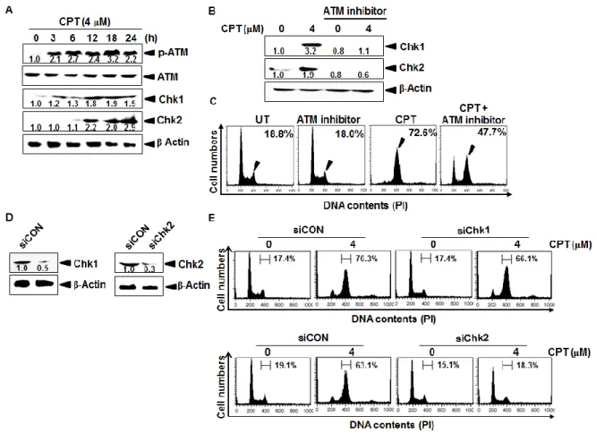
2.1 Introduction
Cell cycle checkpoints are important machineries in eukaryotic cells which accurately serve intact cell division by monitoring defects in the cell cycle [Bertoli et al., 2014]. Thus, the checkpoints permit uncontrolled cells to repair DNA damage or to consequently die by blocking cell division after DNA damage [Hartwell et al., 1989]. In particular, ataxia telangiectasia mutated (ATM), and ATM and Rad3 related (ATR) protein kinases are well known to exert cell cycle delay in the response to genotoxic stress by inducing phosphorylation of a various kinase checkpoint kinase 1 (Chk1) and Chk2 [Mazouzi et al.,
2014]. For instance, Chk1 is phosphorylated at Ser317 and Ser345 by ATM, followed by
autophosphorylation of Ser296 in response to various types of DNA damage which can be
induced by ionizing radiation, and prevents damaged cells from entering into mitosis by inactivating Cdc25 phosphatases or directly abrogate mitotic spindle formation by activating aurora B and BubR1 [Lossaint et al., 2011]. Chk2 is also activated by ATM in response to single or double-strand DNA breaks through dimerization and autophosphorylation, and consequently targets Cdc25C phosphatase by stabilizing p53 responsible for regulating
activation of cyclin-dependent kinase 2 (Cdk2) and cyclin B1 to facilitate the G2/M phase cell
cycle arrest under DNA damage [Stolz et al, 2011; Ito et al., 2007]. Therefore, ATM-mediated Chk1/2 is a main axis by generating reactive oxygen species (ROS) whether the damaged
cells will be repaired or undergo G2/M phase arrest/apoptosis, suggesting that the pathway is
a good therapeutic strategy as a drug target for cancer [Garber et al., 2005].
Nrf2 is a transcription factor that plays a pivotal role in activating an antioxidant response that decreases generation of reactive oxygen species (ROS) and induction of Nrf2-related genes are imperative for defected cells to counteract ROS-mediated oxidative damage [de Vries et al., 2008]. Additionally, disruption of Nrf2 causes the oxidant-mediated acute
17
lung injury and inflammation in mice and Nrf2 knockout mice are greatly predisposed to chemical-induced DNA damage [Reddy et al., 2009]. Previous study also reported that inhibition of Nrf2 by excessive ROS generation caused DNA damage, specifically DNA single or double strand breaks, leading to activating ATM [Chen et al., 2015]. Upon the ROS production, the activation of cell-cycle checkpoints in response to oxidative damage-mediated ATM phosphorylation is also essential for maintenance of genomic integrity and tumor suppression [Kim et al., 2012; Swift et al., 2014]. In particular, several kinds of cyclin and cyclin-dependent kinase (Cdk) complexes are well known to be implicated in DNA single or double strand breaks-induced the checkpoint responsible for cell cycle progression.
Cyclin D and Cdk4/6 complex is active in early G1 phase, whereas cyclin E-Cdk2 is required
for entry into S phase [Asghar et al., 2015]. At oxidative stress involving DNA damage, p21-mediated cell cycle arrest would be also activated to interval for DNA repair and indeed p21 is an inhibitor of Cdk that regulates many cellular processes in a p53-depedent and independent manner [Piccolo et al., 2012]. Recent publication also showed that p21 protected the cells against oxidative stress through upregulation of Nrf2 by competing interacts with the motifs in Nrf2 and thus competes Keap1-Nrf2 binding, compromising ubiquitination of Nrf2 (Asghar et al., 2015). Therefore, ROS-mediated Nrf2 could be an important response to regulate cell cycle and apoptosis as a cell cycle checkpoint.
Camptothecin (CPT) specifically inhibits eukaryotic DNA topoisomerase I (topo I) by trapping a covalent enzyme-DNA intermediate [Chen et al., 2009]. Zeng et al., showed that CPT induced apoptosis in cancer cells through targeting to the 3′ untranslated (UTR) region of Bak1, p53, and Mcl1 by miR-125b mediated mitochondrial pathways [Pommier et al., 2006]. Park et al., reported that CPT induced Cdc2 and cyclin E-associated kinase activities in response to DNA damage [Pommier et al., 2006]. Huang et al., suggested that
CPT-18
induced DNA single-strand breaks are differentially involved in homologous recombination repair by Chk1 and Chk2 [Huang et al., 2008; Zuco et al., 2010]. Nevertheless, no detail
reports emphasize whether CPT induces G2/M phase cell cycle arrest corresponding to the
ROS/Nrf2-mediated cell cycle arrest and autophagy-mediated cytoprotection. In this study,
we investigated that CPT induced an irreversible G2/M phase cell cycle arrest in LNCaP cells
through the ROS-dependent Chk2/Cdc25C and the ATM-dependent Chk2/Cdc25C pathway by activating extracellular-signal regulated kinase (ERK) and c-jun-N-terminal kinase (JNK).
Furthermore, CPT-induced autophagy protects cells from apoptosis and directs to the G2/M
phase cell cycle arrest.
2.2 Materials and methods
Regents and antibodies
CPT, 3-(4,5-dimethylthiazol-2-yl)-2,5-diphenyltetrazolium bromide (MTT), propidium
iodide, glutathione (GSH), N-acetyl-L-cysteine (NAC), MG132, 3-methyladenine (3MA), and
bafilomycin A1 (BAF) were purchased from sigma (St. Louis, MO) and an enhanced chemiluminescence (ECL) kit was purchased from Amersham (Arlington Heights, IL). RPMI 1640 medium, fetal bovine serum (FBS), and antibiotics mixture was purchased from WelGENE (Daegu, Republic of Korea). PD98059, SP600125, and SB239063 were purchased from Calbiochem (San Diego, CA). Antibodies against Cdk2, cyclin B1, p21, β-actin, Nrf2,
nucleolin, phospho (p)-ATM (Ser1981), ATM, Chk1, Chk2, p-Chk2 (Thr68), Cdc25c,
p-Cdc25C (Ser216), ubiquitin, procaspase-3, procaspase-9, Cdk2, Bad, Beclin-1, LC3-II, and
Atg-7 were purchased from Santa Cruz Biotechnology (Santa Cruz, CA). Antibodies against p-histone (H)-3, p-ERK, ERK, p-p38, p38, p-JNK, and JNK were purchased from Cell Signal (Beverly, MA). Peroxidase-labeled donkey anti-rabbit and sheep anti-mouse immunoglobulin
19
were purchased from Koma Biotechnology (Seoul, South Korea).
Cell culture and viability assay
Human prostate cancer cell lines LNCaP and DU145, human hepatoma carcinoma cell line Hep3B, and human leukemia cancer cell line U937 were obtained from the American
Type Culture Collection. Cells were cultured at 37°C in a 5% CO2-humidified incubator and
maintained in RPMI 1640 medium containing 10% heat-inactivated FBS and 1% antibiotics
mixture. The cells were seeded (4 × 10⁴ cells/ml) and then incubated with CPT for 24 h. MTT
assays were performed to determine relative cell viability.
DNA fragmentation assay
After treatment with CPT for 24 h, LNCaP cells were lysed in DNA fragmentation lysis buffer containing 10 mM Tris (pH 7.4), 150 mM NaCl, 5 mM EDTA, and 0.5% Triton X-100 for 1 h on ice. Lysates samples were vortexes and separated centrifugation at 13,000 g for 15 min. Fragmented DNA in the supernatant was extracted with equal volume of phenol:chloroform:isoamyl alcohol (25:24:1) mixture and analyzed electrophoretically on 1.5 % agarose gels.
Flow cytometric analysis
Cells were fixed in 1 U/ml of RNase A (DNase free) and 10 µg/ml of propidium iodide overnight in the dark at room temperature. To assess whether apoptosis had occurred, the cells were incubated with annexin-V (R&D Systems). A FACSCalibur flow cytometer (Becton Dickinson, San Jose, CA) was used to determine the number of apoptotic cells, i.e.,
20 Measurement of ROS
Cells were plated at a density of 5 104 cells/ml, allowed to attach for 24 h, and exposed
to 5 mM NAC alone, 5 mM GSH alone, 4 µM CPT alone, or NAC or GSH plus CPT for 1 h.
The cells were stained with 10 µM H2DCFDA for 10 min at 37°C and flow cytometry was
used to determine the fluorescence intensity.
Western blot analysis
Whole-cell lysates were prepared by PRO-PREP protein extraction solution (iNtRON Biotechnology, Sungnam, Republic of Korea). Cytoplasmic and nuclear protein extracts were prepared using NE-PER nuclear and cytosolic extraction reagents (Pierce, Rockford, IL). The cell lysates were harvested from the supernatant after centrifugation at 13,000 g for 20 min. Total cell proteins were separated on polyacrylamide gels and standard procedures were used the transfer them the nitrocellulose membranes. The membranes were developed using an ECL reagent.
Electrophoretic mobility shift assay (EMSA)
Transcription factor-DNA binding activity assays were carried out with nuclear protein extract. Synthetic complementary anti-oxidant response element (5’- TMANNRTGAYNNGCRWWWW-3’) binding oligonucleotides was 3′-biotinylated utilizing the biotin 3′-end DNA labeling kit (Pierce) according to the manufacturer’s instructions, and annealed for 30 min at 37°C. Samples were loaded onto native 4% polyacrylamide gels pre-electrophoresed for 60 min in 0.5X Tris borate/EDTA (TBE) buffer on ice, in the presence of
transferred onto a positively charged nylon membrane (HybondTM-N+) in 0.5X TBE buffer at
21
membrane at 120 mJ/cm2. Horseradish peroxidase-conjugated streptavidin was utilized
according to the manufacturer′s instructions to monitor the transferred DNA-protein complex.
Small interfering RNA (siRNA)
Cells were seeded on a 24-well plate at a density of 1 × 10⁵ cells/ml and transfected
Nrf2-, Chk1-, and Chk2-specific silencing RNA (siRNA, Santa Cruz Biotechnology) for 24 h. For each transfection, 450 μl growth medium was added to 20 nM siRNA duplex with the transfection reagent G-Fectin (Genolution Pharmaceuticals Inc., Seoul, Republic of Korea) and the entire mixture was added gently to the cells.
Statistical analysis
The images were visualized with Chemi-Smart 2000 (VilberLourmat, Marine, Cedex, France). Images were captured using Chemi-Capt (VilberLourmat) and transported into Photoshop. All bands were shown a representative obtained in three independent experiments and quantified by Scion Imaging software (http://www.scioncorp.com).Statistical analyses were conducted using SigmaPlot software (version 12.0). Values were presented as mean ± standard error (S.E.). Significant differences between the groups were determined using the
unpaired one-way and two-way ANOVA test. Statistical significance was regarded at a and b,
22
2.3 Results
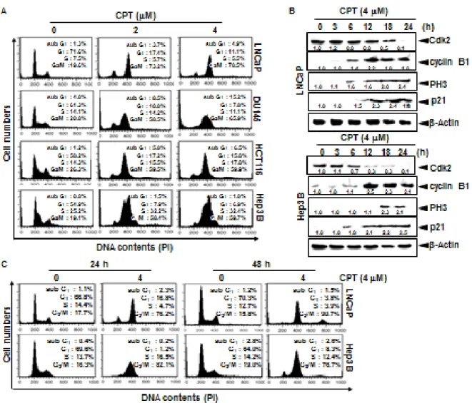
2.3.1 CPT irreversibly induces G2/M phase arrest in various cancer cell lines
CPT was known to inhibit tumor cell growth through the induction of apoptosis via p53 dependent and mitochondrial dependent pathways [Pommier et al., 2006]; however, mechanism by which CPT contributes to cell cycle progression is not known in detail. Therefore, we first investigated the effect of CPT on cell cycle distribution using propidium
iodide. Treatment with CPT resulted in a significant increase in G2/M phase cells at 24 h
which was accompanied by decreased in G0/G1 phase cells in all cell lines studied such as
LNCaP, DU145, HCT116, and Hep3B cells (Fig. 1A). Treatment of 4 μM CPT strongly
induced over 55% G2/M phase arrest in all the cell lines. Additionally, the sub-G1 population
which indicates apoptotic cell death slightly increased in DU145 and HCT116 cell lines. In
order to more clearly evaluate the G2/M phase arrest, the expressional changes of proteins
that control the cell cycle transition were observed in LNCaP and Hep3B cells. As shown in Fig. 1B, gradual decrease of Cdk2 expression suggested that treatment with CPT moves the
cells from G1/S phase to G2/M phase because Cdk2 is most active in the S phase and
decreases in G2/M phase. Our data also confirmed that CPT induced G2/M phase arrest by
inducing p21 and cyclin B1 expression, which functions as tumor suppressor and initiates cell
cycle arrest by inhibiting Cdk activity in G2/M phase in response to DNA damage [Abbas et
al., 2009]. Additionally, treatment with CPT resulted in a significant increase of p-H3 expression which is considered to be a crucial event in onset of mitosis [Hartwell et al.,
1989]. Finally, in order to elucidate whether CPT-induced G2/M phase arrest was irreversible,
the cells were treated with CPT for 24 h and then either checked out cell cycle distribution after exposure to CPT-free fresh media for the indicated times. Treatment with CPT increased
23
phase arrest was sustained by 48 h (Fig. 1C), indicating that CPT irreversibly induces G2/M
phase arrest. Taken together, these results indicate that CPT-induced G2/M arrest was
irreversible.
Fig. 2. Camptothecin (CPT)-induced G2/M phase arrest. Cells were seeded at 1 × 10⁵ cells/ml
and were treated with 4 μM CPT for the indicated times. (A) Cells were harvested, stained with propidium iodine and analyzed for the cell cycle. (B) LNCaP cells and Hep3B cells were treated with 4 μM CPT for the indicated time points. Cell extracts were prepared for western blot analysis for anti-cyclin B1, anti-p-H3, anti-p21, anti-Cdk2, and anti-Cdc2. Cells were treated with 4 μM CPT for 24 h and subsequently the cells were processed for analysis for
24
cell cycle distribution or rest of the cells cultured in drug free culture media for another 24 h and cell cycle distribution was checked. Data from three independent experiments are presented.
2.3.2 ROS are potential initiators of CPT-induced G2/M phase arrest
Recent publication shows that some chemical oxidant-mediated intracellular accumulation of ROS leads to DNA damage and checkpoint activation consequently induces cell cycle arrest [Guo et al., 2014]. Therefore, we monitored ROS generation in CPT-treated
LNCaP cells using H2DCFDA which oxidized with the presence of ROS. CPT significantly
induced the ROS formation in a time-dependent manner (Fig. 2A). Next, we measured the level of ROS generation and the cell cycle progression with the presence of ROS inhibitor
GSH. Treatment with CPT significantly increased the G2/M phase by approximately 80% of
cell population with high ROS generation, whereas pretreatment with GSH reversed
CPT-induced G2/M phase arrest to approximately 55% while increasing the S phase cell portion
with low ROS level, which indicated that GSH could not completely inhibit CPT-induced
G2/M phase arrest, but delayed cell cycle or stop in the S phase (Fig. 2B). These data indicate
25
Fig. 3. Camptothecin (CPT)-induced reactive oxygen species (ROS) mediates G2/M phase
cell cycle arrest. (A) Effect of CPT on ROS production. LNCaP cells were treated with 4 μM
CPT for the indicated time points. H2DCFDA-based fluorescence detection was done through
flow cytometry. (B) LNCaP cells were treated with 5 μM glutathione for 30 min and then incubated with 4 μM CPT for 24 h. Cell cycle distribution was analyzed through flow cytometry after cells stained with propidium iodine. (C) Similar treatment was done and cells
were stained with H2DCFDA-based fluorescence and detected through flow cytometry.
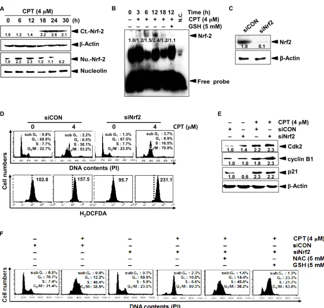
2.3.3 CPT-induced Nrf2 in the early stage delays cell cycle at the S phase
Nrf2 plays a pivotal role in activating an anti-oxidant response that counteracts the ROS to protect against cellular damage [de Vries et al., 2008]. Therefore, we investigated whether
Nrf2 regulates CPT-induced G2/M phase arrest by inhibiting ROS generation. Western blot
26
cytosolic compartment from at 18 h; however, nuclear translocation of Nrf2 significantly increased in the early stages before at 12 h and was suppressed from at 18 h (Fig. 3A). EMSA data also confirmed that CPT gradually induced the specific DNA-binding activity of Nrf2 at the early stage (at 6 h and 12 h) after CPT treatment and then the Nrf2 activity was completely decreased at 18 h (Fig. 3B), which indicated that Nrf2 at the early stages naturally occurred to alleviate ROS-mediated damage; however, Nrf2 at the late stages was completely downregulated by CPT-induced ROS. Similar to CPT-induced Nrf2 downregulation, GSH significantly inhibited the Nrf2-binding activity at the early stage. Next, we investigated
whether CPT-induced Nrf2 at the early stage (at 12 h) influences ROS-mediated G2/M phase
arrest. Nrf2-specific silencing siRNA (siNrf2) significantly reduced the Nrf2 protein level, compared to that of the control siRNA (Fig. 3C). In a parallel experiment, siNrf2
significantly increased CPT-induced G2/M phase arrest (approximately 53% to 70%) with
high levels of ROS compared to those of CPT treatment alone; however, control siRNA (siCON) at 12 h delayed cell cycle at the S phase, suggesting that Nrf2 delays cell cycle at the
S phase to slowly move into G2/M phase (Fig. 3D). As presumed, western blot analysis
showed that siNrf2 sustained CPT-induced cyclin B1 and p21 expression because siNrf2
accelerates into G2/M phase arrest; however, Cdk2 surprisingly sustained at the early stage (at
12 h) under the same condition, resulting from that S phase cell population also increased in
CPT-induced G2/M phase arrest in the presence of siNrf2 (Fig. 3E). In order to in detail
confirm whether CPT/siNrf2-induced G2/M phase arrest directly involved in the ROS
generation, we analyzed the cell cycle in the presence of ROS inhibitors, NAC and GSH. As
shown in Fig. 3F, CPT/siNrf2 significantly increased G2/M phase cell cycle arrest by
approximately 80%; however, NAC and GSH markedly downregulated the G2/M phase arrest