The difference in brain activation related
to the directionality of affective reversal
association between patients with
schizophrenia and healthy controls
Il Ho Park
Department of Medicine
The difference in brain activation related
to the directionality of affective reversal
association between patients with
schizophrenia and healthy controls
Directed by Professor Jae-Jin Kim
The Doctoral Dissertation
submitted to the Department of Medicine,
the Graduate School of Yonsei University
in partial fulfillment of the requirements for the degree
of Doctor of Philosophy
Il Ho Park
This certifies that the Doctoral
Dissertation of Il Ho Park is approved.
---
Thesis Supervisor : Jae-Jin Kim
---
Thesis Committee Member#1 : Dong Goo Kim
---
Thesis Committee Member#2 : Jeong Hoon Kim
---
Thesis Committee Member#3 : Min-Seong Koo
---
Thesis Committee Member#4 : Hae-Jeong Park
The Graduate School
Yonsei University
ACKNOWLEDGEMENTS
I owe my deepest gratitude to my supervisor, professor Jae-Jin Kim for inspiring and guiding me to the enlightening field of neuroscience and neuroimaging. His mentorship was paramount in providing key experiences in the process of neuroscientific research and a role model of a creative and analytic researcher. I am also grateful for his guidance which helped me pursue my academic career. I would like to thank Professor Min-Seong Koo who was considerate and gave me moral support throughout this study. His help was also crucial in recruiting patients and proceeding with this study.
I am also grateful for professor Hae-Jeong Park who has been an invaluable advisor and supporter for providing the knowledge and resources needed to acquire optimal brain imaging data.
I thank professors Dong Goo Kim and Jeong Hoon Kim for providing important and constructive comments and advices that was necessary to be dealt for the publication of this study.
Without the on-site help and hands-on experience of Ji-Won Chun in the fMRI scanning procedures, conducting this research would have been impossible. I also appreciate her prompt and reliable help, and the experiences in problem-solving we have shared. I would like to acknowledge Dr. Seok-Hyeong Kim for her help in recruiting and ensuring that the patients were comfortable with the procedure. I am grateful for professor Suk kyoon An, who gave me the first glimpse in neuroscience, which was an exhilarating experience and made me pursue this field.
None of this would not have been possible without the insightful support of my former mentor, professor Hong Shick Lee, who made it possible for me to meet my supervisor and to study neuroscience and neuroimaging.
Most importantly, I am very grateful of my wife, Hee-Jung for her love and emotional support throughout this study and during my difficult times. I dedicate this work to my parents, Kyung-Ok Suh and Sang-Seek Park who have always stood by my side.
<TABLE OF CONTENTS>
ABSTRACT ··· 1
I. INTRODUCTION ··· 3
II. MATERIALS AND METHODS ··· 6
1. Participants ··· 6
2. Monetary incentive contingency reversal task ··· 8
3. Imaging data acquisition ··· 11
4. fMRI data analysis ··· 11
5. Behavioral data analysis ··· 13
III. RESULTS ··· 13
1. Behavioral results ··· 14
2. Imaging data results ··· 16
3. Correlation with anhedonia and negative symptoms ··· 22
IV. DISCUSSION ··· 25
1. The neural process of contingency reversal, reversal response, and
motivation ··· 25
2. Deficient neural processing of motivation and salience attribution
in patients with Schizophrenia ··· 28
3. Implications and limitations ··· 31
V. CONCLUSION ··· 32
REFERENCES ··· 33
LIST OF FIGURES
Figure 1. Schematic description of the monetary incentive contingency
reversal task ··· 10
Figure 2. Percent correct response of reward-to-punishment (RtoP) and
punishment-to-reward (PtoR) contingency reversals ··· 15
Figure 3. Proportional change of reaction times during
reward-to-punishment (RtoP) and punishment-to-reward (PtoR)
contingency reversals ··· 16
Figure 4. The activation patterns of the amygdala among the region of
interest obtained from the reversal response contrast in healthy controls
and patients with schizophrenia ··· 19
Figure 5. The activation patterns of the putamen among the region of
interest obtained from the reversal response contrast in healthy controls
and patients with schizophrenia ··· 20
Figure 6. . The activation patterns of the anterior cingulate cortex among
the region of interest obtained from the reversal response contrast in
healthy controls and patients with schizophrenia ··· 21
Figure 7. The activation patterns of the lateral orbitofrontal cortex among
the region of interest obtained from the reversal response contrast and
the contingency reversal contrasts in healthy controls and patients with
schizophrenia, respectively ··· 22
Figure 8. Correlations of the physical anhedonia scale scores with
changes of activities in the anterior cingulate cortex during
punishment-to-reward contingency reversal and sustained reward
contingency in the healthy control group ··· 23
Figure 9. Correlations of the physical and social anhedonia scale scores
and the Positive and Negative Syndrome scores with changes of
activities in the lateral orbitofrontal cortex during punishment-to-reward
contingency reversal and sustained reward contingency in patients with
schizophrenia ··· 24
LIST OF TABLES
Table 1. Demographic and clinical characteristics ··· 8
Table 2. The reaction times (ms) and standard deviations before
contingency reversals and during reversal responses. ··· 15
Table 3. Regional activations for contingency reversal contrast ··· 17
ABSTRACT
The difference in brain activation related to the directionality of affective
reversal association between patients with schizophrenia and healthy
controls
Il Ho Park
Department of Medicine
The Graduate School, Yonsei University
(Directed by Professor Jae-Jin Kim)
The dopaminergic mesolimbic pathway, a classical neural system involved in the pathophysiology of schizophrenia, has been implicated as a core neural system for processing motivationally salient information that is either rewarding or aversive. Affective bias in reversal learning, where reattributing appropriate rewarding values is difficult whereas false aversive values is easy, may underlie clinical manifestations of schizophrenia, such as paranoid delusions and avolition. The present study investigated the affective bias in reversal learning and its underlying neural process in the cortico-striato-limbic network in patients with schizophrenia. Fifteen healthy participants and 14 outpatients with schizophrenia underwent an event-related functional magnetic resonance imaging scanning while performing a monetary incentive contingency reversal task. Patients had higher physical and social anhedonia scale score than healthy controls. Both groups showed greater accuracy when reversing from punishment-to-reward contingency than vice versa without group differences. While healthy controls showed unidirectional acceleration in reaction time when reversing from punishment-to-reward contingency, patients showed significantly diminished punishment-to-reward reversal acceleration. In healthy controls, the anterior cingulate cortex was significantly activated and the
amygdala, putamen, and the lateral orbitofrontal cortex activations were also identified during reversal response. In patients with schizophrenia only reversal response-related lateral orbitofrontal cortex activations were identified. Unidirectional punishment-to-reward reversal activations were observed in the lateral orbitofrontal cortex in both groups and in the anterior cingulate gyrus in healthy controls only. Physical anhedonia score correlated with reversal response-related anterior cingulate activity changes in healthy controls, whereas physical and social anhednoia scale scores and PANSS negative symptom scores correlated with the lateral orbitofrontal cortex in the patients. These finding suggest that deficiency in anticipation and engagement in reversing instrumental behavior to obtain reward reflected in the blunted anterior cingulate and compensatory lateral orbitofrontal activity may underlie the neural pathophysiology of anhedonia/avolition in schizophrenia.
---
Key words: contingency reversal; anterior cingulate; orbitofrontal cortex;
negative symptom; anhedonia
The difference in brain activation related to the directionality of affective
reversal association between patients with schizophrenia and healthy
controls
Il Ho Park
Department of Medicine
The Graduate School, Yonsei University
(Directed by Professor Jae-Jin Kim)
I. INTRODUCTION
Schizophrenia is a complex disorder exhibiting both positive symptoms, such as delusions and hallucinations and negative symptoms, such as avolition, anhedonia, and social withdrawal. Fundamental disturbance in emotional learning involving dopamine modulation may underlie these seemingly contrasting core manifestations.1 The dopaminergic mesolimbic pathway, a classical neural system involved in the pathophysiology of schizophrenia, has been implicated as a core neural system for processing motivationally salient information that is either rewarding or aversive.2,3 The process of assigning emotional salience to surrounding stimuli guides approach or withdrawal behaviors that are vital to an individual’s adaptation to his/her environment. It has been proposed that patients with schizophrenia may develop delusion in a cognitive attempt to make sense of one’s “psychotic” experience in which salience is abnormally assigned and may experience the internal representations of aberrant salience as hallucinations. Failure to attribute rewarding salience may lead to anhedonia and amotivation in patients with schizophrenia.4 One remarkable clinical aspect of emotional learning in schizophrenia is an affective
bias in reversal learning where reattributing appropriate rewarding values is difficult and false aversive values easy. Patients with schizophrenia may consider a person as threatening and persistently consider him/her to be persecutory despite displays of goodwill but easily perceive trivial irrelevant responses by that person as a threat. In patients with negative symptoms, motivational values are rarely attained despite pleasurable experience of an activity.
Prior studies have consistently associated reversal learning to the orbitofrontal cortex or ventral prefrontal cortex. Damage to the orbitofrontal cortex, exemplified by the case of Phineas Gage, result in dysfunction in affective regulation and social behavior that has been associated with alterations in flexible stimulus-reward learning and has been demonstrated to show impairment in performance of reversal learning tasks.5,6 Due to its advantage in spatial and temporal resolution, functional magnetic resonance imaging (fMRI) has been extensively used to distinguish the roles of subregions in the orbitofrontal cortex and the subcortical structures, and to examine the process involved in reversal learning. Functional imaging studies have reported the distinct roles of the medial and lateral orbitofrontal cortex, and the involvement of subcortical structures including the striatum and amygdala, and co-activation of the anterior cingulate in reversal learning.7-12 The medial and lateral orbitofrontal activations has been observed during gaining and losing outcomes of a monetary reversal learning task respectively suggesting their roles in reward acquisition and punishing feedback that inhibits prior response during reversal.9 In a study using a probabilistic reversal learning task, the activation in the ventral prefrontal-striatal circuit was observed during feedbacks preceding reversal that was not observed during non-reversal negative feedbacks.10 In another monetary reversal learning fMRI study, the orbitofrontal cortex activities were associated with feedback processing whereas the ventral striatum and ventrolateral prefrontal cortex were specifically associated with reward and
punishment respectively.11 Reversals in fear-conditioning have been shown to activate the striatum and amygdala with striatal activation associated with prediction errors and amygdala activation associated with predicting or retaining the association of aversive outcomes.12 When confounding negative reinforcers from the monetary reversal learning task were excluded from the reversal event, the anterior cingulate was reported to co-activated with the orbitofrontal cortex suggesting the role of the anterior cingulate- orbitofrontal circuit in general reversal learning.7
The impairment of emotional learning in schizophrenia has been reported in studies using gambling tasks.13-15 Recent imaging studies suggest that this impairment in emotional learning is associated with functional and structural abnormality of the ventral striatum and amygdale in schizophrenia. In these studies, the ventral striatum showed impaired neural responses to salient stimuli and inappropriate activations in response to a neutral stimulus and decreased size of the amygdala was associated with impaired emotional learning in patients with schizophrenia.16-18 Neuropsychological studies using a simple reversal learning task and probabilistic reversal learning task have reported a reversal learning deficit in patient with schizophrenia indicating a dysfunction in the orbitofrontal cortex.13,19,20 These findings suggest that dysfunction in the corticolimbic circuit comprised of the orbitofrontal cortex, ventral striatum and amygdala may be associated with impaired reversal learning in schizophrenia. However, whether reversal learning deficit in schizophrenia involve a reversal deficit independent from abnormalities in acquisition of contingency has not been elucidated. In addition, reports of attentional bias to negative affect and deficit in negativity bias, a normal tendency to make negative assessments during simultaneous processing of opposing emotional valence, suggest that patients with schizophrenia may have an affective bias in reversal learning.21-23
The present study investigated the affective bias in reversal learning and its underlying neural process in the cortico-striato-limbic network in patients with
schizophrenia. In order examine affective reversal and to match learning performance between healthy controls and patients, we employed a simplified monetary incentive reversal association paradigm with contingency reversals without probabilistic error trials. The reaction time change during the reversal learning of reward-to-punishment (RtoP) and punishment-to-reward (PtoR) contingency reversals were compared to examine the implicit inclination in affective reversal association. The pattern of neural activities during contingency reversal feedback and reversal response in the orbitofrontal cortex, anterior cingulate gyrus, amygdala, and the striatum according to the direction of affective reversal association were compared between healthy controls and patients with schizophrenia. Patients with schizophrenia were hypothesized to show a greater inclination for RtoP reversal and lesser inclination for PtoR reversal that would be reflected in a distinct pattern of deficient top-down regulation by the prefrontal cortex and a bias in bottom-up salience processing in the subcortical limbic regions.
II. MATERIALS AND METHODS
1. Participants
Eighteen healthy individuals and 16 outpatients with schizophrenia at Myongji Hospital who gave written informed consent to the protocols approved by the Institutional Review Board of Myongji Hospital and Severance Hospital participated in this study. All patients met the DSM-IV-TR criteria24 for schizophrenia without other comorbid psychiatric disorders. Healthy participants with past or present psychiatric illness and any participants with past or present medical or neurological illness, mental retardation according to the Raven’s Progressive Matrices,25 or left-handedness or ambidexterity
according to the Annett Handedness Scale26 were excluded. Trait anhedonia was measured using the Physical and Social Anhedonia Scale27 and the emotional state was measured using the Positive and Negative Affect Scale (PANAS).28 Symptom severity in the patients were assessed using the Positive and Negative Syndrome Scale (PANSS).29
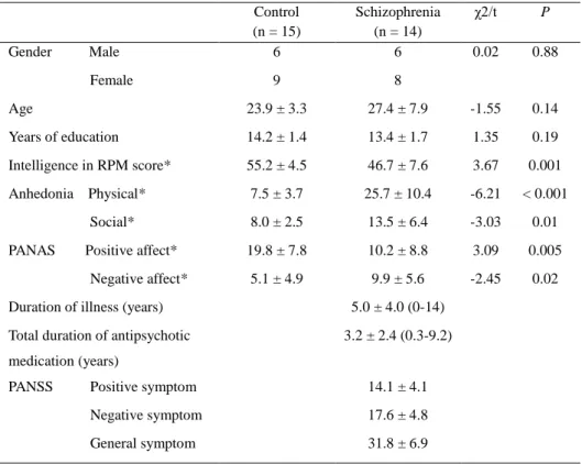
Data from 3 healthy individuals and 2 patients were either missing, had extensive signal loss, had errors due to problems in task presentation, or was an extreme outlier in performance suggesting inattention during task performance. Data from the remaining 15 healthy individuals (6 males; mean age, 23.9 ± 3.3 years; mean years of education, 14.2 ± 1.4) and 14 patients with schizophrenia (6 males; mean age, 27.4 ± 7.9; mean years of education, 27.4 ± 7.9) were analyzed. Both groups were matched for age, gender and years of education (age, χ2 = 0.02, P = 0.88; gender, t = -1.55, P = 0.14; years of education, t = 1.35, P = 0.19). Healthy controls had significantly higher intelligence scores measured by the Raven’s Progressive Matrices (t = 3.67, P = 0.001). Patients had significantly greater Physical and Social Anhedonia Scale scores and Negative Affect scores, and lower Positive affect scores (physical anhedonia, t = -6.21, P < 0.001; social anhedonia, t = -3.03, P = 0.01; negative affect, t = -2.45, P = 0.02; positive affect, t = 3.09, P = 0.005). Patients were ill for less than a year to 14 years (mean = 5.0 years, SD = 4.0) and had taken antipsychotics for 3 month to 9.2 years (mean = 3.2 years, SD = 2.4). Negative symptoms were more dominant in patients with schizophrenia with a mean PANSS positive score of 14.1 (SD = 4.1) and negative score of 17.6 (SD = 4.8) (Table 1). Five patients were on a single serotonin-dopamine receptor antagonist (SDA) (1 on clozapine; 1 on quetiapine; 2 on paliperidol; 1 on risperidone), five patients on a single partial D2 receptor agonist (aripiprazole), two patients on a dopamine receptor antagonist (DRA) and SDAs (1 on haloperidol and quetiapine; 1 on amisulpiride and ziprasidone), one patient on multiple SDAs (quetiapine, paliperidol, ziprasidone), and one patient on a DRA, a partial D2
receptor agonist, and a SDA (haloperidol, aripiprazole, paliperidol).
Table 1. Demographic and clinical characteristics
Control (n = 15) Schizophrenia (n = 14) χ2/t P Gender Male 6 6 0.02 0.88 Female 9 8 Age 23.9 ± 3.3 27.4 ± 7.9 -1.55 0.14 Years of education 14.2 ± 1.4 13.4 ± 1.7 1.35 0.19 Intelligence in RPM score* 55.2 ± 4.5 46.7 ± 7.6 3.67 0.001 Anhedonia Physical* 7.5 ± 3.7 25.7 ± 10.4 -6.21 < 0.001 Social* 8.0 ± 2.5 13.5 ± 6.4 -3.03 0.01
PANAS Positive affect* 19.8 ± 7.8 10.2 ± 8.8 3.09 0.005
Negative affect* 5.1 ± 4.9 9.9 ± 5.6 -2.45 0.02
Duration of illness (years) 5.0 ± 4.0 (0-14)
Total duration of antipsychotic medication (years)
3.2 ± 2.4 (0.3-9.2)
PANSS Positive symptom 14.1 ± 4.1
Negative symptom 17.6 ± 4.8
General symptom 31.8 ± 6.9
RPM, Raven's Progressive Matrices
* Significant difference between groups (P < 0.05)
2. Monetary incentive contingency reversal task (MICRT)
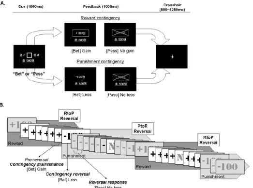
During MRI scanning, healthy controls and patients with schizophrenia performed a task in which players decided to bet a 100 won or pass without betting to a cue that reversed its contingency from or to reward (gain) or punishment (loss) after a pseudo-randomly repeated number of trials. The cue for betting was presented for 1000 ms in which the player had to decide whether to bet or not by pressing a right or left button. If the contingency of the cue was
reward then betting would result in gaining a 100 won. If the contingency of the cue was punishment then betting would result in losing a 100 won. Not betting by passing would result in neither gains nor losses, but not responding within 1 s resulted in a 100 won loss. Feedbacks of the response results along with the correct contingency were presented immediately after the cue presentation for 1000 ms. After each trials a cross hair was presented for 500-4250 ms, jittered in order to obtain optimal hemodynamic response curves in the analysis of rapid
presentation event-related fMRI design30-32 using optseq2
(http://surfer.nmr.mgh.harvard.edu/optseq/). The contingency of the cue was reversed after 6-10 trials of either reward-to-punishment (RtoP) or punishment-to-reward (PtoR) with a total of 36 reversals (Figure 1). The number of reward and punishment contingency trials maintained before reversal, and RtoP and PtoR reversals were counter-balanced. Thirty neutral oddball trials were pseudo-randomly placed 3 continuous betting trials after and before reversal contingency in order to keep the players attentive. The neutral oddball trial in which the player pressed the right or left button according to the direction of a cue triangle within 1000 ms of its presentation and received a 1000 ms feedback showing whether his/her response was correct without any gains or losses.
The participants were instructed to win as much money as possible and to respond as quickly as possible. In order to match correct response rates between the two groups, participants were told that the contingency was maintained for a number of trials then reversed and that they should not try to predict when the contingency was reversed. All participants were told that they would be paid for the amount they won during the task and later given a predetermined amount of money for transportation expenses. The task was conducted in three runs and the assignment of left and right button to betting and passing was counter-balanced across participants. A practice session of the task was conducted before MRI scanning.
Figure 1. Participants performed consecutive trials of the Monetary Incentive Contingency
Reversal Task (MICRT). Trials consisted of rewarding and punishing contingency conditions which repeated and reversed after a pseudo-randomly determined number of trials. Schematic description of the MICRT (A), and the contingency maintenance phases and the reversals of the trial sequence (B) are shown above. A. Participants had to decide and respond whether to bet a 100 won or to pass without betting when a square cue was presented for 1000 ms. Then a feedback on the result of the participant’s response and the contingency of that trial was shown for 1000 ms; During the reward contingency phase, betting resulted in a gain of a 100 won, whereas during the punishment contingency phase, betting resulted in a loss of a 100 won. Passing during both reward and punishment contingency phase resulted in neither a gain nor loss. When participants did not respond, a 100 won was lost. B. During the contingency maintenance phase, the reward or punishment contingency was maintained for 6-10 consecutive trials followed by reward-to-punishment (RtoP) or punishment-to-reward (PtoR) reversals. Neutral oddball trials (N) were pseudo-randomly placed between trials to keep the participants attentive. The last two pre-reversal contingency maintenance trials, the contingency reversal trial and the first two reversal response trials were used for analyses.
3. Imaging data acquisition
Functional and structural MRI were performed using a 3T Philips Intera Achieva scanner (Philips Medical Systems, Best, The Netherlands). The functional images were acquired using a T2*-weighted gradient echo echo-planar imaging (FEEPI) sequence (39 slices of 3mm thickness and no gaps, repetition time [TR] = 2500 ms, echo time [TE] = 30 ms, flip angle [FA] = 90°, image matrix = 128 x 128, field of view [FOV] = 220mm) with an in-plane resolution of 1.719 mm x 1.719 mm. In order to minimize signal loss in the orbitofrontal cortex, imaging slices were obtained at tilted angle of 30° from the anterior commissure-posterior commissure line33,34. Because of the short TR, high spacial resolution was maintained by limiting the range of imaging acquisition below the superior parietal area which was not included in the regions of interest. Structural images with a resolution of 0.859 mm x 0.859 mm x 1.2 mm were acquired using a 3D T1-weighted gradient echo (T1TFE) sequence (170 slices, TR = 9.692 ms, TE = 4.59 ms, image matrix = 256 x 256).
4. fMRI data analysis
Functional MRI data were analyzed using SPM8
(http://www.fil.ion.ucl.ac.uk/spm/software/spm8/). The first five volumes of each functional time series were discarded to remove non-steady-state effect caused by T1 saturation. All image volumes were adjusted for slice timing differences, realigned to the mean volume for motion correction, coregistered to the mean image of each individual’s T1 images, spatially normalized to the standard Montreal Neurological Institute (MNI) template and then smoothed with a 8-mm full-width-at-half-maximum Gaussian kernel.
On the 1st-level analysis, two pre-reversal contingency conditions (reward and punishment), a contingency reversal condition and two reversal response conditions (punishment and reward) were designed and modeled separately as
explanatory variable convolved with the canonical hemodynamic response function. The pre-reversal contingency conditions consisted of two trials before contingency reversal representing contingency maintenance and the reversal response conditions consisted of two trials following contingency reversal representing the initial reversal responses. Individual realignment parameters were entered as regressors to control for movement-related variance.
Contingency reversal and reversal response contrast maps were generated by subtracting the pre-reversal contingency conditions from the contingency reversal conditions and reversal response condition respectively. These contrast maps were combined for a 2nd-level random effect analysis. Separate one sample t-tests were conducted for the control group and the patient group to identify the apriori hypothesized regions of interest and to explore differences in the pattern of regional activations. A mask containing regions of interest in the inferior orbitofrontal cortex, putamen, anterior cingulate cortex and the amygdala defined by the Automated Anatomical Labeling (AAL) map based on the MNI average brain35 and the ventral striatum defined as a sphere with 10-mm radius around the center (x = ±11, y = 11, z = -2) based on a prior study by Knutson et al.36 were generated using the PickAtlas SPM tool.37 Images were initially thresholded at a cluster-size of more than 10 voxels with a peak-level uncorrected P < 0.001 and the regions of interest mask was applied to correct for multiple testing using the familywise error rate (FWE) method at a significance level of P < 0.05.
The percent blood-oxygen-level-dependent (BOLD) signal changes related to pre-reversal contingency and reversal response during RtoP and PtoR reversals were obtained using MarsBaR version 0.41 (http://marsbar.sourceForge.net/) in the clusters identified by the ROI analyses. Post-hoc comparison of the patterns of activity changes during RtoP and PtoR reversal responses and contingency maintenance between the two groups were conducted by paired t-tests with a Bonferroni-corrected significance level of P < 0.025. Correlation analyses were
performed to examine the relationship between neural activity changes related contingency reversal and maintenance and physical and social anhedonia scores and PANSS negative symptom scores across healthy controls and patients with schizophrenia at a significance level of P < 0.05.
5. Behavioral data analysis
Correct RtoP or PtoR reversal was defined as correctly responding according to the prior contingency in the last trial before and during the contingency reversal and correctly responding according to the reversed contingency in two consecutive trials immediately after contingency reversal. Average reaction times of the correct reversals during the last two trials before reversing responses representing pre-reversal response during contingency maintenance, and the two trials following contingency reversal representing reversal response were calculated. In order to compare the change in reaction times between RtoP and PtoR reversals, difference in baseline reaction times between reward and punishment contingency was controlled by dividing the difference in reaction time between the pre-reversal and reversal response trials by the reaction time during pre-reversal response trials.
The percent correct responses between the two groups and between RtoP and PtoR reversals were compared using independent sample t-test and paired t-test at a Bonferroni-corrected significance level of P < 0.025. Between group and within group comparison of the reaction times and the adjusted reaction time changes were conducted using Multivariate Analysis of Variance (MANOVA) and repeated measures MANOVA at a significance level of P < 0.05
1. Behavioral results
The healthy control group and the patients group did not show significant difference in the percent correct responses during both gain and loss contingency trials of the gambling task (Gain: control 98.9%, SD = 1.6, schizophrenia 97.2%, SD = 3.7, t = 1.63, P = 0.12; Loss: control 97.7%, SD = 2.5, schizophrenia 95.9%, SD = 5.2, t = 1.19, P = 0.25). In addition, both group showed no significant difference in the percent correct responses during the neutral oddball trials (control 88.2%, SD = 8.0, schizophrenia 78.1%, SD = 17.6, t = 1.98, P = 0.06) suggesting that attention during task performance was not different between the two groups.
Both healthy controls and patients with schizophrenia showed significantly greater correct responses during loss-to-gain than gain-to-loss contingency reversals (control, gain-to-loss mean = 88.4%, SD = 3.1, loss-to-gain mean = 92.9%, SD = 1.3, t = 6.21, P < 0.001; patient, gain-to-loss mean = 84.9%, SD = 8.4, loss-to-gain mean = 92.3%, SD = 6.5, t = 4.61, P < 0.001). However, no significant differences in the percent correct responses of gain-to-loss or loss-to-gain contingency reversals were found between the two groups (gain-to-loss, t = 1.53, P = 0.14; loss-to-gain, t = 1.05, P = 0.31) (Figure 2) Reaction times before contingency reversals and during reversal responses were not significantly different between the two groups (Table 2). However, the proportional decreases in reaction time during loss-to-gain reversals were significantly greater than gain-to-loss reversals only in the control group (F = 7.52, P = 0.01; t = 2.97, P = 0.01). In addition, the proportional decrease in reaction time during loss-to-gain reversals were significantly smaller in the patients than the healthy controls (F = 19.08, P = 0.005; t = 3.05, P = 0.005) (Figure 3).
In the healthy controls, the higher social anhedonia scores significantly correlated with slower reaction times of reversal responses during both
directions (gain-to-loss, r = 0.58, P = 0.02; loss-to-gain, r = 0.59, P = 0.02) and smaller proportional decrease in reaction times during loss-to-gain reversals ( r = 0.55, P = 0.03)
Figure 2. The percent correct responses of the Reward-to-Punishment (RtoP) and
Punishment-to-Reward (PtoR) contingency reversals in healthy controls (n = 15) and patients with schizophrenia (n = 14). Both healthy controls and patients with schizophrenia showed significantly greater percent correct response of the RtoP than the PtoR contingency reversals. No statistical differences between groups in the percent correct responses of the RtoP and the PtoR reversals were observed. *Significant difference at P < 0.025.
Table 2. The mean reaction times (ms) and standard deviations before contingency reversals and during reversal responses in healthy controls and patients with schizophrenia. No significant differences either between or within group were found.
Control (n = 15) Schizophrenia (n=14) Reward-to-Punishment Pre-reversal (Reward) 384.6 ± 34.6 388.4 ± 69.5
Reversal response (Punishment) 365.6 ± 38.2 382.7 ± 61.7
Punishment-to-Reward
Pre-reversal (Punishment) 392.1 ± 36.3 380.0 ± 57.1
Figure 3. The proportional changes of reaction time during the Reward-to-Punishment (RtoP) and
the Punishment-to-Reward (PtoR) contingency reversals in healthy controls (n = 15) and patients with schizophrenia (n = 14). Healthy controls showed significantly greater decreases in the proportional changes in reaction time during the PtoR than the RtoP reversals, whereas patients with schizophrenia did not show any difference in proportional changes in reaction times. Decreases in the proportional changes in reaction time during the PtoR reversal were significantly greater in healthy controls than patients with schizophrenia. *Results of repeated measures Multivariate Analysis of Variance with significant difference at P < 0.05.
2. Imaging data results
Among the regions of interest, the orbitofrontal cortex (i.e. right lateral orbitofrontal cortex (Brodmann area 47), anterior cingulate cortex, amygdala, and the putamen were identified from the contingency reversal contrast in the control group at significance level of clusters with more than 10 contiguous voxels with an uncorrected P < 0.001.
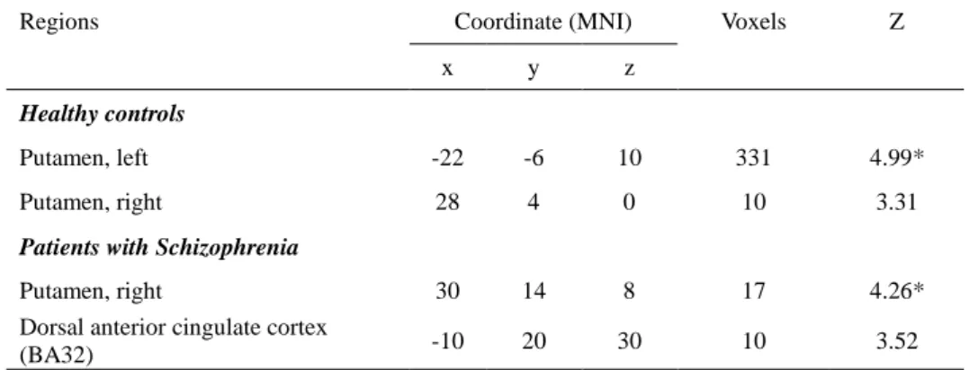
During contingency reversal, activations of bilateral putamens in healthy controls and the right putamen and the dorsal anterior cingulate cortex in patients with schizophrenia were identified among regions of interest. The healthy controls showed a larger area of activations in the putamen than patients with schizophrenia. Among these regional activities, the healthy controls showed significant activations in the right putamen (Z = 4.99, FWE-corrected P = 0.002) whereas patients with schizophrenia showed significant activations in
the left putamen (Z = 4.26, FWE-corrected P = 0.04) (Table 3).
Table 3. Regional brain activations identified from the contingency reversal contrasts. Healthy controls (n = 15) and patients with schizophrenia (n=14) showed significant contingency reversal activations in the left and right putamen, respectively.
Regions Coordinate (MNI) Voxels Z
x y z
Healthy controls
Putamen, left -22 -6 10 331 4.99*
Putamen, right 28 4 0 10 3.31
Patients with Schizophrenia
Putamen, right 30 14 8 17 4.26*
Dorsal anterior cingulate cortex
(BA32) -10 20 30 10 3.52
Results with activation clusters of more than 10 contiguous voxels with a peak uncorrected P < 0.001 using the regions of interest mask are shown.
BA: Brodmann area.
*Significant at P < 0.05 corrected for familywise error rate
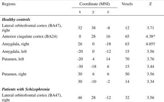
During reversal responses, the anterior cingulate cortex showed significant activation (Z = 3.71, FWE-corrected P = 0.02) and the amygdala was activated at a trend significance level (Z = 4.05, FEW-corrected P = 0.06). In the reversal contrasts of the patient group, only the right lateral orbitofrontal cortex activation was identified at significance level of clusters with more than 10 contiguous voxels with an uncorrected P < 0.001 (Table 4).
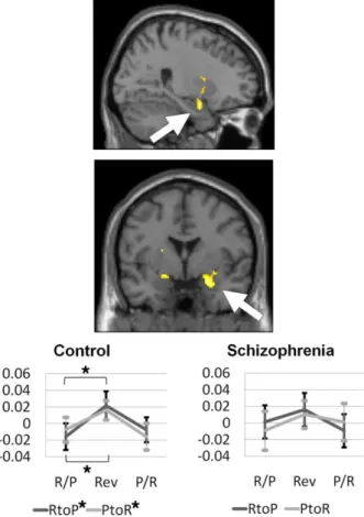
Post-hoc analyses revealed biased neural activations according to the direction of contingency reversal response. In the healthy control group, bidirectional RtoP and PtoR reversal response was significant in the amygdala (RtoP, t = 4.21,
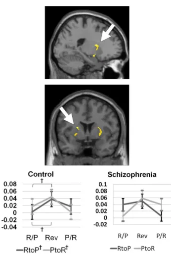
P = 0.001; PtoR, t = 3.05, P = 0.009) (Figure 4) and a trend significant in the
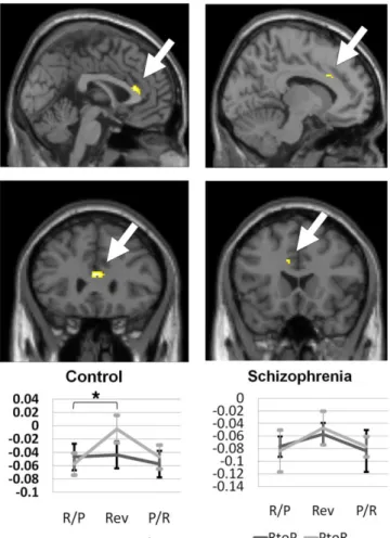
putamen (RtoP, t = 2.47, P = 0,03; PtoR, t = 2.32, P = 0.04) (Figure 5). Unidirectional PtoR reversal responses were observed in the anterior cingulate
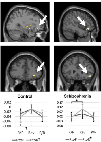
cortex at a significant level (RtoP, t = 0.25, P = 0.80; PtoR, t = 3.45, P = 0.004) (Figure 6) and in the lateral orbitofrontal cortex at a trend level of significance (RtoP, t = 1.30, P = 0.22; PtoR, t = 2.24, P = 0.04) (Figure 7). Patients with schizophrenia showed significant reversal response activations only in the lateral orbitofrontal cortex which was a unidirectional PtoR reversal response (RtoP, t = 1.37, P = 0.19; PtoR, t = 3.62, P = 0.003) (Figure 7).
Table 4. Regional brain activations identified from the reversal response contrasts. Only the lateral orbitofrontal cortex was associated with reversal response activation in patients with schizophrenia (n = 14). In healthy controls (n =15), the anterior cingulate cortex and the amygdala showed reversal response related activations at a statistically significant and trend significance level, respectively.
Regions Coordinate (MNI) Voxels Z
x y z
Healthy controls
Lateral orbitofrontal cortex (BA47),
right 32 38 -8 12 3.71
Anterior cingulate cortex (BA24) 0 28 16 65 4.38*
Amygdala, right 26 0 -18 63 4.05† Amygdala, left -20 0 -12 15 3.56 Putamen, left -20 4 14 70 3.76 -30 -18 6 15 3.44 Putamen, right 30 6 6 50 3.56 30 -10 -2 14 3.34
Patients with Schizophrenia Lateral orbitofrontal cortex (BA47),
right 46 28 -12 32 3.56
Results with activation clusters of more than 10 contiguous voxels with a peak uncorrected P < 0.001 using the regions of interest mask are shown.
BA: Brodmann area.
*Significant at P < 0.05 corrected for familywise error rate,
Figure 4. The activation patterns of the amygdala among the region of interest obtained from the
reversal response contrast in healthy controls (n = 15) and patients with schizophrenia (n=14). Only healthy controls showed significant activations during the Reward-to-Punishment (RtoP) and the Punishment-to-Reward (PtoR) contingency reversals. Percent signal changes of the pre-reversal Reward or Punishment association (R/P or P/R), reversal response (Rev), and the next pre-reversal Punishment or Reward association (P/R or R/P) trials clusters during the RtoP and the PtoR reversals are shown. *Significant at P < 0.025
Figure 5. The activation patterns of the putamen among the region of interest obtained from the
reversal response contrast in healthy controls (n =15) and patients with schizophrenia (n = 14). Only healthy controls showed trend level of significant activations during the Reward-to-Punishment (RtoP) and the Punishment-to-Reward (PtoR) contingency reversals. Percent signal changes of the pre-reversal Reward or Punishment association (R/P or P/R), reversal response (Rev), and the next pre-reversal Punishment or Reward association (P/R or R/P) trials clusters during the RtoP and the PtoR reversals are shown. †Trend level of significance at P < 0.05
Figure 6. The activation patterns of the anterior cingulate cortex among the region of interest
obtained from the reversal response contrast in healthy controls (n = 15) and patients with schizophrenia (n = 14). Only healthy controls showed significant activations during the Punishment-to-Reward (PtoR) contingency reversals. Percent signal changes of the pre-reversal Reward or Punishment association (R/P or P/R), reversal response (Rev), and the next pre-reversal Punishment or Reward association (P/R or R/P) trials clusters during the Reward-to-Punishment (RtoP) and the PtoR reversals are shown. *Significant at P < 0.025
Figure 7. The activation patterns of the lateral orbitofrontal cortex among the region of interest
obtained from the reversal response contrast and the contingency reversal contrasts in healthy controls (n = 15) and patients with schizophrenia (n = 14), respectively. Healthy controls and patient with schizophrenia showed the Punishment-to-Reward (PtoR) contingency reversal activations at a trend significance and significant level, respectively. Percent signal changes of the pre-reversal Reward or Punishment association (R/P or P/R), reversal response (Rev), and the next pre-reversal Punishment or Reward association (P/R or R/P) trials clusters during the Reward-to-Punishment (RtoP) and the PtoR reversals are shown. *Significant at P < 0.025. † Trend level of significance at P < 0.05
3. Correlations with anhedonia and negative symptoms
Among the regions of interest related to reversal response, the anterior cingulate activity changes significantly correlated with the physical anhedonia
scale scores in the healthy control group. Greater physical anhedonia was correlated with smaller activity increases during PtoR contingency reversal and greater activity decreases during sustained gain contingency after reversal in the anterior cingulate cortex (Figure 8).
Figure 8. Correlations of the physical anhedonia scale scores with changes of activities in the
anterior cingulate cortex (Brodmann area 24) during the Punishment-to-Reward (PtoR) contingency reversal and during sustained reward contingency after reversal in the healthy controls (n = 15).
In patients with schizophrenia, activity changes in the lateral orbitofrontal cortex significantly correlated with both physical and social anhedonia scale scores and PANSS negative symptom scores. Greater increases in the lateral orbitofrontal activity during PtoR contingency reversal correlated with greater severity of physical and social anhedonia and negative symptoms (r = -0.53, P = 0.04). Whereas smaller decreases in the lateral orbitofrontal activity during sustained reward contingency after reversal correlated with greater physical anhedonia and severity of negative symptoms (r = 0.64, P = 0.01) (Figure 9). Among the negative symptoms items of the PANSS, greater passive/apathetic social withdrawal (N4) and lack of spontaneity and flow of conversation (N6) significantly correlated with greater increase in activities during PtoR
contingency reversal (N4, Spearman’s rho = 0.59, P = 0.03; N6, Spearman’s rho = 0.56, P = 0.04) and smaller decrease in activities during sustained reward contingency after reversal (N4, Spearman’s rho = 0.59, P = 0.03; N6, Spearman’s rho = 0.61, P = 0.02) in the lateral orbitofrontal cortex.
Figure 9. Correlations of the physical and social anhedonia scale scores and the Positive and
Negative Syndrome Scale (PANSS) scores with changes of activities in the lateral orbitofrontal cortex (Brodmann area 42) during the Punishment-to-Reward (PtoR) contingency reversal and during the sustained reward contingency after reversal in patients with schizophrenia (n = 14).
IV. DISCUSSION
The present study demonstrates that patients with schizophrenia have an affective bias in reversal association where implicit inclination for goal-directed engagement to reverse association from PtoR contingency is diminished with concurrent blunted reversal response in the anterior cingulate gyrus. Reversal response-related activations were identified in the anterior cingulate cortex, amygdala, putamen and the right lateral orbitofrontal cortex in healthy controls. Nucleus accumbens activity was not identified as hypothesized probably because the task paradigm used in this study did not involve prediction error learning. Among these regions, the anterior cingulate cortex and the lateral orbitofrontal cortex showed a unidirectional PtoR reversal response. Among the reversal response-related regions, patients with schizophrenia showed blunted bidirectional reversal response in the anterior cingulate gyrus, amygdala, and the putamen during reversal response and a unidirectional PtoR reversal activation in the right lateral orbitofrontal cortex. In the healthy controls, physical anhedonia was associated with smaller reversal response-related activations and greater decline in activity after reversal as contingency was maintained in the anterior cingulate cortex suggesting its role in the acquisition and maintenance of rewarding salience. Whereas physical anhedonia and
negative symptom severity was associated with greater reversal
response-related activations and smaller post-reveral decline during contingency maintenance in the lateral orbitofrontal cortex in patient with schizophrenia reflecting a compensatory regulation of blunted reward reversal-related activities.
motivation
In the healthy controls, the putamen activation was involved in contingency reversal as well as the following reversal response. During contingency reversal trials, participants, not knowing when reversal takes place, were most likely to expect pre-reversal contingency to continue and received unexpected feedback about their response. Therefore the observed bilateral putamen activation was most likely evoked by the feedback of contingency reversal. In the following reversal response trials, the participant is prepared reverse their response to the cue and receives expected response according to the reversed contingency. Findings of bilateral putamen activations during bidirectional reversal response trials with concurrent accelerations of reaction time suggest its role in linking reversal feedback to a prepared response. In a study using a probabilistic error reversal learning paradigm associated the role of putamen with the process of stimulus-action-reward association, whereas the caudate nucleus and ventral striatum were associated with reward prediction error.38 In addition, putamen activation was observed in anticipation of responding for both certain and uncertain reward suggesting its role in instrumental response.39 Findings of the putamen’s involvement in regulating performance have been reported in a prior study associating the putamen activation to an advantage in reaction time during implicit sequence learning40 and another study relating greater error-related putamen activation to smaller error rates.41
In the healthy controls, the unidirectional PtoR reversal activations in the anterior cingulate activation was observed in the context of greater correct reversal responses and accelerated reaction time. Assuming this neural activation occurred in response to the response reversal cue following the feedback of contingency reversal, improved efficiency of reversal response is consistent with prior studies indicating the role of anterior cingulate activation in response anticipation which is suggested to include anticipation attention, motivation, and motor preparation.42,43 In addition, the anterior cingulate
activity has been reported to play a role in adjustments in performance.44,45 However, the anterior cingulate activation during response reversal was not bidirectional, but selective to PtoR contingency reversal. Since the reversal response trials included both cue and feedback, one may argue that the anterior cingulate activated in response to rewarding novel feedback that may have a stronger impact than repeated feedbacks of reward. However, a prior study of reward-based decision making showed that the anterior cingulate cortex was activated by reduced reward and response switch feedbacks that signaled both to switch response leading to an expected reward.46 Therefore, it is more likely that the ACG activation during reversal response trials were related to reward expectation than the reward itself. It has been proposed that using error commission feedback from the striatum and the mesencephalic dopamine system, the anterior cingulate implements error-based reinforcement learning and improve performance by modifying the strength of stimulus-response mappings using dopaminergic input.47 In addition, recent studies have implicated the anterior cingulate cortex and the putamen in the computation and analysis of effort versus benefit in pursuing rewarding behavior.48,49 The correlations between the anterior cingulate activations and physical anhedonia also indicate that greater anticipatory response related to engagement of behavioral change and maintaining this anticipatory response underlies the trait for experiencing pleasure.
In this study, the amygdala showed bidirectional reversal response activations and the orbitofrontal cortex showed a unidirectional PtoR reversal response activation that suggests their respective roles in detecting contingency reversal and modulating goal-directed response. The amygdala and the orbitofrontal cortex have direct interconnections and have been consistently associated with associative learning.7,50,51 Previous studies have shown that the amygdala and orbitofrontal cortex have a distinct role in encoding motivational significance of stimuli and using these motivation significance to guide behavior. From animal
to human studies, the amygdala has been consistently implicated in the emotional process of appraising salience to stimuli that are either rewarding or aversive.52 Prior animal studies also reported the activation in the amygdala after contingency reversal and during the attribution of both positive and negative values to visual stimuli suggesting the role of amygdala in encoding the motivational significance of cues.51,53 In studies using instrumental conditioning procedures where a certain cue paired with a specific response leads to a delivery of a reinforcer, the amygdala showed cue-onset activations and the orbitofrontal cortex, response-onset activations suggesting their specific roles in expectancies. Holland and Gallagher (2004)50 has suggested that the orbitofrontal cortex generate responses on the basis of the cue-reinforcer expectancies by suppressing response based on competing but less desirable expected consequence. Studies of patients with orbitofrontal damage also suggested that the lateral orbitofrontal cortex is most likely involved in suppressing previously acquired stimulus-reward associations during reversal learning.54,55 A number of recent studies have shown that activities in the right lateral orbitofrontal cortex have a U-shaped relationship with reward value where activation is highest in response to high rewards received or omitted while performing a monetary incentive task which did not include a choice of loss avoidance.56,57 The unidirectional reversal response in the lateral orbitofrontal cortex in this study is probably related to the relative difference in the reward value received by reward approach and loss avoidance. A choice of action that can result in a reward is more motivationally engaging than a choice action to avoid loss. If one was given an alternate choice that can provide an avoidance of loss and receipt of reward simultaneously than the alternate choice may be equally motivationally engaging as reward choice.
2. Deficient neural processing of motivation and salience attribution in patients with schizophrenia
Contingency reversal feedback-related activations in patients with schizophrenia were not found in the dorsal anterior cingulate cortex and unilaterally in the right putamen. Activations in these regions were not observed during reversal response. Interestingly, the healthy controls showed a relatively larger area of activations in the left than right putamen. In a prior study, the left putamen showed relatively greater activations in response to rewarding monetary reinforcers.57 Therefore, lack of reversal feedback-related activation in the left putamen as well as absence of putamen activations during reversal response may reflect a deficiency in positive reinforcement feedback transferring to response modulation. The dorsal anterior cingulate gyrus has been associated with conflict monitoring at different levels of cognitive processing including stimulus evaluation, presentation and response, and the anterior cingulate impairment in conflict-monitoring has been consistently reported in patients with schizophrenia.44,58,59 In contrast, our findings show that patients with schizophrenia activated the dorsal anterior cingulate gyrus to reversal feedback that was not observed in the healthy controls and showed comparable accuracy in performance. This may be explained by the use of a simplified association reversal paradigm in this study that was cognitively less burdening to the patients. However, the recruitment of the dorsal anterior cingulate cortex in the patients may reflect their cognitive efforts in order to compensate for the deficient striatal functioning in processing reversal feedback information.
One important finding of this study is the diminished acceleration of correct reaction time with the absence of PtoR reversal feedback activations in the anterior cingulate cortex patients with schizophrenia. This finding suggests that the motivation to readily respond to reward contingency was weak in patients with schizophrenia. Prior studies examining amotivation in schizophrenia using the reward prediction or reinforcement learning model have mainly focused on the dysfunction in the ventral striatum.60 In the present study, the task paradigm
was designed to exclude components of reward prediction and reinforcement learning resulting in a matched correct response performance between the patients and the healthy controls. Anticipation in this study was reflected by how response to gain reward was readily engaged by the initial cue of reversed contingency rather than prediction ability. Only a few studies have examined the neural processing of anticipation in schizophrenia. One study reported a diminished anterior cingulate activation while anticipating to respond and another recent study using a delayed incentive paradigms with monetary rewards, but no losses, has shown that reward expectation-related activations were diminished in patients with schizophrenia.56,61 The concurrent absence of putamen activations during reversal response in the present finding may reflect the restricted effort put into acting that could be partially responsible for the lack of rapid engagement to reward contingency in the patients. The present finding demonstrates that only anticipation to reward-approaching response is deficient while anticipation to loss-avoidance response is intact in patients with schizophrenia.
Another interesting finding in the patients with schizophrenia was the absence of bidirectional amygdala and putamen activations during reversal response. The deficient activations of amygdala to both reward and loss-reversed cues corresponds to prior studies that reported mesiotemporal volume reduction in chronic and drug-naïve first-episode schizophrenia, and reduction in amygdala/medial prefrontal activity in response to fear stimuli in patients with paranoid schizophrenia.62-64 With regard to the amygdala-orbitofrontal network, reversal learning functioning of the orbitofrontal cortex may seem impossible when amygdala response to salient stimuli is deficient. However, animal studies have reported that amygdala lesions do not affect the acquisition and use of expectancies of relatively neutral events and may in fact abolish OFC-dependent reversal impairments.50,65 Because of the easy difficulty level of the task, patients with schizophrenia were able to performance well in accuracy
despite anterior cingulate, amygdala, and putamen dysfunction. However, failure to recruit this limbic-striatum network contributed to the disadvantage in reaction time in the patients.
Paradoxically, patients with schizophrenia showed greater lateral orbitofrontal cortex activations during PtoR reversal response that were associated with greater physical and social anhedonia, and negative symptom severity. This finding points to the role of the lateral orbitofrontal cortex in a compensatory top-down control for the disadvantage caused by the deficient processing of salience signals, reward anticipation and instrumental response by the amygdala, anterior cingulate cortex, and the putamen
3. Implications and limitations
Clinically, patients with schizophrenia with negative symptoms enjoy activities that give immediate pleasure, such as eating or watching television, but do not engage in activities that require effort to obtain a rewarding outcome. Findings of this study suggest that the neural pathophysiology of these clinical aspects of avolition involve the deficient activations of the anterior cingulate gyrus and putamen that may underlie effort or engagement to pursue rewarding reinforcers.
A number of recent pharmacologic studies have examined the effects of antipsychotics on the reward learning pathways. Olanzapine has been demonstrated to decrease activity in the anterior cingulate, inferior frontal cortex and ventral striatum, but not putamen and diminish acceleration of reaction times when higher rewards were expected during a monetary incentive delay task in healthy subjects.66 Haloperidol also was shown to diminish prediction error-related activities including the ventral striatum, putamen, insula, and the anterior cingulated.67 These reports point to the possible involvement of antipsychotics in negative symptoms and the need to examine neural correlates of negative symptoms proper. Inclination in reversal from reward to punishment
in patients with schizophrenia was not observed in this study as hypothesized probably because positive symptoms were stabilized by antispychotics. Therefore, future studies examining antipsychotic-free patients may be warranted.
V. CONCLUSION
In conclusion, this study demonstrated the deficient neural responses in the anterior cingulate, amygdala and the putamen during reversal association with a unidirectional reversal response in the anterior cingulate gyrus and concurrent decrease in acceleration of reaction time while engaging in reversal response from avoidance to reward approach in schizophrenia. These findings indicate that deficiency in the neural correlates of anticipation and engagement to instrumental behavior may be involved in negative symptoms of anhedonia and avolition in schizophrenia.
REFERENCES
1. Laviolette SR. Dopamine Modulation of Emotional Processing in Cortical and Subcortical Neural Circuits: Evidence for a Final Common Pathway in Schizophrenia? Schizophr Bull. 2007;33:971-81.
2. Horvitz JC. Mesolimbocortical and nigrostriatal dopamine responses to salient non-reward events. Neuroscience. 2000;96:651-6.
3. Berridge KC, Robinson TE. What is the role of dopamine in reward: hedonic impact, reward learning, or incentive salience? Brain Res Rev. 1998;28:309-69.
4. Kapur S. Psychosis as a State of Aberrant Salience: A Framework Linking Biology, Phenomenology, and Pharmacology in Schizophrenia. Am J Psychiatry. 2003;160:13-23.
5. Rolls ET, Hornak J, Wade D, McGrath J. Emotion-related learning in patients with social and emotional changes associated with frontal lobe damage. J Neurol Neurosurg Psychiatr. 1994;57:1518-24.
6. Fellows LK, Farah MJ. Ventromedial frontal cortex mediates affective shifting in humans: evidence from a reversal learning paradigm. Brain. 2003;126:1830-7.
7. Kringelbach ML. The human orbitofrontal cortex: linking reward to hedonic experience. Nat Rev Neurosci. 2005;6:691-702.
8. Ernst M, Nelson EE, Jazbec S, McClure EB, Monk CS, Leibenluft E, et al. Amygdala and nucleus accumbens in responses to receipt and omission of gains in adults and adolescents. Neuroimage. 2005;25:1279-91.
9. O'Doherty J, Kringelbach ML, Rolls ET, Hornak J, Andrews C. Abstract reward and punishment representations in the human orbitofrontal cortex. Nat Neurosci. 2001;4:95-102.
10. Cools R, Clark L, Owen AM, Robbins TW. Defining the Neural Mechanisms of Probabilistic Reversal Learning Using Event-Related Functional Magnetic Resonance Imaging. J Neurosci. 2002;22:4563-7.
11. Remijnse PL, Nielen MMA, Uylings HBM, Veltman DJ. Neural correlates of a reversal learning task with an affectively neutral baseline: An event-related fMRI study. Neuroimage. 2005;26:609-18.
12. Schiller D, Levy I, Niv Y, LeDoux JE, Phelps EA. From Fear to Safety and Back: Reversal of Fear in the Human Brain. J Neurosci. 2008;28:11517-25. 13. Lee Y, Kim Y-T, Seo E, Park O, Jeong S-H, Kim SH, et al. Dissociation of
emotional decision-making from cognitive decision-making in chronic schizophrenia. Psychiatry Res. 2007;152:113-20.
14. Ritter LM, Meador-Woodruff JH, Dalack GW. Neurocognitive measures of prefrontal cortical dysfunction in schizophrenia. Schizophr Res. 2004;68:65-73.
15. Shurman B, Horan WP, Nuechterlein KH. Schizophrenia patients demonstrate a distinctive pattern of decision-making impairment on the Iowa Gambling Task. Schizophr Res. 2005;72:215-24.
16. Exner C, Boucsein K, Degner D, Irle E, Weniger G. Impaired emotional learning and reduced amygdala size in schizophrenia: a 3-month follow-up. Schizophr Res. 2004;71:493-503.
17. Jensen J, Willeit M, Zipursky RB, Savina I, Smith AJ, Menon M, et al. The Formation of Abnormal Associations in Schizophrenia: Neural and Behavioral Evidence. Neuropsychopharmacology. 2007;33:473-9.
18. Taylor SF, Luan Phan K, Britton JC, Liberzon I. Neural Response to Emotional Salience in Schizophrenia. Neuropsychopharmacology. 2005;30:984-95. 19. Murray GK, Cheng F, Clark L, Barnett JH, Blackwell AD, Fletcher PC, et al.
Reinforcement and Reversal Learning in First-Episode Psychosis. Schizophr Bull. 2008;34:848-55.
20. Waltz JA, Gold JM. Probabilistic reversal learning impairments in schizophrenia: Further evidence of orbitofrontal dysfunction. Schizophr Res. 2007;93:296-303.
21. Strauss GP, Allen DN, Duke LA, Ross SA, Schwartz J. Automatic affective processing impairments in patients with deficit syndrome schizophrenia. Schizophr Res. 2008;102:76-87.
22. Moritz S, Lauden A. Attention bias for paranoia-relevant visual stimuli in schizophrenia. Cognit Neuropsychiatry. 2007;12:381-90.
23. Seok JH, An SK, Lee E, Lee HS, Lee YJ, Jeon JH, et al. Behavioral evidence of blunted and inappropriate affective responses in schizophrenia: Lack of a
negativity bias. Psychiatry Res. 2006;142:53-66.
24. American Psychiatric Association. Diagnostic and statistical manual of mental disorders, fourth edition, text revision. Washington, DC: American Psychiatric Association; 2000.
25. Raven JC, Sourt J, Raven J. Raven-matrizen-test standard progressive matrices. Wenheim: Beltz Test Gesellschaft; 1988.
26. Annett M. A classification of hand preference by association analysis. Br J Psychol. 1970;61:303-21.
27. Chapman LJ, Chapman JP, Raulin ML. Scale for physical and social anhedonia. J Abnorm Psychol. 1976;85:374-82.
28. Watson D, Clark LA, Tellegen A. Development and validation of brief measures of positive and negative affect: The PANAS scales. J Pers Soc Psychol. 1988;54:1063-70.
29. Kay SR, Fiszbein LA, Opler LA. The positive and negative syndrome scale (PANSS) for schizophrenia. Schizophr Bull. 1987;13:261-76.
30. Buckner RL, Goodman J, Burock M, Rotte M, Koutstaal W, Schacter D, et al. Functional-Anatomic Correlates of Object Priming in Humans Revealed by Rapid Presentation Event-Related fMRI. Neuron. 1998;20:285-96.
31. Burock MA, Dale AM. Estimation and detection of event-related fMRI signals with temporally correlated noise: a statistically efficient and unbiased approach. Hum Brain Mapp. 2000;11:249-60.
32. Miezin FM, Maccotta L, Ollinger JM, Petersen SE, Buckner RL. Characterizing the Hemodynamic Response: Effects of Presentation Rate, Sampling Procedure, and the Possibility of Ordering Brain Activity Based on Relative Timing. Neuroimage. 2000;11:735-59.
33. Deichmann R, Gottfried JA, Hutton C, Turner R. Optimized EPI for fMRI studies of the orbitofrontal cortex. Neuroimage. 2003;19:430-41.
34. Kim H, Adolphs R, O'Doherty JP, Shimojo S. Temporal isolation of neural processes underlying face preference decisions. Proc Natl Acad Sci U S A. 2007;104:18253-8.
35. Tzourio-Mazoyer N, Landeau B, Papathanassiou D, Crivello F, Etard O, Delcroix N, et al. Automated Anatomical Labeling of Activations in SPM
Using a Macroscopic Anatomical Parcellation of the MNI MRI Single-Subject Brain. Neuroimage. 2002;15:273-89.
36. Knutson B, Wimmer GE. Splitting the Difference: How does the brain code reward episodes? Ann N Y Acad Sci. 2007;1104:54-69.
37. Maldjian JA, Laurienti PJ, Kraft RA, Burdette JH. An automated method for neuroanatomic and cytoarchitectonic atlas-based interrogation of fMRI data sets. Neuroimage. 2003;19:1233-9.
38. Haruno M, Kawato M. Different neural correlates of reward expectation and reward expectation error in the putamen and caudate nucleus during stimulus-action-reward association learning. J Neurophysiol. 2006;95:948-59. 39. Bjork JM, Hommer DW. Anticipating instrumentally obtained and
passively-received rewards: A factorial fMRI investigation. Behav Brain Res. 2007;177:165-70.
40. Rauch SL, Whalen PJ, Savage CR, Curran T, Kendrick A, Brown HD, et al. Striatal recruitment during an implicit sequence learning task as measured by functional magnetic resonance imaging. Hum Brain Mapp. 1997;5:124-32. 41. Polli FE, Barton JJS, Thakkar KN, Greve DN, Goff DC, Rauch SL, et al.
Reduced error-related activation in two anterior cingulate circuits is related to impaired performance in schizophrenia. Brain. 2008;131:971-86.
42. Fan J, Kolster R, Ghajar J, Suh M, Knight RT, Sarkar R, et al. Response anticipation and response conflict: An event-related potential and functional magnetic resonance imaging study. J Neurosci. 2007;27:2272-82.
43. Fassbender C, Foxe JJ, Garavan H. Mapping the functional anatomy of task preparation: Priming task-appropriate brain networks. Hum Brain Mapp. 2006;27:819-27.
44. Kerns JG, Cohen JD, MacDonald AW, III, Johnson MK, Stenger VA, Aizenstein H, et al. Decreased conflict- and error-related activity in the anterior cingulate cortex in subjects with schizophrenia. Am J Psychiatry. 2005;162:1833-9.
45. Kerns JG, Cohen JD, MacDonald AW, III, Cho RY, Stenger VA, Carter CS. Anterior cingulate conflict monitoring and adjustments in control. Science. 2004;303:1023-6.