DOI: https://doi.org/10.3339/jkspn.2020.24.2.83 ISSN 2384-0250 (online)
Association of Neutrophil Gelatinase associated
Lipocalin and Leukocyte Differential Count in Children
with Febrile Urinary Tract Infections
Purpose: To investigate the association between urinary neutrophil gelatinase-associated lipocalin (uNGAL) and leukocyte differential count in children with urinary tract infections (UTIs).
Methods: A retrospective chart review was performed in children undergoing uNGAL measurements between June 2018 and September 2019. Patients with suspected or diagnosed UTIs were included. The relationship between uNGAL and blood leukocyte differential count was investigated in children.
Results: A total of 197 children were included in this study, 119 of whom (60%) had UTIs. The non-UTI patients (n=78) were diagnosed with pneumonia, acute gastroenteritis, viral upper respiratory infection, and others. After adjusting for age, gender, and fever duration, the leukocyte count, monocyte count, and uNGAL levels were higher in the UTI group than in the non-UTI group (P<0.05). uNGAL showed positive correlations with neutrophil counts, monocyte counts, the neutrophil-to-lymphocyte ratio, and the monocyte-to-lymphocyte ratio in the UTI group (P<0.05). uNGAL levels were only associated with the neutrophil-to-lymphocyte ratio in the non-UTI group (P<0.05). In a multivariable logistic regres-sion analysis, only uNGAL was associated with the presence of UTI (P<0.05). The area under the receiver operating characteristic curves for uNGAL and monocyte counts to identify UTI were 0.89 (95% confidence interval (CI): 0.824–0.939; P= 0.025) and 0.7 (95% CI: 0.627–0.774; P=0.038), respectively.
Conclusions: In children with UTIs, uNGAL levels may be associated with blood leukocyte differential counts. uNGAL measurements and monocyte counts can be helpful in children with suspected UTIs.
Key words: Biomarkers, Monocytes, Urinary tract infections
Ji Won Jang, M.D.
Hyung Eun Yim, M.D., Ph.D.
Kee Hwan Yoo, M.D., Ph.D.
Department of Pediatrics, Korea University College of Medicine, Seoul, Korea
Corresponding author:
Hyung Eun Yim, M.D., Ph.D.
Department of Pediatrics, Korea University Ansan Hospital, Korea University College of Medicine, 123 Jeokgeum-ro, Danwon-gu, Ansan 15355, Republic of Korea
Tel: +82-31-412-5096 Fax: +82-31-405-8591 E-mail: [email protected] Received: 16 June 2020 Revised: 17 September 2020 Accepted: 21 September 2020
This is an open-access article distributed under the terms of the Creative Commons Attribu tion Non-Commercial License (http:// crea tivecom mons.org/licenses/by-nc/4.0/) which permits unrestricted non-commercial use, distribution, and reproduction in any medium, provided the original work is properly cited.
Copyright © 2020 The Korean Society of Pediatric Nephrology
Introduction
Urinary tract infection (UTI) is one of the most common febrile infectious diseases in children1). Early detection of UTI and proper management are
critical since UTI can lead to permanent kidney damage, hypertension, chronic kidney disease (CKD), and ultimately end-stage renal disease2).
How-ever, UTIs are not easy to recognize because the symptoms and signs may be ambiguous, especially in young children3). Besides, there are some other
dif-ficulties in diagnosing UTI in young children, such as delayed urine sampling and frequent over-diagnosis from contaminated urine specimens4). There is
also a practical difficulty in accurately interpreting the number of bacteria in the urine, and distinguishing bet-ween asymptomatic bacteriuria and a true UTI5,6).
There-fore, other markers for diagnosing UTI are needed. And several studies for new markers such as procalcitonin and leukocyte esterase have been conducted recently4,7).
Neutrophil gelatinase-related lipocalin (NGAL) is a member of the lipocalin superfamily8). It is a 25-kDa
pro-tein covalently linked to gelatinase in human neutrophils and highly expressed in neutrophils, monocytes, and ma-crophages8). As an iron carrier protein, NGAL plays a role
in innate immunity. It binds bacterial siderophores and thus, limits bacterial growth9). It has been reported that
NGAL could be a useful predictor for differentiating acute bacterial infections from viral ones during acute infections in children10). Several studies have also demonstrated the
diagnostic utility of urinary and plasma NGAL in detecting UTIs experimentally and clinically8,11,12).
Leukocyte differential counts have long been used to determine the cause of infection13-17). When an infection
occurs, neutrophils and monocytes move to the peripheral site, which is essential for defending against pathogens18).
As the infection progresses, the number of neutrophils and monocytes increases rapidly, and the cells migrate to the inflamed tissue18). Several studies have revealed a link
bet-ween monocytes, infections, and immune mechanisms19- 21). Another paper reported that neutrophils and monocytes
had an effect on combating microbial pathogens22).
Mono-cytes defend bacteria by providing macrophages and dend-ritic cell precursors to tissues22). A previous study found
that the neutrophil-to-lymphocyte ratio (NLR) and mono-cyte-to-lymphocyte ratio (MLR) may help in determining which of the patients with fever are more likely to have bac-terial infections and should be considered for antibiotic treatment13).
Considering these facts, we hypothesized that NGAL relates to leukocyte differential count in blood in children with UTIs. Therefore, in this study, we aimed to investigate the association between urinary NGAL (uNGAL) and leu-kocyte differential counts in children with febrile UTIs. The diagnostic accuracy of uNGAL and leukocyte diffe-rential counts for identifying UTIs was also examined.
Materials and methods
1. Patients and inclusion criteriaA retrospective chart review was performed in children undergoing urinary NGAL measurements between June 2018 and September 2019. The data from 417 pediatric pa-tients with suspected UTIs who were admitted to the Korea University Guro Hospital were included. And the age of the patient was composed from minimum 1 month to maxi-mum 12 years. The diagnosis of first febrile UTI was as follows : (1) fever of ≥38℃, (2) pyuria (≥5 white blood cells (WBC)s per high-power field), (3) positive urine culture collected from a midstream urine (defined as the growth of a single organism with ≥100,000 colony-forming units/ mL or ≥50,000 colony-forming units/mL with clinical sign of infection such as fever) or catheterized specimen (defined as the growth of a single organism with ≥ 50,000 colony-forming units/mL)23) and (4) no previous history of UTI.
Patients with recurrent UTIs, acute kidney injury (AKI), CKD, systemic disease, and known congenital anomalies of the kidney and the urinary tract (CAKUT) were excluded.
2. Measurements
First, we investigated children who were tested for uNGAL for suspected UTI between June 2018 and September 2019. A chart review of children who met the inclusion criteria was conducted retrospectively in the electronic medical records. The children were divided into the UTI group and the non-UTI group. Clinical data, such as age, sex, and du-ration of fever were collected. Also, the results of blood tests performed prior to the initiation of antibiotic treatment, including complete blood cell count (CBC), leukocyte, neu-trophils, lymphocytes, monocyte counts, and C-reactive protein (CRP) results were collected. The NLR and MLR were then calculated using the neutrophil, lymphocyte, and monocyte counts. Urine samples for urinalysis, urine culture, and the uNGAL test were also collected before anti-biotic treatment. uNGAL test was performed using the ARCHITECT Urine NGAL assay. It uses NGAL mouse monoclonal anti-NGAL antibody-coated microparticles and arcidine-related anti-NGAL antibodies24). Clinical
data including age, sex, and fever duration, and laboratory data including uNGAL measurement, leukocyte count, neutrophil count, and monocyte count were compared
between the UTI and non-UTI groups. The association between uNGAL and the leukocyte differential count in children with or without febrile UTI was investigated. The relationship between serum CRP levels and the leukocyte differential count was additionally examined.
3. Statistics
IBM SPSS ver. 20.0 (IBM Co., Armonk, NY, USA) was used to compare the variables. The continuous variables are presented as mean±standard deviation or standard error of the mean and the categorical variables as number (%). The Mann-Whitney U test was used for continuous vari-ables, such as age, leukocyte count, absolute neutrophil count (ANC), lymphocyte count, monocyte count, NLR, MLR, and uNGAL. The Chi-squared test was used for cate-gorical variables such as gender. To control for the effects of age, gender, and fever duration, analysis of covariance (ANCOVA) was used. Univariable and multivariable logi-stic regression analyses were also performed to find the predictive factors for UTI. The odds ratio (OR) of each vari-able for UTI was calculated using logistic regression lysis. Variables with a P-value of ≤0.20 in univariable ana-lyses were included in the multivariable model. Correlation analysis was carried out to determine the relationship bet-ween two variables by Spearman’s correlation test. The discriminative ability of each biomarker for UTI was evalu-ated by plotting ROC curves for the biomarkers. The area under the ROC curve (AUC) with a 95% confidence interval (CI) was calculated and compared between the variables. All differences were considered significant at a P-value of <0.05.
Results
1. Patient characteristics
Among 417 children with suspected UTIs, a total of 197 patients were included in this study. One hundred nineteen patients were diagnosed with UTIs. The patients with non- UTI (n=78) were diagnosed with pneumonia (n=18), acute gastroenteritis (n=12), viral upper respiratory infection (n=30), and others (n=18) (Fig. 1). The proportion of female patients was higher in the non-UTI group than in the UTI group (P<0.05). However, there were no differences in age
and fever duration (>72 hours) between the two groups. Children with UTIs showed higher monocyte counts, uNGAL levels, and blood urea nitrogen (BUN)-to-creati-nine (Cr) ratios compared to the non-UTI group (P<0.05). Serum Cr concentration, WBC count, and CRP level were not different between the two groups (Table 1). When the age, gender, and fever duration-adjusted predicted means were compared between the UTI and non-UTI groups,
Table 1. Patient Characteristics and Clinical Findings between the UTI and Non-UTI Groups
Non-UTI UTI P-value
Age (month) 32.5±30.6 20.1±41.0 0.774* Female, n (%) 45/78 (58%) 40/119 (34%) 0.024† Fever duration Total (>72 hrs), n (%) 25/78 (32.1%) 31/119 (26.1%) 0.079† WBC count (/mm3) 11,073±6,110 13,517±5,024 0.36* ANC 6,124±5,157 7,089±4,110 0.146* Lymphocyte count (/mm3 ) 3,857±2,366 4,706±2,378 0.867* Monocyte count (/mm3) 953±577 1,463±836 0.004* Neutrophil-to-lymphocyte ratio 2.28±2.64 3.13±7.62 0.151* Monocyte-to-lymphocyte ratio 0.24±0.33 0.55±2.09 0.1* uNGAL (ng/mL) 23.1±41.8 286.2±1,506.4 <0.001* Serum Cr (mg/dL) 0.15±0.21 0.15±0.17 0.119* BUN-to-Cr ratio 36.9±13.6 42.8±18.7 0.015* CRP (mg/L) 27.8±37.0 32.8±36.0 0.564*
The values are presented as mean±standard deviation or number (%). Abbreviations: UTI, urinary tract infection; WBC, white blood cell; ANC, absolute neutrophil count; uNGAL, urinary neutrophil gelatinase-associ-ated lipocalin; Cr, creatinine; BUN, blood urea nitrogen; CRP, C-reactive protein.
*Mann-Whitney U test. †Chi-square test.
Fig. 1. Study flow diagram. NGAL, urinary neutrophil gelatinase-associated lipocalin.
UTI, Urinary tract infection; AKI, acute kidney injury; CKD, chronic kidney disease; CAKUT, congenital anomalies of the kidney and urinary tract.
WBC counts, monocyte counts, and uNGAL levels were significantly higher in the UTI group (P<0.05) (Table 2).
2. Association between uNGAL and leukocyte differential counts
Paired sample correlations were conducted by Spearman’s correlation analysis with an adjustment for age, gender, and fever duration. In the UTI group, uNGAL levels showed positive correlations with ANC, monocyte counts, NLR, and MLR (P<0.05). In the non-UTI group, uNGAL levels were associated only with the NLR (P<0.05). In contrast, serum CRP concentrations were positively correlated with ANC, monocyte counts, NLR, and MLR in both the UTI and non-UTI groups (P<0.05) (Table 3).
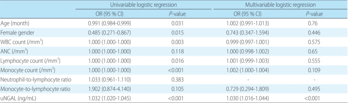
3. Univariable and multivariable logistic regression analyses
Univariable and multivariable logistic regression ana-lyses were performed to test each parameter as an indepen-dent predictor of UTI. Univariable logistic regression analysis showed that age, female gender, WBC count, lym-phocyte count, monocyte count, and uNGAL levels were significant predictors of UTI (P<0.05). However, in a multi-variable logistic regression analysis, only uNGAL levels independently predicted the presence of UTI (OR=1.030; 95% CI: 1.016–1.044; P<0.001) (Table 4).
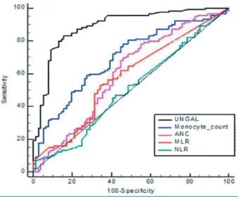
4. ROC analyses
The diagnostic properties of various markers were
exa-Table 2. Age, Gender, Fever Duration-adjusted Predicted Means between the UTI and Non-UTI Groups
Non-UTI UTI P-value
WBC count (/mm3) 11,272±617 13,436±490 0.007* ANC (/mm3) 7,169±411 5,900±521 0.06* Lymphocyte count (/mm3) 4,590±203 4,115±256 0.153* Monocyte count (/mm3) 1,013±82 1,428±65 <0.001* Neutrophil-to-lymphocyte ratio 3.16±0.57 2.19±0.72 0.302* Monocyte-to-lymphocyte ratio 0.53±0.15 0.26±0.19 0.277* uNGAL (ng/mL) 26.6±136.8 284.0±108.7 <0.001* Serum Cr (mg/dL) 0.16±0.02 0.13±0.02 0.317* BUN-to-Cr ratio 42.3±1.6 37.7±2.0 0.074* CRP (mg/L) 33.4±3.2 26.7±4.2 0.213*
The values are presented as predicted mean±standard error.
Abbreviations: UTI, urinary tract infection; WBC, white blood cell; ANC, absolute neutrophil count; uNGAL, urinary neutrophil gelatinase-asso-ciated lipocalin; Cr, creatinine; BUN, blood urea nitrogen; CRP, C-reactive protein.
*ANCOVA (analysis of covariance).
Table 3. Associations between uNGAL or Serum CRP and Leuko-cyte Differential Counts, Adjusted by Age, Gender, and Fever Duration in UTI and Non-UTI Groups
N uNGAL (ng/mL) CRP (mg/L) SPCC* P-value SPCC P-value UTI 119 ANC (/mm3) 0.333 <0.001 0.343 <0.001 Monocyte count (/mm3) 0.206 0.027 0.457 <0.001 NLR 0.317 <0.001 0.21 0.024 MLR 0.227 0.015 0.277 0.003 Non-UTI 78 ANC (/mm3) 0.208 0.077 0.568 <0.001 Monocyte count (/mm3 ) -0.096 0.422 0.443 <0.001 NLR 0.257 0.028 0.664 <0.001 MLR 0.052 0.661 0.655 <0.001 Abbreviations: UTI, urinary tract infection; uNGAL, urinary neutrophil gelatinase-associated lipocalin; CRP, C-reactive protein; ANC, absolute neutrophil count; NLR, neutrophil-to-lymphocyte ratio; MLR, monocyte-to-lymphocyte ratio.
*Spearman’s partial correlation coefficient.
Table 4. Univariable and Multivariable Logistic Regression Analysis for the UTI Group
Univariable logistic regression Multivariable logistic regression
OR (95 % CI) P-value OR (95 % CI) P-value
Age (month) 0.991 (0.984-0.999) 0.031 1.002 (0.991-1.013) 0.76 Female gender 0.485 (0.271-0.867) 0.015 0.743 (0.347-1.594) 0.446 WBC count (/mm3) 1.000 (1.000-1.000) 0.003 0.999 (0.997-1.001) 0.575 ANC (/mm3 ) 1.000 (1.000-1.000) 0.118 1.000 (0.998-1.002) 0.65 Lymphocyte count (/mm3) 1.000 (1.000-1.000) 0.016 1.001 (0.999-1.003) 0.555 Monocyte count (/mm3) 1.000 (1.000-1.000) <0.001 1.002 (1.000-1.004) 0.109 Neutrophil-to-lymphocyte ratio 1.033 (0.961-1.110) 0.383 - - Monocyte-to-lymphocyte ratio 1.902 (0.874-4.140) 0.105 0.729 (0.294-1.809) 0.495 uNGAL (ng/mL) 1.032 (1.020-1.045) <0.001 1.030 (1.016-1.044) <0.001
Abbreviations: OR, odds ratio; CI, confidence interval; WBC, white blood cell; ANC, absolute neutrophil count; uNGAL, urinary neutrophil gelatinase-associ-ated lipocalin.
mined for their predictability of UTI using ROC curves (Fig. 2). The area under the ROC curve was 0.89 for uNGAL (95% CI: 0.824–0.939; P=0.025), 0.7 for monocyte count (95% CI: 0.627–0.774; P=0.038), 0.6 for ANC (95% CI: 0.518– 0.687; P=0.043), 0.58 for MLR (95% CI: 0.500–0.663; P= 0.042), and 0.51 for NLR (95% CI: 0.428–0.594; P=0.042). The best cutoff value of uNGAL for detecting UTI was 34.8 ng/mL, with a sensitivity of 83.2% and a specificity of 84.6 %, and that of monocyte counts was 932/mm3, with a
sen-sitivity of 72.3% and a specificity of 60.3% (Table 5).
Discussion
In this study, we investigated the association of uNGAL levels with leukocyte differential counts in blood and the usefulness of leukocyte differential counts and uNGAL as
biomarkers of UTI in children with fever. While serum CRP was correlated with leukocyte differential counts in both children with and without UTIs, uNGAL levels were correlated with ANC, monocyte counts, and MLR only in children with febrile UTIs. Using univariable and multi-variable logistic analyses, we showed that only uNGAL levels were an independent predictor of UTI. The AUCs of monocyte counts and uNGAL levels were considerably elevated for detecting UTI in children with suspected UTIs. These findings suggest that uNGAL levels could be related to leukocyte differential counts in the blood of children with febrile UTIs and monocyte counts as well as uNGAL levels may enhance the diagnostic accuracy for detecting UTIs in children with suspected UTIs.
Several studies have correlated leukocyte differential counts, especially neutrophil counts or the NLR, with bac-terial infections13,25,26). Also, lymphocytopenia has been
reported to help predict bacterial infections27,28). While
significant research on monocyte counts or the MLR and infections are lacking, some studies have revealed a link between the MLR and bacterial infection. Naess et al. re-ported that the NLR and MLR were higher in the bacterial infection group, suggesting that they could be used for diagnosing bacterial infection13). Also, Wang et al. recently
found that the MLR is an important, independent factor in the diagnosis of severe Klebsiella pneumonia29). Even
compared to the NLR, the MLR appeared to be a better indicator for Klebsiella pneumonia evaluation, and also correlated with disease severity29). In contrast, in critically
ill and injured patients, the MLR was meaningful as a se-condary marker for non-bacterial infections in patients with peritonitis30). These results showed that the association
between the MLR and bacterial infection has not been established. The link between monocytes and bacterial infection has been rarely studied, and, to the best of our knowledge, there has been no clinical research on the
cli-Table 5. Predictive Value of Biomarkers for UTI using the ROC Curve
AUC 95% CI P-value Cutoff value Sensitivity (%) Specificity (%)
uNGAL (ng/mL) 0.89 0.824–0.939 0.025 34.8 83.2 84.6
Monocyte count (/mm3) 0.7 0.627–0.774 0.038 932 72.3 60.3
ANC (/mm3) 0.6 0.518–0.687 0.043 4,572 69.8 54
MLR 0.58 0.500–0.663 0.042 0.21 52.9 66.7
NLR 0.51 0.428–0.594 0.042 1.51 16 75.6
Abbreviations: AUC, area under the curve; CI, confidence interval; uNGAL, urinary neutrophil gelatinase-associated lipocalin; NLR, neutrophil-to-lymphocyte ratio; MLR, monocyte-to-lymphocyte ratio; ANC, absolute neutrophil count.
Fig. 2. Receiver operating characteristic curve of uNGAL levels, monocyte counts, the ANC, NLR, and MLR for predicting UTIs in children with fevers.
uNGAL, urinary urinary neutrophil gelatinase-associated lipo-calin; ANC, absolute neutrophil count; NLR, neutrophil-to-lym-phocyte ratio; MLR, monocyte-to-lymneutrophil-to-lym-phocyte ratio.
nical utility of monocyte counts in the evaluation of UTI. When an infection occurs, neutrophils and monocytes move to the peripheral site, which is essential for defending against pathogens18). As the infection progresses, the
num-ber of neutrophils and monocytes developing in the bone marrow increases rapidly and the cells migrate to the in-flamed tissue18). CCL2/CCR2-dependent inflammatory
monocytes are released from the bone marrow. Eventually, these monocytes migrate to tissues and differentiate into macrophages or dendritic cells31). Monocytes are also
in-volved in tissue healing, the removal of pathogens and dead cells, and the onset of adaptive immunity20). Macrophages
differentiated from monocytes are under the mucous mem-brane of the urinary tract, and, in the case of infection, more macrophages are recruited to this site21). When
acti-vated due to infection, macrophages produce important cytokines and chemokines that regulate their activity. In addition, other nearby immune cells are also activated, affecting the inflammatory response32). In the present study,
monocyte counts in addition to uNGAL levels were higher in the UTI group compared to the non-UTI group, even after adjustment for age, gender, and fever duration. The ANC, NLR, and MLR were not different between patients with UTI and non-UTI. In the UTI group, there was a correlation between uNGAL levels and ANC, monocyte counts, and the MLR. The NLR showed a positive relation-ship with uNGAL levels both in the UTI and non-UTI groups. In contrast, there was no relationship between uNGAL levels and monocyte counts or the MLR in the non-UTI group. Therefore, monocyte counts seem to be more specific than any other leukocyte differential count in the detection of UTIs in this clinical setting.
NGAL, which is highly expressed in monocytes/macro-phages as well as neutrophils, serves as a mediator of the innate immune response9,33). As an iron carrier protein, it
plays a significant role in limiting bacterial growth by bin-ding bacterial siderophores9). Many studies including ours
have shown that uNGAL measurements could be helpful in the prediction of UTIs in children11,34,35). Previously, we
identified the uNGAL and uNGAL/Cr were promising evaluations to diagnose and manage febrile UTIs in child-ren. The AUC for detecting a UTI in children was 0.89 for uNGAL levels and 0.9 for uNGAL/Cr34). Lubell et al.
de-monstrated that in a total of 260 children with suspected
UTIs, uNGAL had higher sensitivity than Gram stains, the combination of leukocyte esterase activity, or nitrite positivity for the detection of UTI11). The AUC for detecting
a UTI was 0.871 for Gram stain, with a sensitivity of 74.3% and a specificity of 100%11). The AUC was 0.858 for nitrite
positivity, with a sensitivity of 20.0% and a specificity of 100 %11). At a cutoff level of 39.1 ng/mL, the AUC for uNGAL
was 0.978, with a sensitivity of 97.1% and a specificity of 95.6%11). Jagadesan et al. also reported that uNGAL levels
could be more sensitive and specific than urine WBCs in identifying UTIs35). The AUC for detecting a UTI was 0.70
for urine WBCs, with a sensitivity of 70.6% and a specificity of 53%35). uNGAL showed higher AUC value of 0.76, with
a sensitivity of 79.4% and a specificity of 68.2%35).
Consi-stent with these findings, we also confirmed that uNGAL levels had a high discriminating ability for UTIs in children with suspected UTIs. The AUC to predict UTI was 0.89 for uNGAL, with the best cutoff value of 34.8 ng/mL (sensiti-vity 83.2%, specificity 84.6%). Additionally, in the ROC analysis, monocyte counts were helpful for detecting UTIs. At a cutoff monocyte count of 932/mm3, the AUC for
iden-tifying UTI was 0.70 (sensitivity 72.3%, specificity 60.3%). These findings suggest that elevated uNGAL levels in child-ren with UTIs may be associated with increased monocyte counts in blood. And the monocyte counts, as well as uNGAL levels, may be helpful in detecting UTIs in children with suspected UTIs. However, in a multivariable logistic regression analysis, only uNGAL levels independently predicted the presence of a UTI. Serum monocyte counts were not retained as a predictor of UTI in children with fevers. Although monocyte counts are not as high in AUC, sensitivity and specificity as uNGAL, it was confirmed that monocyte counts were related to uNGAL in the UTI group. In addition, monocyte counts are highly effective as a simple blood test routinely performed in patients.
CRP is an inflammatory marker long used as an indi-cator of acute infection36-38). CRP and WBC counts,
inclu-ding the ANC, have been reported to increase in acute bac-terial infections and positively correlate with each other38).
However, in our clinical setting, CRP did not seem to be specific in predicting UTIs in children with suspected UTIs. Serum CRP showed positive correlations with the ANC, monocyte count, NLR, and MLR in both the UTI and non- UTI groups. Further, the CRP levels were not different in
children with or without UTI, even after adjustment for age, gender, and fever duration. Therefore, the usefulness of serum CRP may be limited for predicting UTI in children with fevers.
This study had some limitations. First, the sample size was relatively small because it was a single-center retro-spective study. Multicenter, proretro-spective studies involving a large number of patients are needed to overcome these limitations. Second, we could not completely confirm the viral cause of illness in the non-UTI group since we did not perform viral cultures in all children in the non-UTI group. Third, we only classified children with fever into UTI group and non-UTI group, and there was no control group with-out fever. Fourth, only uNGAL was analyzed in this study. Since plasma NGAL is a more characteristic marker for systemic inflammatory conditions39), studies are needed to
compare the relationship between plasma NGAL levels and leukocyte differential counts.
In conclusion, uNGAL levels may be associated with blood leukocyte differential counts in children with febrile UTIs. Both uNGAL measurements and monocyte counts can help in identifying UTI in a child with a suspected UTI, although uNGAL seems to be most valuable in predicting UTI. Further studies involving a large number of patients are needed to clarify the clinical significance of leukocyte differential counts, especially monocyte in children with suspected UTIs.
Conflicts of interest
No potential conflict of interest relevant to this article was reported.
Patient consent
This study was approved by the Institutional Review Board (IRB), and the consent was waived due to the nature of the retrospective study [IRB number 2019GR0237].
ORCID IDs
Ji Won Jang https://orcid.org/0000-0003-4959-2396 Hyung Eun Yim https://orcid.org/0000-0001-9805-9278 Kee Hwan Yoo https://orcid.org/0000-0001-6490-4293
References
1. Lee SJ. Clinical guideline for childhood urinary tract infection (second revision). Child Kidney Dis 2015;19:56-64.
2. Strohmeier Y, Hodson EM, Willis NS, Webster AC, Craig JC. Anti-biotics for acute pyelonephritis in children. Cochrane Database Syst Rev, 2014;28;CD003772.
3. National Collaborating Centre for Women’s and Children’s Health. Urinary Tract Infection in Children: Diagnosis, Treatment and Long Term Management. London, England: RCOG Press; 2007. 4. Han SY, Lee IR, Park SJ, Kim JH, Shin JI. Usefulness of
neutrophil-lymphocyte ratio in young children with febrile urinary tract infection. Korean J Pediatr 2016;59:139-44.
5. Tullus K. Difficulties in diagnosing urinary tract infections in small children. Pediatr Nephrol 2011;26:1923-6.
6. Tullus K. Low urinary bacterial counts: do they count? Pediatr Nephrol 2016;31:171-4.
7. Masajtis-Zagajewska A, Nowicki M. New markers of urinary tract infection. Clinica Chimica Acta 2017;471:286-91.
8. Yim HE. Neutrophil gelatinase-associated lipocalin and kidney diseases. Child Kidney Dis 2015;19:79-88.
9. Ichino M, Kuroyanagi Y, Kusaka M, Mori T, Ishikawa K, Shiroki R, et al. Increased urinary neutrophil gelatinase associated lipocalin levels in a rat model of upper urinary tract infection. J Urol 2009; 181:2326-31.
10. Fjaertoft G, Foucard T, Xu S, Venge P. Human neutrophil lipocalin (HNL) as a diagnostic tool in children with acute infections: a study of the kinetics. Acta Paediatr 2005;94:661-6.
11. Lubell TR, Barasch JM, Xu K, Ieni M, Cabrera KI, Dayan PS. Urinary neutrophil gelatinase-associated lipocalin for the diagnosis of urinary tract infections. Pediatrics 2017;140:e20171090.
12. Skowron B, Baranowska A, Dobrek L, Ciesielczyk K, Kaszuba-Zwo-inska J, Wiecek G, et al. Urinary neutrophil gelatinase-associated lipocalin, kidney injury molecule-1, uromodulin, and cystatin C concentrations in an experimental rat model of ascending acute kidney injury induced by pyelonephritis. J Physiol Pharmacol 2018;69:625-37.
13. Naess A, Nilssen SS, Mo R, Eide GE, Sjursen H. Role of neutrophil to lymphocyte and monocyte to lymphocyte ratios in the diagnosis of bacterial infection in patients with fever. Infection 2017;45:299-307.
14. Loonen AJ, de Jager CP, Tosserams J, Kusters R, Hilbink M, Wever PC, et al. Biomarkers and molecular analysis to improve
blood-stream infection diagnostics in an emergency care unit. PloS One 2014;9:e87315.
15. Riley LK, Rupert J. Evaluation of patients with leukocytosis. AM Fam Phys 2015;92:1004-11.
16. Terradas R, Grau S, Blanch J, Riu M, Saballs P, Castells X, et al. Eosinophil count and neutrophil-lymphocyte count ratio as prognostic markers in patients with bacteremia: a retrospective cohort study. PloS One 2012;7:e42860.
17. Meshaal MS, Nagi A, Eldamaty A, Gaber M, Rizk HJTEHJ. Neu-trophil-to-lymphocyte ratio (NLR) and platelet-to-lymphocyte ratio (PLR) as independent predictors of outcome in infective endocarditis (IE). Egypt Heart J 2019;71:13.
18. Serbina NV, Hohl TM, Cherny M, Pamer EG. Selective expansion of the monocytic lineage directed by bacterial infection. J Im-munol 2009;183:1900-10.
19. Dixit A, Bottek J, Beerlage AL, Schuettpelz J, Thiebes S, Brenzel A, et al. Frontline science: proliferation of Ly6C(+) monocytes during urinary tract infections is regulated by IL-6 trans-signaling. J Leukoc Biol 2018;103:13-22.
20. Ingersoll MA, Platt AM, Potteaux S, Randolph GJ. Monocyte traf-ficking in acute and chronic inflammation. Trends Immunol 2011; 32:470-7.
21. Abraham SN, Miao Y. The nature of immune responses to urinary tract infections. Nat Rev Immunol 2015;15:655-63.
22. Bieber K, Autenrieth SE. Insights how monocytes and dendritic cells contribute and regulate immune defense against microbial pathogens. Immunobiology 2015;220:215-26.
23. Pérez RP, Ortega MJC, Álvarez JA, Baquero-Artigao F, Rico JCS, Zúñiga RV, et al. Recommendations on the diagnosis and treat-ment of urinary tract infection]. Anales de Pediatría 2019;90:400. e1-9.
24. Krzeminska E, Wyczalkowska‐Tomasik A, Korytowska N, Paczek L. Paczek comparison of two methods for determination of NGAL levels in urine: ELISA and CMIA. J Clin Lab Anal 2016;30:956-60. 25. Lowsby R, Gomes C, Jarman I, Lisboa P, Nee PA, Vardhan M, et al.
Neutrophil to lymphocyte count ratio as an early indicator of blood stream infection in the emergency department. Emerg Med J 2015;32:531-4.
26. Turak O, Özcan F, İşleyen A, Başar FN, Gül M, Yilmaz S, et al. Useful-ness of neutrophil-to-lymphocyte ratio to predict in-hospital outcomes in infective endocarditis. Can J Cardiol 2013;29:1672-8. 27. de Jager CP, van Wijk PTL, Mathoera RB, de Jongh-Leuvenink J,
van der Poll T, Wever PC. Lymphocytopenia and neutrophil-lymphocyte count ratio predict bacteremia better than
conven-tional infection markers in an emergency care unit. Crit Care 2010;14:R192-R20021034463.
28. Wyllie DH, Bowler ICJW, Peto TEA. Relation between lympho-penia and bacteraemia in UK adults with medical emergencies. J Clin Pathol 2004;57:950-5.
29. Wang Jl, Lu Xy, Xu Xh, Zhang Kj, Gong H, Lv D, et al. Predictive role of monocyte-to-lymphocyte ratio in patients with Klebsiella pneumonia infection: a single-center experience. Medicine (Bal-timore) 2019;98:e17215.
30. Djordjevic D, Rondovic G, Surbatovic M, Stanojevic I, Udovicic I, Andjelic T, et al. Neutrophil-to-lymphocyte ratio, monocyte-to-lymphocyte ratio, platelet-to-monocyte-to-lymphocyte ratio, and mean platelet volume-to-platelet count ratio as biomarkers in critically ill and injured patients: which ratio to choose to predict outcome and nature of bacteremia?. Mediators Inflamm 2018;15:2018:3758068. 31. Serbina NV, Pamer EG. Monocyte emigration from bone marrow
during bacterial infection requires signals mediated by chemo-kine receptor CCR2. Nature Immunol 2006;7:311-7..
32. Duell BL, Carey AJ, Dando SJ, Schembri MA, Ulett GC. Human bladder uroepithelial cells synergize with monocytes to promote IL-10 synthesis and other cytokine responses to uropathogenic Escherichia coli. PLoS ONE 2013;8:e78013.
33. Cai L, Rubin J, Han W, Venge P, Xu S. The origin of multiple mole-cular forms in urine of HNL/NGAL. Clin J Am Soc Nephrol 2010;5: 2229-35.
34. Yim HE, Yim H, Bae ES, Woo SU, Yoo KH. Predictive value of uri-nary and serum biomarkers in young children with febrile uriuri-nary tract infections. Pediatr Nephrol 2014;29:2181-9.
35. Jagadesan I, Agarwal I, Chaturvedi S, Jose A, Sahni RD, Fleming JJ. Urinary neutrophil gelatinase associated lipocalin - a sensitive marker for urinary tract infection in children. Indian J Nephrol 2019;29:340-4.
36. Zarkesh M, Sedaghat F, Heidarzadeh A, Tabrizi M, Moghadam KB, Ghesmati S. Diagnostic Value of IL-6, CRP, WBC, and Absolute Neutrophil Count to Predict Serious Bacterial Infection in Febrile Infants. Acta Medica Iranica 2015;53.
37. Sproston NR, Ashworth JJ. Ashworth Role of C-reactive protein at sites of inflammation and infection. Front Immunol 2018;9:754. 38. Shaikh S, Salim E, Ram PV, Memon SS, Zubairi A, Khawaja SA, et
al. Correlation of C-reactive protein and total leukocyte count in acute infections. Pak J Surg 2019;35:271-4.
39. Forster CS, Devarajan P. Neutrophil gelatinase-associated lipoca-lin: utility in urologic conditions. Pediatric Nephrology 2017;32: 377-81.