A THESIS
FOR THE DEGREE OF MASTER OF SCIENCE
Sulforaphane suppresses TNF-α-mediated
activation of NF-
kB and induces apoptosis
through activation of reactive oxygen
species-dependent caspase-3
Sang-Hyuck Kang
Department of Marine Life Science
GRADUATE SCHOOL
CHEJU NATIONAL UNIVERSITY
Sulforaphane suppresses TNF-α-mediated
activation of NF-kB and induces apoptosis
through activation of reactive oxygen
species-dependent caspase-3
Sang-Hyuck Kang
(Supervised by professor Gi-Young Kim)
A thesis submitted in partial fulfillment of the requirement for the
degree of Master of Science
Department of Marine Life Science
GRADUATE SCHOOL
CHEJU NATIONAL UNIVERSITY
1
Contents
Contents
··· 1
Abstract
··· 4
List of Figures
··· 6
. Introduction
Ⅰ
··· 7
. Material and Methods
Ⅱ
··· 10
1. Reagents ··· 10
2. Antibodies ··· 11
3. Cell Lines ··· 11
4. Cell viability and growth ··· 12
5. Determination of caspase activity ··· 12
6. Flow cytometric analysis ··· 13
7. Western blot analysis ··· 14
8. Electrophoretic mobility shift assays (EMSAs) ··· 15
9. Immunofluorescence and nuclear staining ··· 16
2
11. Statistical analysis ··· 17
. Results
Ⅲ
1. Combined treatment with SFN and TNF-α significantly reduced cell viability in
human leukemia cells ··· 18
2. A sub-toxic dose of SFN sensitized TNF-α-induced apoptosis in human
leukemia THP-1 cells ··· 20
3. SFN attenuated TNF-α-induced-NF-κB activity in THP-1 cells ··· 24
4. Inhibition of inducible NF-κB activation by SFN was not cell type specific ··· 28
5. SFN inhibited TNF-α-dependent IκBα degradation and phosphorylation/nuclear
translocation of p65 ··· 30
6. SFN repressed TNF-α-induced NF-κB-dependent gene product expression ··· 32
7. NAC pre-treatment protected against the combined treatment-induced apoptosis
through suppression of ROS generation ··· 34
8. ROS-dependent caspase-3 was a major target for combined treatment-mediated
apoptosis ··· 36
3
. Discussion
Ⅳ
··· 38
.
Ⅴ 국문요약
··· 41
. References
Ⅵ
··· 43
4
Abstract
Sulforaphane (SFN) is a biologically active compound extracted from cruciferous
vegetables, and possessing potent anti-cancer and anti-inflammatory activities. Here, we
show that tumor necrosis factor-α (TNF-α), in combination with a sub-toxic dose of
SFN, significantly triggered apoptosis in TNF-α-resistant leukemia cells (THP-1, HL60,
U937, and K562), which was associated with caspase activity and poly
(ADP-ribose)-polymerase cleavage. We also report that SFN non-specifically inhibited
TNF-α-induced NF-κB activation through the inhibition of IκBα phosphorylation, IκBα
degradation, and p65 nuclear translocation. This inhibition correlated with the
suppression of NF-κB-dependent genes involved in anti-apoptosis (IAP-1, IAP-2, XIAP,
Bcl-2, and Bcl-xL), cell proliferation (c-Myc, COX-2, and cyclin D1), and metastasis
(VEGF and MMP-9). These effects suggest that SFN inhibits TNF-α-induced NF-κB
activation through the suppression of IκBα degradation, leading to reduced expression
of NF-κB-regulated gene products. Combined treatment with SFN and TNF-α was also
accompanied by the generation of reactive oxygen species (ROS). Pre-treatment with
N-acetyl-L-cysteine significantly attenuated the combined treatment-induced ROS
generation and caspase-3-dependent apoptosis, implying the involvement of ROS in this
5
type of cell death. In conclusion, the results of the present study indicate that SFN
suppresses TNF-α-induced NF-kB activity and induces apoptosis through activation of
ROS-dependent caspase-3.
6
List of Figures
Fig. 1. Combined treatment with SFN and TNF-α decreased cell viability in
human leukemia cells.
··· 19
Fig. 2. SFN sensitized monocytic leukemia THP-1 cells to
TNF-α-mediated apoptosis.
··· 22
Fig. 3. SFN suppressed TNF-α-induced NF-κB activation.
··· 26
Fig. 4. Inhibition of inducible NF-κB activation by SFN was not cell type
specific.
··· 29
Fig. 5. SFN inhibited TNF-α-induced IκBα degradation and nuclear
translocation of NF-κB.
··· 31
Fig. 6. SFN inhibited the expression of NF-κB-dependent gene product
induced by TNF-α.
··· 33
Fig. 7. NAC pretreatment protected against the combined
treatment-induced apoptosis through suppression of ROS generation.
··· 35
Fig. 8. ROS-dependent caspase-3 was a major target for combined
7
Ⅰ
. Introduction
The nuclear transcription factor-κB (NF-κB) has been constitutively and inducibly activated in most leukemia cells and is implicated in the regulation of proliferation, survival, angiogenesis, apoptosis, and differentiation in these cells [1–5]. In the most cells, NF-κB is usually sequestered by IκB complexes in the cytoplasm [6–8]. Rapid phosphorylation and degradation of IκBα allows for the translocation of the NF-κB complex into the nucleus [9,10]. In the nucleus, the binding of the NF-κB complex to a specific sequence in the promoter region of genes triggers the transcriptional activation of NF-κB-regulated genes, which include anti-apoptosis (e.g., IAPs, XIAP, Bcl-2, and Bcl-xL), cell proliferation (e.g., c-Myc, COX-2, and cyclin D1), and metastasis (e.g., COX-2, MMP-9, and VEGF) genes [11–14]. Therefore, recent studies have suggested that chemopreventive agents capable of suppressing NF-κB activity may be potentially useful in the prevention and management of various types of cancers [1–6]. NF-κB can be also activated by a wide variety of stimuli such as tumor necrosis factor (TNF)-α, interleukin-1 (IL-1), LPS, UV light, and reactive oxygen species (ROS) [15,16]. Of particular note are some reports showing that ROS contribute to cell transformation through the protection of tumor cells from apoptosis through NF-κB activation [17,18]. In contrast, some studies have reported that chemotherapeutic agents or radiation-induced ROS can lead to an induction of
8
apoptosis and a consequent suppression of tumorigenesis [19,20]. The discrepancy among these studies reflects the lack of knowledge concerning ROS-induced apoptosis and survival in tumor cells.
Sulforaphane (SFN; 1-isothiocyanato-4-(methylsulfinyl)-butane), a naturally occurring member of the isothiocyanate family of chemopreventive agents, has been shown to possess anti-cancer properties in several cancer cell lines [21–23]. Recent studies also showed that SFN inhibited osteoclastogenesis and inflammation by inhibiting direct NF-κB binding to DNA or interaction with redox regulators [24,25]. Additionally, Xu et al. reported that SFN suppresses NF-κB activity and NF-κB-related VEGF, cyclin D1, and Bcl-xL gene expression through the inhibition of IκBα phosphorylation and degradation, as well as the decrease of nuclear translocation of NF-κB subunit p65 in human prostate cancers [26]. These findings indicate that SFN may induce apoptotic and anti-inflammatory effects through the suppression of NF-κB activity. Additional data supported the postulation that SFN-induced cell death in human prostate cancer cells was initiated by ROS accumulation [27], and combined treatment with SFN and TNF-α-related apoptosis-inducing ligand (TRAIL) sensitized TRAIL-resistant hepatoma cells through ROS-mediated upregulation of death receptor 5 (DR5) [28]. All of the above data implies that SFN-induced apoptosis is mediated by ROS accumulation and NF-κB down-regulation. However the relationship between the occurrence of ROS and NF-κB activity
9
has not yet been elucidated until now. Kim et al. reported that TNF-α-mediated apoptosis was not sensitized in hepatoma cells by combined treatment with SFN [28]. Therefore, further studies are warranted to determine whether combined treatment with SFN and TNF-α sensitizes apoptosis in other cancer cells through ROS-dependent NF-κB regulation.
Our findings suggest that SFN sensitizes ROS generation, leading to rapid induction of TNF-α-mediated cell death. Moreover, SFN strongly reduced TNF-α-induced NF-κB activity and NF-κB-related gene expression. The ROS generation and apoptosis that was induced by combined treatment with SFN and TNF-α was significantly attenuated upon treatment with N-acetyl-L-cysteine (NAC). These results suggest that the combined treatment with SFN and TNF-α might sensitize cell death in TNF-TNF-α-resistant cells through activation of ROS-dependent caspase-3 and suppresses activation of NF-κB.
10
Ⅱ
. Materials and methods
1. Reagents
SFN (purity >99%), NAC, propidium iodide (PI), 4,6-diamidino-2-phenylindole (DAPI), and 3-(4,5-dimethyl-2-thiazolyl)-2,5-diphenyl-2H-tetrazolium bromide (MTT) were obtained from Sigma (St. Louis, MO). SFN was dissolved in dimethyl sulfoxide (DMSO) as a stock solution at 20 mM concentration and stored in aliquots at -20oC. DMSO (0.1%) was used as a
vehicle control. Hydroethidine (HE) and 6-carboxy-2’,7’-dichlorodihydrofluorescein diacetate (H2DCFDA) were from Molecular Probes (Eugene, OR). Human recombinant TNF-α was
purchased from KOMA Biotechnology (Seoul, Republic of Korea) and prepared in PBS at stock concentrations of 10 µg/ml. The caspase activity assay kit was obtained from R&D Systems (Minneapolis, MN). Caspase-3 inhibitor 1 (z-DEVD-fmk) was obtained from Calbiochem (San Diego, CA). The Lightshift EMSA Optimization kit was purchased from Pierce (Rockford, IL). An enhanced chemiluminescence kit was purchased from Amersham (Arlington Heights, IL). All other chemicals not specifically cited here were purchased from Sigma.
11
2. Antibodies
Antibodies against IAP-1, IAP-2, XIAP, Bcl-2, Bcl-xL, c-Myc, COX-2, cyclin D1, VEGF, MMP-9, and poly (ADP-ribose) polymerase (PARP) were purchased from Santa Cruz Biotechnology (Santa Cruz, CA). Antibodies against p65, p50, phospho (p)-IκBα, and IκBα were obtained from Cell Signaling (Beverly, MA), and the antibody against β-actin was obtained from Sigma. Peroxidase-labeled anti-rabbit and anti-mouse immunoglobulins were purchased from KOMA Biotechnology.
3. Cell lines
Cell lines used in our studies included human monocytic leukemia (U937 and THP-1), human acute myeloblastic leukemia (HL60), human erythroid chronic myeloid leukemia (K562), human prostate carcinoma (PC-3), human breast carcinoma (MCF-7), and human hepatoma (HepG2). All cell lines were obtained from the American Type Culture Collection (Manassas, VA) and cultured in RPMI 1640 with 10% fetal bovine serum. All media were supplemented with 100 units/ml penicillin and 100 μg/ml streptomycin.
12
4. Cell viability and growth
Cells were seeded at 2 ´ 105 cells/ml and treated with the indicated concentrations of SFN,
TNF-α, or combined treatment (SFN+TNF-α). Following treatment, viability and cell number were determined by MTT assays and trypan blue exclusion assays, respectively.
5. Determination of caspase activity
The activity of caspase-like proteases was measured using the caspase activation kit according to the manufacturer’s protocol. This assay is based on spectrophotometric detection of the color reporter molecule p-nitroanaline (pNA) that is linked to the end of the caspase-specific substrate. The cleavage of the peptide by the caspase releases the chromophore pNA, which can be quantified spectrophotometrically at a wavelength of 405 nm. Ac-DEVD-pNA (for caspase-3), Ac-IETD-pNA (for caspase-8), and LEHD-pNA (for caspase-9) are used as the substrates.
13
6. Flow cytometric analysis
Cell cycle status was analyzed by flow cytometry of PI-stained cells. Cells (1 ´ 106) were
fixed in 70% ethanol overnight at 4°C. The cells were washed in phosphate-buffered saline (PBS) with 0.1% BSA. Cells were incubated with 1 U/ml of RNase A (DNase free) and 10 μg/ml of PI overnight at room temperature in the dark. Cells were analyzed by using a FACSCalibur flow cytometer (Becton Dickenson; San Jose, CA). The levels of apoptotic cells with sub-G1 DNA were determined as a percentage of the total number of cells. For annexin V
staining, live cells were washed in PBS and incubated with annexin V-fluorescein isothiocyanate (R&D Systems). Cells were analyzed using the FACSCalibur flow cytometer.
14
7. Western blot analysis
For extraction of total protein, cells were lysed in lysis buffer (137 mM NaCl, 15 mM EGTA, 0.1 mM sodium orthovanadate, 15 mM MgCl2, 0.1% Triton X-100, 25 mM MOPS, 100
μM phenylmethylsulfonyl fluoride, and 20 μM leupeptin adjusted to pH 7.2) for 30 mins on ice and clarified by centrifugation. To determine the levels of protein expression in the cytoplasm and the nucleus, extracts were prepared using NE-PER nuclear and cytosolic extraction reagents (Pierce). Protein concentration was quantified using the Bio-Rad detergent-compatible protein assay reagent (Bio-Rad Laboratories; Hercules, CA) and 50 μg of proteins were separated on 10% polyacrylamide gels and transferred to nitrocellulose membranes using standard procedures. The nitrocellulose was blocked in 5% powdered milk in Tris-buffered saline containing 0.1% (v/v) Tween 20 (TBST) and incubated with primary antibodies overnight at 4ºC. Blots were washed three times for 10 mins in TBST and probed with mouse or rabbit secondary antibodies for 1 h. Membranes were washed three times for 10 mins in TBST and then developed using ECL. Gels were stripped and re-probed with antibodies against β-actin or nucleolin, as a loading control.
15
8. Electrophoretic mobility shift assays (EMSAs)
DNA-protein binding assays were carried out using nuclear extracts. Synthetic complementary NF-κB binding oligonucleotides (5’-AGT TGA GGG GAC TTT CCC AGG C-3’) (Santa Cruz Biotechnology) were 3’-biotinylated using the biotin 3’-end DNA labeling kit (Pierce) according to the manufacturer’s instructions, and annealed for 1 h at room temperature. Binding reactions were carried out for 20 mins at room temperature in the presence of 50 ng/μl poly (dI-dC), 0.05% Nonidet P-40, 5 mM MgCl2, 10 mM EDTA, and 2.5% glycerol in 1×
binding buffer (LightShiftTM chemiluminescent EMSA kit; Pierce) using 20 fmol of
biotin-end-labeled target DNA and 10 µg of nuclear extract. Assays were loaded onto native 4% polyacrylamide gels pre-electrophoresed for 60 mins in 0.5 × Tris borate/EDTA and electrophoresed at 100 V before being transferred onto a positively charged nylon membrane (HybondTM-N+) in 0.5 × Tris borate/EDTA at 100 V for 30 mins. Transferred DNA samples
were cross-linked onto the membrane at 120 mJ/cm2 and detected using horseradish
peroxidase-conjugated streptavidin (LightShiftTM chemiluminescent EMSA kit) according to the
16
9. Immunofluorescence and nuclear staining
After treatment with SFN and TNF-α for 30 mins, the cells were harvested, washed in ice-cold PBS, fixed with 3.7% paraformaldehyde and then permeabilized with 0.2% Triton-X 100. The fixed cells were washed with PBS and then incubated with anti-p65 antibody. The nuclei were stained with DAPI solution and the anti-p65 antibody was detected using anti-mouse IgG conjugated with Alexa Fluor 488 (Molecular Probes; Eugene, OR). Nuclear morphology and p65-detection were evaluated by fluorescence microscopy.
10. Measurement of ROS
Intracellular ROS generation was measured by flow cytometry following staining with HE and H2DEFDA, which have been shown to be specific for the detection of O2- and H2O2. Briefly,
5 ´ 104 cells were seeded in 60-mm dishes and exposed to the indicated compounds. After a 3
h-treatment, the cells were stained with 2 μM HE or 5 μM H2DCFDA for 30 mins at 37°C. The
cells were collected, and the fluorescence was analyzed using flow cytometer. In a parallel experiment, the cells were pre-treated with NAC prior to SFN, TNF-α, or SFN+TNF-α exposure and analyzed ROS generation.
17
11. Statistical analysis
All data from MTT assays, trypan blue exclusion assays, FACS analyses, Western blot analyses, and caspase activity assays were derived from at least three independent experiments with a similar pattern. The images were visualized with Chemi-Smart 2000 (Vilber Lourmat; Cedex, France). Images were captured using Chemi-Capt (Vilber Lourmat) and transported into Adobe Photoshop (version 8.0). All data are presented as mean ± SD. Significant differences between the groups were determined using the unpaired Student’s t-test. A value of *p < 0.05 was accepted as an indication of statistical significance.
18
Ⅲ
. Results
1. Combined treatment with SFN and TNF-α significantly reduced cell viability in human leukemia cells
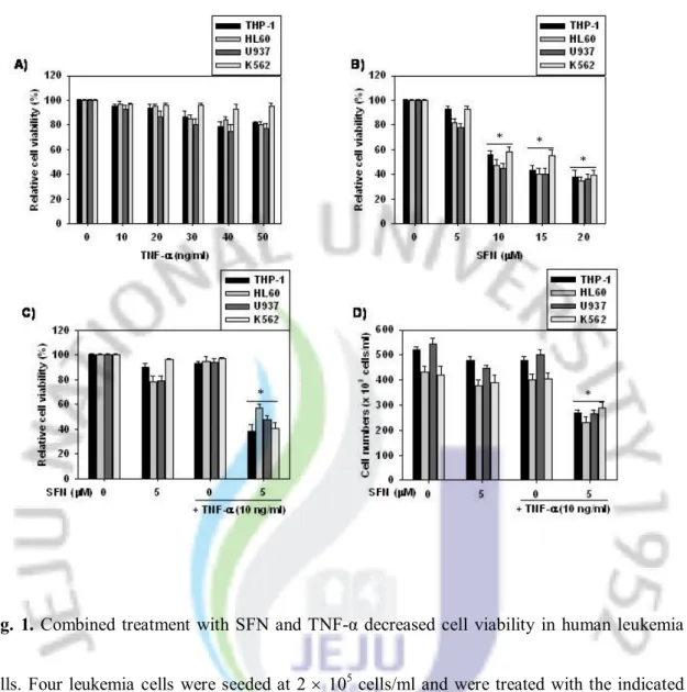
Although TNF-α has been used in anti-cancer therapy, many tumor cells are resistant to TNF-α largely due to increased NF-κB activation. Therefore, we asked the question as to whether SFN enhanced TNF-α-induced apoptosis by repression of NF-κB, thereby enhancing the killing of tumor cells. In an attempt to address this question, we first investigated the effects of SFN, TNF-α, or their combined treatment (SFN+TNF-α) on cell viability. Leukemia cells were treated with the indicated agents and subjected to MTT assays and cell counting. As shown in Fig. 1A, four leukemia cell lines tested were resistant to TNF-α-induced cell death at concentrations ranging between 10 and 50 ng/ml. Treatment with SFN at concentrations greater than 10 μM resulted in a dose-dependent decrease of cell viability in all leukemia cells tested, as determined by MTT assay (Fig. 1B). Notably, treatment of these cells with a combination of 10 ng/ml TNF-α and a sub-toxic dose of 5 μM SNF resulted in a synergistic anti-proliferation about 50%, as determined by MTT assay (Fig. 1C) and cell counting assay (Fig. 1D), respectively. Thus, SFN was found to sensitize TNF-α-resistant leukemia cells to anti-proliferation.
19
Fig. 1. Combined treatment with SFN and TNF-α decreased cell viability in human leukemia
cells. Four leukemia cells were seeded at 2 ´ 105 cells/ml and were treated with the indicated
concentrations of TNF-α (A) or SFN (B) alone for 24 h. Cell viability was determined by MTT assays. In a parallel experiment, cells were pre-incubated with 5 µM SFN for 2 h and then treated with 10 ng/ml TNF-α for 24 h. Cell viability was determined by MTT assays (C) and cell counting assays (D), respectively. Each point represents the mean ± SD of three independent experiments. The significance was determined by Student’s t-test (*p < 0.05 vs. vehicle control).
20
2. A sub-toxic dose of SFN sensitized TNF-α-induced apoptosis in human leukemia THP-1 cells
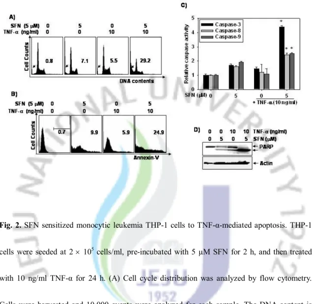
In order to obtain a quantitative measure of the induction of apoptosis, we next investigated the percentage of sub-G1 DNA content in THP-1 cells using flow cytometric analysis. As shown
in Fig. 2A, combined treatment of the cells with 5 μM SFN and 10 ng/ml TNF-α resulted in significant accumulation of sub-G1 DNA content (28 ± 3%). In order to further assess apoptosis,
we examined the exposure of phosphatidylserine on the cell surface using annexin V-staining. Flow cytometric analysis revealed that the percentage of annexin V-staining cells increased with combined treatment (25 ± 2%) (Fig. 2B). Treatment of the cells with SFN or TNF-α alone only slightly increased the percentage of cells undergoing apoptosis. These results suggested that SFN significantly stimulated TNF-α-mediated-apoptosis in TNF-α-resistant THP-1 cells.
Caspases are important mediators of apoptosis and contribute to the overall apoptotic morphology by cleavage of various cellular substrates. In order to elucidate the role of caspase activity in TNF-α-induced apoptotic cell death, we analyzed the activation of caspase-3 and upstream caspases-8 and -9 in THP-1 cells. As shown in Fig. 2C, treatment of the cells with either 5 μM SFN or 10 ng/ml TNF-α alone for 24 h resulted in only a slight increase in caspase-3, -8, and -9 activity. However, the cells treated with a combination of TNF-α and SFN had
21
significantly greater caspase activity relative to cells treated with TNF-α or SFN alone. Caspase-3 activity was especially affected, exhibiting a 4.2 fold increase relative to the control group. As shown in Fig. 2D, Western blot analysis revealed that treatment of the cells with TNF-α or SFN alone only slightly increased the cleavage of PARP, a known caspase substrate. However, combined treatment induced much greater PARP cleavage. These results indicated that combined treatment of cells with SFN and TNF-α induced apoptotic death, at least in part, through a caspase-3-dependent pathway.
22
Fig. 2. SFN sensitized monocytic leukemia THP-1 cells to TNF-α-mediated apoptosis. THP-1
cells were seeded at 2 ´ 105 cells/ml, pre-incubated with 5 µM SFN for 2 h, and then treated
with 10 ng/ml TNF-α for 24 h. (A) Cell cycle distribution was analyzed by flow cytometry. Cells were harvested and 10,000 events were analyzed for each sample. The DNA content is represented on the x-axis and the number of cells counted is represented on the y-axis. The percentages of sub-G1 phase were presented. (B) Flow cytometric analysis of apoptotic cells
was performed using annexin V-FITC. The percentages of annexin V-FITC+ cells were
represented. (C) Caspase (-3, -8, and -9) activity was determined using a caspase assay kit. (D) Equal amounts of cell lysates (50 µg) were resolved by SDS-PAGE, transferred to nitrocellulose,
23
and probed with specific antibodies to PARP. Actin was used as a loading control. Each point represents the mean ± SD of three independent experiments. The significance was determined by Student's t-test (*p < 0.05 vs. vehicle control).
24
3. SFN attenuated TNF-α-induced-NF-κB activity in THP-1 cells
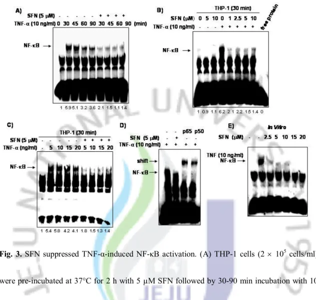
Recent evidence demonstrated that NF-κB can be activated by TNF-α and that NF-κB is a negative regulator of TNF-α-induced apoptosis [28]. Therefore, we tested whether SFN blocked TNF-α-induced NF-κB activity using EMSA analysis. THP-1 cells were stimulated with 10 ng/ml TNF-α in the presence or absence of 5 µM SFN and nuclear extracts were prepared at various time intervals from 0 to 90 mins. As shown in Fig. 3A, TNF-α strongly induced DNA binding of the NF-κB at the 30-min time point, but pre-treatment of the cells with 5 μM SFN significantly inhibited TNF-α-induced NF-κB binding activity at all time points tested. Less than 5 μM SFN also significantly attenuated TNF-α-induced NF-κB activation (Fig. 3B). SFN inhibited NF-κB activation by over 70% at a dose of 5 μM. We also examined the effects of combined treatment of the cells with 5 μM of SFN and varying concentrations of TNF-α (5, 10, 15, or 20 ng/ml). As shown in Fig. 3C, SFN completely suppressed NF-κB-DNA binding activity regardless of TNF-α concentration. The heterodimer of p50 and p65 is one of the most frequent complexes of NF-κB associated with tumorigenesis [29]. Therefore, we tested whether SFN-mediated inhibition of NF-κB activity was due to decreased p50/p65 heterodimer formation. When nuclear extracts from TNF-α-activated cells were incubated with antibodies to p50 or p65, the resulting bands were shifted to higher molecular masses (Fig. 3D), suggesting
25
that p50 and p65 were associated with TNF-α-activated complex. Moreover, when we incubated nuclear extracts from TNF-α-treated cells with SFN in vitro, the results of EMSA showed that SFN had a direct effect on the binding of NF-κB to DNA (Fig. 3E).
26
Fig. 3. SFN suppressed TNF-α-induced NF-κB activation. (A) THP-1 cells (2 ´ 105 cells/ml)
were pre-incubated at 37°C for 2 h with 5 µM SFN followed by 30-90 min incubation with 10 ng/ml TNF-α. Nuclear extracts were then prepared and assayed for NF-κB, as described in the experimental procedure section. (B) THP-1 cells were pre-incubated at 37°C for 2 h with the indicated concentrations of SFN followed by 30 mins incubation with 10 ng/ml TNF-α and then tested for NF-κB activation. (C) After treatment with 5 µM SFN for 2 h, the cells were treated with TNF-α at the indicated concentrations for 30 mins and assayed for NF-κB. (D) Nuclear extracts were prepared from THP-1 cells that were treated with 10 ng/ml TNF-α, 5 µM SFN, or a combination of both agents. Extracted nuclear proteins were incubated for 2 h with antibodies
27
against p65 and p50 and then assayed for NF-κB by EMSA. (E) To investigate the direct effect of SFN on the NF-κB complex, nuclear extracts were prepared from THP-1 cells that were untreated or treated with 10 ng/ml TNF-α, incubated for 30 mins with SFN at the indicated concentrations, and then assayed for NF-κB activation using EMSA.
28
4. Inhibition of inducible NF-κB activation by SFN was not celltype specific
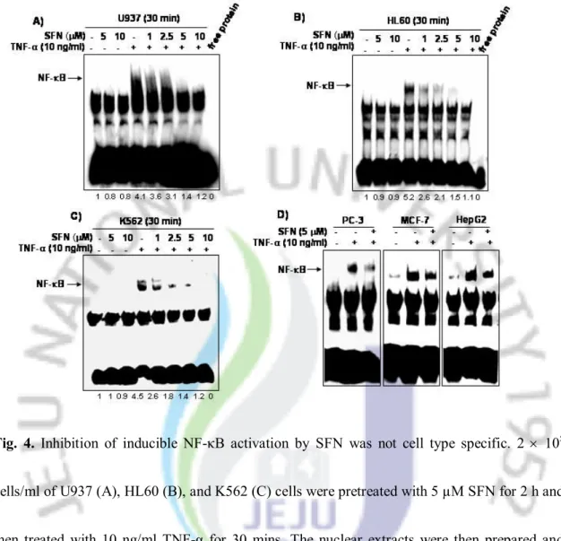
Since the signal transduction pathway leading to NF-κB activation may vary in different cell types [30,31], we investigated whether SFN effectively suppressed TNF-α-induced activation of NF-κB in other human cell lines derived from a variety of tumors, including U937, HL60, K562, PC-3, MCF-7, and HepG2. We exposed these cells to TNF-α for 30 mins in the presence or absence of SFN and then tested them for NF-κB activation using EMSA. SFN was shown to substantially suppress TNF-α-induced NF-κB activation in all six cell lines tested, indicating that the effect of SFN was not cell type specific (Fig. 4A–4D).
29
Fig. 4. Inhibition of inducible NF-κB activation by SFN was not cell type specific. 2 ´ 105
cells/ml of U937 (A), HL60 (B), and K562 (C) cells were pretreated with 5 µM SFN for 2 h and then treated with 10 ng/ml TNF-α for 30 mins. The nuclear extracts were then prepared and analyzed for NF-κB using EMSA as described in the experimental procedure section. (D) 2 ´ 105 cells/ml human prostate (PC-3), human breast (MCF-7), and human hepatoma (HepG2)
cancer cells were exposed to 5 µM SFN for 2 h and then cells were treated with 10 ng/ml TNF-α for 30 mins. Nuclear extracts were prepared and assayed for NF-κB using EMSA.
30
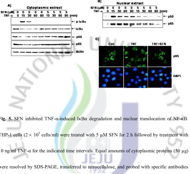
5. SFN inhibited TNF-α-dependent IκBα degradation and phosphorylation/nuclear translocation of p65
NF-κB activation by TNF-α is mediated via the interaction of the TNFR1 withNIK and IKK, resulting in phosphorylation of IκBα [32]. Therefore, we investigated the effect of SFN on TNF-α-regulating proteins for NF-κB. Western blot analysis with an antibody that specifically detected the serine-phosphorylated form of IκBα indicated that SFN completely suppressed TNF-α-induced IκBα phosphorylation (Fig. 5A). Fig. 5A also showed that SFN blocked the TNF-α-dependent IκBα degradation and sequestered both p65 and p50 in cytoplasm. As shown in Fig. 5B, Western blot analysis also indicated that SFN significantly inhibited TNF-α-induced nuclear translocation of p65 and p50. Immunohistochemistry further revealed that SFN inhibited TNF-α-induced nuclear translocation of p65. These results indicated that SFN suppressed TNF-α-induced NF-κB activation by preventing IκBα degradation and NF-κB nuclear translocation.
31
Fig. 5. SFN inhibited TNF-α-induced IκBα degradation and nuclear translocation of NF-κB.
THP-1 cells (2 ´ 105 cells/ml) were treated with 5 µM SFN for 2 h followed by treatment with
10 ng/ml TNF-α for the indicated time intervals. Equal amounts of cytoplasmic proteins (50 µg) were resolved by SDS-PAGE, transferred to nitrocellulose, and probed with specific antibodies (A: anti-p-IκBα, anti-IκBα, anti-p50, and anti-p65). β-Actin was used as a loading control. (B) Nuclear extracts were prepared, resolved by 10% SDS-PAGE, and electrotransferred onto a nitrocellulose membrane. Western blot analysis was conducted with antibodies against p65 and p50. (C) THP-1 cells were incubated with 10 ng/ml TNF-α for 30 min in the presence or absence of SFN for 2 h, and then subjected to immunocytochemistry.
32
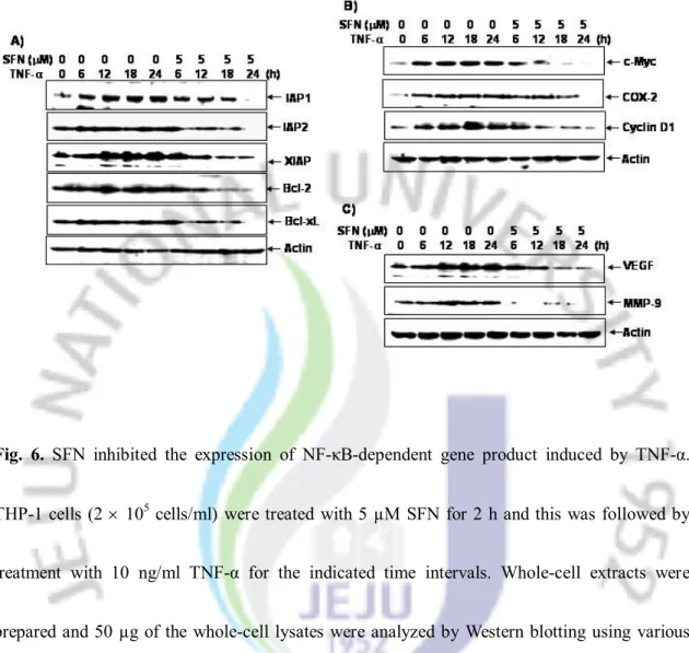
6. SFN repressed TNF-α-induced NF-κB-dependent gene product expression
NF-κB has been known to regulate the expression of the anti-apoptotic, proliferative, and metastatic gene products [11–14]. Therefore, we investigated whether SFN regulated the expression of these gene products following TNF-α treatment. As shown in Fig. 6A, TNF-α exposure induced the expression of these anti-apoptotic proteins in a time-dependent manner, whereas SFN suppressed it. Our results also showed that SFN abolished the TNF-α-induced expression of proliferative gene products such as c-Myc, cyclin D1, and COX-2 (Fig. 6B) as well as metastatic gene products such as VEGF and MMP-9 (Fig. 6C). These results supported our postulate that SFN blocked TNF-α-induced NF-κB-regulated gene expression.
33
Fig. 6. SFN inhibited the expression of NF-κB-dependent gene product induced by TNF-α.
THP-1 cells (2 ´ 105 cells/ml) were treated with 5 µM SFN for 2 h and this was followed by
treatment with 10 ng/ml TNF-α for the indicated time intervals. Whole-cell extracts were prepared and 50 µg of the whole-cell lysates were analyzed by Western blotting using various antibodies (A: IAP-1, IAP-2, XIAP, Bcl-2, and Bcl-xL; B: c-Myc, cyclin D1, and COX-2; C: VEGF and MMP-9).
34
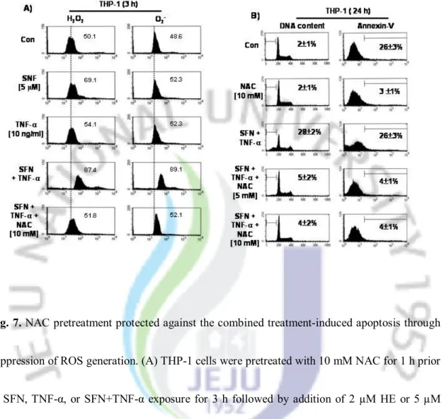
7. NAC pre-treatment protected against the combined treatment-induced apoptosis through suppression of ROS generation
Singh et al. [27] recently reported that ROS initiates SFN-induced cell death in prostate cancer cells. In an attempt to confirm whether ROS contributed to the cell death caused by the combined treatment, we examined the effect of NAC, a known antioxidant, on ROS generation and apoptosis induction. As shown in Fig. 7A, H2DCFDA-based FACS detection revealed that
intracellular H2O2 levels increased slightly in cells that were treated with SFN or TNF-α alone
after 3 h without O2- generation (Fig. 7A). Combined treatment of cells with SFN and TNF-α
significantly exhibited an approximate 2-fold increase of both H2O2 and O2- as compared to the
vehicle control, which was reduced on pre-treatment of the cells with NAC. TNF-α (10 ng/ml) alone, or NAC (10 mM) alone, showed no effect on intracellular H2O2 and O2- generation.
Treatment of cells with NAC also significantly reduced the populations of sub-G1 phase and
annexin V+ cells increased by combined treatment of the cells with SFN and TNF-α (Fig. 7B).
Taken together, these results suggest that ROS generation is an important target in apoptosis by combined treatment with SFN and TNF-α.
35
Fig. 7. NAC pretreatment protected against the combined treatment-induced apoptosis through
suppression of ROS generation. (A) THP-1 cells were pretreated with 10 mM NAC for 1 h prior to SFN, TNF-α, or SFN+TNF-α exposure for 3 h followed by addition of 2 µM HE or 5 µM H2DCFDA for 30 mins at 37°C and subsequent FACS analysis for intracellular accumulation of
ROS. In parallel experiments (B and C), cell cycle distribution (left) and annexin V-FITC+
(right) were analyzed by flow cytometry. Cells were harvested and 10,000 events were analyzed for each sample. The fluorescence intensity was plotted on the X-axis and the relative number of cells was plotted on the Y-axis.
36
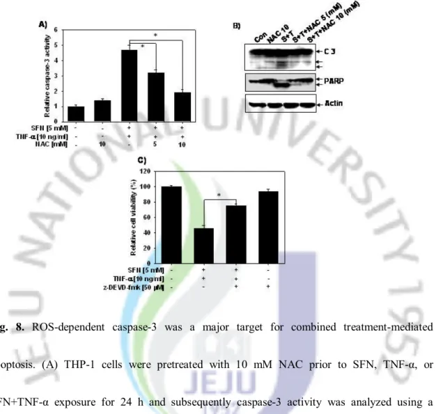
8. ROS-dependent caspase-3 was a major target for combined treatment-mediated apoptosis
To confirm activation of ROS-dependent caspase-3 in THP-1 cells, we investigated effects of combined treatment in the presence of NAC. As shown in Fig. 8A, caspase-3 was activated following combined treatment (4.5-fold), however the caspase activity was significantly attenuated in the presence of NAC. Additionally, we also evaluated the activation of caspase-3 and cleavage of PARP using Western blot analysis. Combined treatment induced the cleavage of PARP and caspase-3, whereas NAC pre-treatment significantly inhibited the cleavage of PARP and caspase-3 (Fig. 8B). Combined treatment-mediated cell death was also suppressed by z-DEVD-fmk (Fig. 8C), indicating that the TNF-α-induced apoptosis by SFN was mediated through caspase-3 activation. These results showed that combined treatment of cells with SFN and TNF-α induced cell death through ROS-dependent capase-3 activation.
37
Fig. 8. ROS-dependent caspase-3 was a major target for combined treatment-mediated
apoptosis. (A) THP-1 cells were pretreated with 10 mM NAC prior to SFN, TNF-α, or SFN+TNF-α exposure for 24 h and subsequently caspase-3 activity was analyzed using a caspase assay kit. (B) Equal amounts of cell lysates (50 µg) were resolved by SDS-PAGE, transferred to nitrocellulose and probed with specific antibodies against caspase-3 and PARP. β-Actin was used as a loading control. (C) Cells were incubated with SFN+TNF-α for 24 h in the presence or absence of z-DEVD-fmk (50 µM). Cells viability was measured using MTT assay. Each point represents the mean ± SD of three independent experiments. The significance was determined by Student's t-test (*p < 0.05 vs. vehicle control).
38
Ⅳ
. Discussion
TNF-α has previously been used as an anti-cancer agent due to its broad cytotoxic effects against a number of tumor cells [33,34]. However, the clinical use of TNF-α has been largely limited because it can induce pro-inflammatory and anti-apoptotic gene transcription that can cause resistance to chemotherapy in many types of tumors [35,36]. Sensitizing agents capable of overcoming this resistance may facilitate the establishment of TNF-α-mediated apoptosis for the purposes of improved treatment against cancer cells [37,38]. Therefore, we investigated whether combined treatment with sub-toxic doses of SFN and TNF-α triggered apoptosis in leukemia cells that are normally resistant to α alone. Our findings indicated that SFN triggers TNF-α-induced apoptosis through suppression of NF-kB activity and ROS-dependent caspase-3.
Specific NF-κB inhibitors have been used in combination with TNF-α in order to promote apoptosis in tumor cells [1–6]. However, these agents also cause harmful side effects that limit their use. Therefore, many studies have attempted to identify agents that can induce apoptosis in cancer cells with limited cytotoxicity to normal cells. Here, we showed that a sub-toxic dose of SFN attenuated TNF-α-induced NF-κB activity, thereby sensitizing TNF-α-resistant leukemia cells to TNF-α-mediated apoptosis. Previous studies have reported that treatment with SFN inhibits osteoclastogenesis and inflammation by inhibition of NF-κB activity in vitro [24,25],
39
and SFN was shown to be administered at doses of 50 μM without deleterious side effects to normal tissues in rat [39]. Kim et al. also reported that neither TNF-α- nor Fas-mediated apoptosis was sensitized in hepatoma cells by combined treatment with SFN [28]. Therefore, we tested whether SFN suppressed TNF-α-induced NF-κB activation in other types of cancer cells such as leukemia, hepatoma, breast carcinoma, and prostate carcinoma. It was shown that SFN non-specifically inhibited NF-κB activation by suppressing IκBα phosphorylation and degradation stimulated with TNF-α, and down-regulated the expression of NF-κB-regulated gene products involved in cellular proliferation (c-Myc, cyclin D1, and COX-2), metastasis (VEGF and MMP-9), and anti-apoptosis (IAP-1, IAP-2, XIAP, Bcl-2, and Bcl-xL). These results suggested that SFN induced TNF-α-mediated apoptosis with suppression of NF-κB activity and NF-κB-related gene products.
NF-κB is activated by a variety of proinflammatory agents, including TNF-α, phorbol esters, and many growth factors [15,16]. Of these agents, ROS has been deeply implicated in TNF-α-mediated NF-κB activation through phosphorylation of IκBα and p65 [40]. In contrast, some researchers reported that ROS demonstrated opposing effects to induce TNF-α cooperative cytotoxicity and proliferation through the suppression of TNF-α-induced NF-κB activity and NF-κB-dependent gene expression [41,42]. These discrepancies may be due to dual effects of ROS varying between cell toxicity and viability. The present study has revealed that the initial
40
signal for SFN-induced apoptosis is derived from ROS in many cancer cells and that SFN also suppresses NF-κB activity by various stimuli. The involvement of ROS in NF-κB activation is well established, based on the observations that ROS-inducing agents activate NF-κB activation [43,44]. However Hayakawa and his colleagues reported that endogenous ROS production did not mediate NF-κB activity, but instead lowered the magnitude of its activation [45]. Our results indicated that the combined treatment of cells with SFN and TNF-α triggered apoptosis through suppression of NF-κB activity and ROS-dependent caspase-3 activation.
In conclusion, a combined treatment with SFN and TNF-α may offer a good strategy for the treatment of a variety of human cancers that are resistant to TNF-α treatment alone. Taken together, the use of TNF-α in combination with sub-toxic doses of SFN may provide an effective therapeutic strategy for safely treating certain resistant types of leukemia through suppression of NF-kB activity and activation of ROS-dependent caspase-3.
41
Ⅴ
. 국문 요약
Sulforaphane (SFN) 은 cruciferous vegetables에서 추출한 물질로 항암 · 항염증 효 능을 가지고 있다고 알려져 있다.
이 논문에서는 tumor necrosis factor-α (TNF- α) 와 sub-toxic 농도의 SFN을 함께 처리하였을 때, TNF-α-resistant 인체 백혈병 세포주인 THP-1, HL60, U937과 K562에 세포사멸을 효과적으로 유도함을 확인하였다. 또한, SFN의 처리는 TNF-α 에 의해 유도되는 NF-κB의 활성을 억제하였다. 이러한 현상은 IκBα의 인산화 억제, IκBα의 분해와 p65의 핵으로의 이동을 억제함으로써 나타난 결과였다. 또한 SFN은 NF-κB 에 의해 유도되는 anti-apoptosis proteins (IAP-1, IAP-2, XIAP, Bcl-2와 Bcl-xL), cell proliferation proteins (c-Myc, COX-2와 cyclin D1)과 metastasis proteins (VEGF와 MMP9) 의 발현을 억제하였다. 그리고 SFN과 TNF-α의 동시처리는 reactive oxygen species (ROS)을 생성시켰다. 항산화제인 N-acetyl-L-cystein의 전 처리시 SFN과 TNF-α 에 의
해 유도되는 ROS의 생성과 caspase-3 의존적인 apoptosis가 상당히 감소하는 것을 확 인하였다.
결과를 종합해 보면, SFN은 IκBα의 분해를 억제 함으로서 TNF-α 에 의해 유도 되는 NF-κB의 활성을 억제한다고 보여지며, 이를 통해 NF-κB에 의해 조절 받는 유 전자 산물의 발현량을 감소시키고, ROS 의존적인 caspase-3의 활성화를 통해 세포사 멸을 유도한다고 사료된다.
42
Ⅵ
. References
[1] A.C. Bharti, N. Donato, S. Singh, B.B. Aggarwal, Curcumin (diferuloylmethane) down-regulates the constitutive activation of nuclear factor-κB and IκBα kinase in human multiple myeloma cells, leading to suppression of proliferation and induction of apoptosis, Blood 101 (2003) 1053–1062.
[2] A.C. Bharti, S. Shishodia, J.M. Reuben, D. Weber, R. Alexanian, S. Raj-Vadhan, Z. Estrov, M. Talpaz, B.B. Aggarwal, Nuclear factor-κB and STAT3 are constitutively active in CD138+ cells derived from multiple myeloma patients, and suppression of these
transcription factors leads to apoptosis, Blood 103 (2004) 3175–3184.
[3] Z. Estrov, S.K. Manna, D. Harris, Q. Van, E.H. Estey, H.M. Kantarjian, M. Talpaz, B.B. Aggarwal, Phenylarsine oxide blocks interleukin-1β-induced activation of the nuclear transcription factor NF-κB, inhibits proliferation, and induces apoptosis of acute myelogenous leukemia cells, Blood 94 (1999) 2844–2853.
[4] Z. Estrov, S. Shishodia, S. Faderl, D. Harris, Q. Van, H.M. Kantarjian, M. Talpaz, B.B. Aggarwal, Resveratrol blocks interleukin-1β-induced activation of the nuclear transcription factor NF-κB, inhibits proliferation, causes S-phase arrest, and induces apoptosis of acute myeloid leukemia cells, Blood 102 (2003) 987–995.
43
[5] S. Shishodia, B.B. Aggarwal, Nuclear factor-κB: a friend or a foe in cancer, Biochem. Pharmacol. 68 (2004) 1071–1080.
[6] Y. Ben-Neriah, Regulatory functions of ubiquitination in the immune system, Nat. Immunol. 3 (2002) 20–26.
[7] M. Karin, A. Lin, NF-κB at the crossroads of life and death, Nat. Immunol. 3 (2002) 221– 227.
[8] N. Silverman, T. Maniatis, NF-κB signaling pathways in mammalian and insect innate immunity, Genes Dev. 15 (2001) 2321–2342.
[9] S. Yamaoka, G. Courtois, C. Bessia, S.T. Whiteside, R. Weil, F. Agou, H.E. Kirk, R.J. Kay, A. Israel, Complementation cloning of NEMO, a component of the IκB kinase complex essential for NF-κB activation, Cell 93 (1998) 1231–1240.
[10] E. Zandi, D.M. Rothwarf, M. Delhase, M. Hayakawa, M. Karin, The IκB kinase complex (IKK) contains two kinase subunits, IKKα and IKKβ, necessary for IκB phosphorylation and NF-κB activation, Cell 91 (1997) 243–252.
[11] H. Chan, D.P. Bartos, L.B. Owen-Schaub, Activation-dependent transcriptional regulation of the human Fas promoter requires NF-κB p50-p65 recruitment, Mol. Cell. Biol. 19 (1999) 2098–2108.
44
[12] J.Z. Qin, V. Chaturvedi, M.F. Denning, D. Choubey, M.O. Diaz, B.J. Nickoloff, Role of NF-κB in the apoptotic-resistant phenotype of keratinocytes, J. Biol. Chem. 274 (1999) 37957–37964.
[13] D.C. Guttridge, C. Albanese, J.Y. Reuther, R.G. Pestell, A.S. Baldwin Jr., NF-κB controls cell growth and differentiation through transcriptional regulation of cyclin D1, Mol. Cell. Biol. 19 (1999) 5785–5799.
[14] M. Hinz, D. Krappmann, A. Eichten, A. Heder, C. Scheidereit, M. Strauss, NF-κB function in growth control: regulation of cyclin D1 expression and G0/G1-to-S-phase transition,
Mol. Cell. Biol. 19 (1999) 2690–2698.
[15] Y. Takada, S. Singh, B.B. Aggarwal, Identification of a p65 peptide that selectively inhibits NF-κB activation induced by various inflammatory stimuli and its role in down-regulation of NF-κB-mediated gene expression and up-down-regulation of apoptosis, J. Biol. Chem. 279 (2004) 15096–15104.
[16] G.F. Vile, A. Tanew-Ilitschew, R.M. Tyrrell, Activation of NF-κB in human skin fibroblasts by the oxidative stress generated by UVA radiation, Photochem. Photobiol. 62 (1995) 463– 468.
45
[17] A. Dhar, M.R. Young, N.H. Colburn, The role of AP-1, NF-κB and ROS/NOS in skin carcinogenesis: the JB6 model is predictive, Mol. Cell. Biochem. 234–235 (2002) 185– 193.
[18] E.C. Vaquero, M. Edderkaoui, S.J. Pandol, I. Gukovsky, A.S. Gukovskaya, Reactive oxygen species produced by NAD(P)H oxidase inhibit apoptosis in pancreatic cancer cells, J. Biol. Chem. 279 (2004) 34643–34654.
[19] Y.H. Ling, L. Liebes, Y. Zou, R. Perez-Soler, Reactive oxygen species generation and mitochondria dysfunction in the apoptotic response to Bortezomib, a novel proteasome inhibitor, in human H460 non-small cell lung cancer cells, J. Biol. Chem. 278 (2003) 33714–33723.
[20] I.V. Lebedeva, Z.Z. Su, D. Sarkar, R.V. Gopalkrishnan, S. Waxman, A. Yacoub, P. Dent, P.B. Fisher, Induction of reactive oxygen species renders mutant and wild-type K-ras pancreatic carcinoma cells susceptible to Ad.mda-7-induced apoptosis, Oncogene 24 (2005) 585–596.
[21] L. Gamet-Payrastre, P. Li, S. Lumeau, G. Cassar, M.A. Dupont, S. Chevolleau, N. Gasc, J. Tulliez, F. Terce, Sulforaphane, a naturally occurring isothiocyanate, induces cell cycle arrest and apoptosis in HT29 human colon cancer cells, Cancer Res. 60 (2000) 1426–1433.
46
[22] A. Herman-Antosiewicz, D.E. Johnson, S.V. Singh, Sulforaphane causes autophagy to inhibit release of cytochrome c and apoptosis in human prostate cancer cells, Cancer Res. 66 (2006) 5828–5835.
[23] G. Parnaud, P. Li, G. Cassar, P. Rouimi, J. Tulliez, L. Combaret, L. Gamet-Payrastre, Mechanism of sulforaphane-induced cell cycle arrest and apoptosis in human colon cancer cells, Nutr. Cancer 48 (2004) 198–206.
[24] S.J. Kim, S.Y. Kang, H.H. Shin, H.S. Choi, Sulforaphane inhibits osteoclastogenesis by inhibiting nuclear factor-κB, Mol. Cells 20 (2005) 364–370.
[25] E. Heiss, C. Herhaus, K. Klimo, H. Bartsch, C. Gerhauser, Nuclear factor kappa B is a molecular target for sulforaphane-mediated anti-inflammatory mechanisms, J. Biol. Chem. 276 (2001) 32008–32015.
[26] C. Xu, G. Shen, C. Chen, C. Gelinas, A.N. Kong, Suppression of NF-κB and NF-κB-regulated gene expression by sulforaphane and PEITC through IκBα, IKK pathway in human prostate cancer PC-3 cells, Oncogene 24 (2005) 4486–4495.
[27] S.V. Singh, S.K. Srivastava, S. Choi, K.L. Lew, J. Antosiewicz, D. Xiao, Y. Zeng, S.C. Watkins, C.S. Johnson, D.L. Trump, Y.J. Lee, H. Xiao, A. Herman-Antosieqicz, Sulforaphane-induced cell death in human prostate cancer cells is initiated by reactive oxygen species, J. Biol. Chem. 280 (2005) 19911–19924.
47
[28] H. Kim, E.H. Kim, Y.W. Eom, W.H. Kim, T.K. Kwon, S.J. Lee, K.S. Choi, Sulforaphane sensitizes tumor necrosis factor-related apoptosis-inducing ligand (TRAIL)-resistant hepatoma cells to TRAIL-induced apoptosis through reactive oxygen species-mediated up-regulation of DR5, Cancer Res. 66 (2006) 1740–1750.
[29] S. Ghosh, M.J. May, E.B. Kopp, NF-κB and Rel proteins: evolutionarily conserved mediators of immune responses, Annu. Rev. Immunol. 16 (1998) 225–260.
[30] G. Bonizzi, J. Piette, M.P. Merville, V. Bours, Distinct signal transduction pathways mediate nuclear factor-κB induction by IL-1β in epithelial and lymphoid cells, J. Immunol. 159 (1997) 5264–5272.
[31] S. Ghosh, M. Karin, Missing pieces in the NF-κB puzzle, Cell 109 (2002) Suppl.:S81–S96. [32] H. Hsu, H.B. Shu, M.G. Pan, D.V. Goeddel, TRADD-TRAF2 and TRADD-FADD interactions define two distinct TNF receptor 1 signal transduction pathways, Cell 84 (1996) 299–308.
[33] N. Watanabe, Y. Niitsu, H. Sone, H. Neda, I. Urushizaki, A. Yamamoto, M. Nagamuta, Y. Sugawara, Therapeutic effect of endogenous tumor necrosis factor on ascites Meth A sarcoma, J. Immunopharmacol. 8 (1986) 271–283.
48
[34] A. Tomita, Y. Fuchino, K. Otsuka, T. Shinohara, S.N. Tanaka, T. Umeno, S. Ikeda,Clinical effects of exogenous/endogenous TNF therapy on metastatic lesions of 34 colorectal cancer patients, Anticancer Res. 18 (1998) 3937–3939.
[35] A.A. Beg, D. Baltimore, An essential role for NF-κB in preventing TNF-α-induced cell death, Science 274 (1996) 782–784.
[36] C.Y. Wang, D.C. Guttridge, M.W. Mayo, A.S. Baldwin Jr., NF-κB induces expression of the Bcl-2 homologue A1/Bfl-1 to preferentially suppress chemotherapy-induced apoptosis, Mol. Cell. Biol. 19 (1999) 5923–5929.
[37] M.L. Neale, N. Matthews, Development of tumour cell resistance to tumour necrosis factor does not confer resistance to cytotoxic drugs, Eur. J. Clin. Oncol. 25 (1989) 133– 137.
[38] N. Watanabe, Y. Niitsu, N. Yamauchi, Y. Ohtsuka, H. Sone, H. Neda, M. Maeda, I. Urushizaki, Synergistic cytotoxicity of recombinant human TNF and various anti-cancer drugs, Immunopharmacol. Immunotoxicol. 10 (1988) 117–127.
[39] R. Hu, V. Hebbar, B.R. Kim, C. Chen, B. Winnik, B. Buckley, P. Soteropoulos, P. Tolias, R.P. Hart, A.N. Kong, In vivo pharmacokinetics and regulation of gene expression profiles by isothiocyanate sulforaphane in the rat, J. Pharmacol. Exp. Ther. 310 (2004) 263–271.
49
[40] Y. Takada, A. Mukhopadhyay, G.C. Kundu, G.H. Mahabeleshwar, S. Singh, B.B. Aggarwal, Hydrogen peroxide activates NF-κB through tyrosine phosphorylation of IκBα and serine phosphorylation of p65: evidence for the involvement of IκBα kinase and Syk protein-tyrosine kinase, J. Biol. Chem. 278 (2003) 24233–24241.
[41] I. Jaspers, W. Zhang, A. Fraser, J.M. Samet, W. Reed, Hydrogen peroxide has opposing effects on IKK activity and IκBα breakdown in airway epithelial cells, Am. J. Respir. Cell. Mol. Biol. 24 (2001) 769–777.
[42] I. Ginis, J.M. Hallenbeck, J. Liu, M. Spatz, R. Jaiswal, E. Shohami, Tumor necrosis factor and reactive oxygen species cooperative cytotoxicity is mediated via inhibition of NF-κB, Mol. Med. 6 (2000) 1028–1041.
[43] S.S. Brar, T.P. Kennedy, A.R. Whorton, A.B. Sturrock, T.P. Huecksteadt, A.J. Ghio, J.R. Hoidal, Reactive oxygen species from NAD(P)H:quinine oxidoreductase constitutively activate NF-κB in malignant melanoma cells, Am. J. Physol. Cell. Physiol. 280 (2001) C659–C676.
[44] R. Schreck, P. Rieber, P.A. Baeuerle, Reactive oxygen intermediates as apparently widely used messengers in the activation of the NF-κB transcription factor and HIV-1, EMBO J. 10 (1991) 2247–2258.
50
[45] M. Hayakawa, H. Miyashita, I. Sakamoto, M. Kitagawa, H. Tanaka, H. Yasuda, M. Karin, K. Kikugawa, Evidence that reactive oxygen species do not mediate NF-κB activation, EMBO J. 22 (2003) 3356–3366.
51
감사의 글
시간이 흘러 석사 2년 과정이 끝나고 졸업 논문을 작성하였습니다. 사람은 늙기 쉽고 배움은 이루기 어려우니, 찰나의 시간을 아끼며 소중히 사용하라는 말을 마음속에 새겨 생활하려고 노력했지만, 2년의 기간을 돌아보니 언제나 부족했다는 생각만 떠오릅니다. 이렇게 너무나도 부족한 저를 제자로 받아주시고 항상 친자식처럼, 때론 아버지처럼 이끌어주시며 믿어주시고 제게 이러한 기회를 주신 김기영 교수님께 더 좋은 모습과 결과를 드리지 못해 죄송하다는 마음과 더불어 마음속 깊이 감사의 글을 올립니다. 바쁘신 와중에도 저와 저희 실험실에 항상 많은 관심과 격려를 아끼지 않으시고, 졸업논문을 검토해주신 이경준 교수님, 이제희 교수님, 허문수 교수님께 깊은 감사의 인사를 드립니다. 석사과정에서 많은 가르침과 관심을 가져주셨던 해양생명과학과 이기완 교수님, 최광식 교수님, 정준범 교수님께 깊은 감사의 인사를 드리고, 학부과정에서 보다 학문에 관심을 가지고 정진할 수 있도록 도와주신 수산생명의학과 송춘복 교수님, 여인규 교수님, 전유진 교수님께도 감사의 인사 드립니다. 지도교수님과의 인연으로 멀리 떨어져 있어도 항상 관심을 가져주신 동의대 한의과대학 최영현 교수님께도 감사의 인사 드립니다. 지난 2년의 석사과정 동안 부족한 후배를 친동생처럼 아껴주고 격려하고 이끌러 주며, 많은 가르침을 주었던 동오형과 인연이란 이름으로 알게되어 1년이란 시간동안 부족한 모습만 보여주었지만 많은 도움과 웃음을 주었던 창희, 항상 웃음으로 대해주며 많은 시간을 실험실에서 보낸 부산대 박사학위과정의 문옥씨에게도 감사의 글을 전하고 싶습니다. 또한, 지금은 같이 없지만 멀리서 우릴 지켜보고 있을 문현식 학우와 같은52 시기를 같이 고생하며 서로에게 큰 힘이 되었던 동기들 경용, 봉규, 영득, 석천, 성명, 윤범, 형철, 민석, 진이와 언제나 친동생처럼 아껴준 철홍이형, 영건이형, 만철이형, 현성이형, 상우형, 성표형, 주상이형, 송헌이형과 학과를 위해 고생하는 조교선생님 경주형과 진우와 많은 실험실 후배들과 외국인 유학생들에게도 고맙다는 말을 하고 싶습니다. 그리고, 학부시절 처음 실험실 생활을 하며 아무것도 모르던 철부지 같은 저를 도와주고 가르침을 주었던 용욱이형과 항상 바쁘다고 투덜대도 대학원 생활한다고 이해해주고 고교생활의 인연으로 만나 10여 년을 같이 지내온 우리 YC친구들, 선배에게도 쓴소리를 아끼지 않았던 친동생과 같은 형진이에게도 고맙다는 말을 하고 싶습니다. 마지막으로 부모님, 항상 투덜대고 짜증만 내는 아들이지만 공부하고 싶다는 자식을 위해 고생하시는 아버지와 어머니에게 고맙다는 말보단 사랑한다는 말을 하고 싶습니다. 그리고, 부족한 제게 언제나처럼 곁에 있어주고 모든 것을 같이 할수 있는 기쁨을 준 창수에게도 고맙다는 말을 전합니다.