저작자표시-비영리-변경금지 2.0 대한민국 이용자는 아래의 조건을 따르는 경우에 한하여 자유롭게 l 이 저작물을 복제, 배포, 전송, 전시, 공연 및 방송할 수 있습니다. 다음과 같은 조건을 따라야 합니다: l 귀하는, 이 저작물의 재이용이나 배포의 경우, 이 저작물에 적용된 이용허락조건 을 명확하게 나타내어야 합니다. l 저작권자로부터 별도의 허가를 받으면 이러한 조건들은 적용되지 않습니다. 저작권법에 따른 이용자의 권리는 위의 내용에 의하여 영향을 받지 않습니다. 이것은 이용허락규약(Legal Code)을 이해하기 쉽게 요약한 것입니다. Disclaimer 저작자표시. 귀하는 원저작자를 표시하여야 합니다. 비영리. 귀하는 이 저작물을 영리 목적으로 이용할 수 없습니다. 변경금지. 귀하는 이 저작물을 개작, 변형 또는 가공할 수 없습니다.
Effect of Isoflurane Post-treatment on
tPA-exaggerated Brain Injury in a Rat Ischemic Stroke
Model
by
Eun Jung Kim
Major in Medicine
Department of Anesthesiology
Effect of Isoflurane Post-treatment on
tPA-exaggerated Brain Injury in a Rat Ischemic Stroke
Model
by
Eun Jung Kim
A Dissertation Submitted to The Graduate School of Ajou
University in Partial Fulfillment of the Requirements for The
Degree of Ph.D. in Medicine
Supervised by
Jin Soo Kim, M.D., Ph.D.
Major in Medicine
Department of Anesthesiology
The Graduate School, Ajou University
This certifies that the dissertation
ofEun Jung Kim is approved.
SUPERVISORY COMMITTEE
________________________
Sook Young Lee
________________________
Jin Soo Kim
________________________
Sang-Kee Min
________________________
Bon-Nyeo Koo
________________________
Sung-Yong Park
The Graduate School, Ajou University
June, 18, 2015
ACKNOWLEDGEMENTS
I would like to express my sincere gratitude to my advisor Professor
Jin Soo Kim for his valuable guidance, and consistent encouragement
throughout the doctoral program. I would also like to thank my
committee members, Professor Sook Young Lee, Professor
Sang-KeeMin, and Professor Sung-Yong Park for serving as my committee
members even at hardship. I also want to thank Professor Bon-Nyeo Koo,
who had been such a tremendous mentor for me. I would like to thank
her for encouraging my works and for allowing me to take part in her
wonderful research.
I am deeply thankful to my family for their love, support, and
sacrifices. Without them, it would never have been possible. I dedicate
this thesis to the memory of my father, Dr. Seung-Ho Kim, whose role in
my life was, and remains, immense. May he rest in peace.
i
- ABSTRACT –
Effect of Isoflurane Post-treatment on tPA-exaggerated Brain Injury
in a Rat Ischemic Stroke Model
Intravenous tissue-type plasminogen activator (tPA) is recognized as the standard treatment for ischemic stroke. However, its narrow therapeutic window and association with an increased risk of intracranial hemorrhage have required caution when used. In this context, several approaches are required to deal with the shortcomings of such a double-edged drug. Anesthetics are known to protect against ischemic reperfusion injury, and their protective role in ischemic post-conditioning is crucial for reducing ischemia-related injury. The aim of this study was to assess the effect of isoflurane post-treatment on intracranial hemorrhage and cerebral infarction after tPA treatment for transient cerebral ischemia.
Cerebral ischemia was modeled in male Sprague-Dawley rats (n = 32) by occluding the right middle cerebral artery for 1 hour (h), followed by intravenous tPA administration. Rats were randomly divided into control and isoflurane post-treatment group, and isoflurane post-treatment group was post-treated by administering 1.5% isoflurane for 1 h from the start of reperfusion. 24 h after reperfusion, neurobehavioral changes were assessed. The extent of cerebral infarction and intracranial hemorrhage were also assessed by quantification of infarction volume and cerebral hemoglobin concentration from brain tissue, respectively.
Neurobehavioral testing showed better functional outcomes in the isoflurane post-treatment group than the control group. The extent of cerebral infarction and intracranial hemorrhage were both reduced in isoflurane post-treatment group compared to control group.
Isoflurane post-treatment may mitigate infarction volume and intracranial hemorrhage in tPA-exaggerated brain injury. Our findings provide an encouraging novel approach for enhancing clinical outcomes in tPA-exaggerated brain injury.
__________________________________________________________________________
Keywords: Tissue Plasminogen Activator, Intracranial Hemorrhages, Isoflurane, Ischemic
ii
TABLE OF CONTENTS
ABSTRACT ··· i TABLE OF CONTENTS ··· ii LIST OF FIGURES ··· iv LIST OF TABLES ··· v I. INTRODUCTION ··· 1II. MATERIALS AND METHODS ··· 2
A. MATERIALS ··· 2
1. Animals ··· 2
B. METHODS ··· 2
1. Animal Preparation ··· 2
2. Middle Cerebral Artery Occlusion Model and Grouping ··· 2
3. Neurobehavioral Assessment ··· 3
4. Measurement of Infarct Volume and Hemorrhagic Transformation ··· 4
5. Statistical Analysis ··· 5
III. RESULTS ··· 6
iii
V. CONCLUSION ··· 13
REFERENCES ··· 14
iv
LIST OF FIGURES
Fig. 1.The effects of post-treatment with isoflurane toward neurobehavioral function using the duration in the rota-rod test and neurologic score ··· 6
Fig. 2.TTC (2,3,5-triphenyltetrazolium chloride) stained images of brain sections from MCAO rats···8
Fig. 3.The intracranial bloodvolume quantified by extracting hemoglobin from the intracranial hemisphere of rats··· 9
v
LIST OF TABLES
1
I.INTRODUCTION
Stroke is one of the leading causes of death and disability worldwide, and its incidence is continually growing. (Feigin et al, 2014) Tissue-type plasminogen activator (tPA) is a thrombolytic agent that degrades fibrin clots through activation of plasminogen to plasmin. It is considered to be the most effective intervention for the emergency treatment of stroke when given within three hours of the onset of stroke symptoms. (Eissa et al, 2012; Wardlaw et al, 2014) However, there are many potential complications of tPA treatment, including risks of hemorrhagic transformation, neurotoxicity, and cerebral edema. (Eissa et al, 2012; Yepes et al, 2009) Such adverse effects, along with a short therapeutic time window, have stimulated various efforts to both increase the efficiency of tPA and decrease the required doses in order to extend the therapeutic time window. (Zhu et al, 2010)
Isoflurane is an inhaled anesthetic that is commonly used in clinical practice. Many studies have demonstrated the neuroprotective role of anesthetics, especially isoflurane, and their clinical importance in ischemia-related cerebral injury. (McMurtrey and Zuo, 2010) However, inquiries regarding to the effects of isoflurane on intracranial hemorrhage and the blood-brain barrier (BBB) integrity after tPA treatment are still lacking.
This study uses a rat model to investigate the effects of isoflurane on the extent of infarct volume and hemorrhagic transformation following tPA treatment for transient cerebral ischemia. The aim of this study was to assess the effect of isoflurane post-treatment on intracranial hemorrhage and cerebral infarction after tPA treatment for transient cerebral ischemia.
2
II. MATERIALS AND METHODS
A. MATERIALS 1.Animals
A total of 32 adult male Sprague-Dawley rats weighing 280-320 g (Orientbio Inc., Seongnam, Korea) were used and allowed free access to food and water before and after experimentation.
B. METHODS
1. Animal Preparations
All animal procedures were achieved according to a protocol authorized by the Animal Care and Use Committee, and were in accordance with the National Institutes of Health guidelines for care and use of laboratory animals.Anesthesia was induced with intraperitoneal injection of a mixture of 30 mg/kg zoletil (Virbac Lab., Carros, France) and 10 mg/kgxylazine (Bayer Korea Ltd., Seoul, Korea).Zoletil is a combination of tiletaminehypochloride and zolazepamhypochloride, which are dissociative anesthetic and benzodiazepine, respectively.Xylazine is an alpha-2 adrenergic agonist and acts on presynpatic and postsynaptic receptors of the central and peripheral nervous systems. After tracheal intubation, rats were mechanically ventilated with 50% oxygen to achieve normocapnia. Rats were placed supine on a heated pad, with body temperature maintained at 37 ± 0.5℃, according to a rectal thermometer. A polyethylene catheter (PE-50, Becton Dickinson, Sparks, MD, USA) was placed into the right femoral artery for blood pressure measurement and arterial blood gas samplings.
2. Middle Cerebral Artery Occlusion Model and Grouping
The experimental middle cerebral artery occlusion (MCAO) model was generated, as previously described. (Zheng et al, 2008) Under an operating microscope, the right common carotid, external carotid (EC), and internal carotid (IC) arteries were exposed. The EC was cut down proximal to the lingual and maxillary artery branches, after proper ligation and
3
coagulation of other EC branches. All other branches of the EC were coagulated and transected. The IC and the vagus nerves were isolated carefully to avoid neurologic damage. A 4-0 monofilament nylon suture (Dermalone; United States Surgical, CT, USA) with a flame-rounded head was inserted through the IC, using a small incision in the EC stump. The distance between the bifurcation of the common carotid artery and the tip of the suture was nearly 18.5 mm in all rats, which is coherent with published descriptions of the MCAO model. Cerebral blood flow was monitored using laser Doppler flowmetry (LDF; Omega flow, FLO-C1, Neuroscience, Tokyo, Japan), with a flexible probe placed in cortical areas supplied by the MCA (2 mm posterior and 6 mm lateral to the bregma). When the MCA was occluded by thread insertion, rats with less than a 70% reduction in cerebral blood flow were excluded from the experiment. (Xing et al, 2008) After 1 hour (h) of occlusion, the thread was withdrawn, the skin properly sutured, and rats were allowed to recover. tPA (Genentech Inc., San Francisco, CA, USA) was administered intravenously at 10 mg/㎏, or the same volume of solvent with a 10% bolus and 90% continuous infusion over 30 minutes. Rats were randomly divided into two groups: the control (TPA) group (n = 17) received fresh gas (FiO2 : 0.3) after reperfusion, and the isoflurane post-treatment (TPA + ISO) group (n = 15) received 1.5% isoflurane with fresh gas for 1 h from the start of reperfusion. All rats were sacrificed 24 h after reperfusion.
3. Neurobehavioral Assessment
Twenty-four h after reperfusion, animals were examined for neurologic deficits by an investigator who was blind to the groups. Neurologic function was quantified using a 5-point score, as described previously: 0 = no deficit; 1 = failure to fully extend left forepaw; 2 = circling to the left; 3 = falling to the left; 4 = unable to walk spontaneously. (Longa et al, 1989) After the neurologic assessment, the rota-rod test was performed to evaluate the recovery of impaired motor function after MCAO (n = 8 in TPA group, n = 8 in TPA + ISO group). The accelerating rota-rod test (ENV-577; Med Associates Inc., Geordia, VT, USA) was performed as described by Hunter et al. (Hunter et al, 2000), with a subtle adjustment. Exercise time was recorded as the time an animal remained on the accelerating rota-rod
4
cylinder. Speed was increased from 4 to 40 revolutions/minute (rpm) over 5 minutes (min). The trial ended when the animal either fell off the rungs, gripped the device, or spun around for two consecutive revolutions without attempting to walk on the rungs.
4. Measurement of Infarct Volume and Hemorrhagic Transformation
Animals were anesthetized with a mixture of Zoletil and Xylazine, and were decapitated following the completion of a neurologic assessment and rota-rod test at 24 h post-reperfusion. Brains were quickly isolated and sectioned into 2-mm-thick serial coronal slices (n = 5 per rat). Brain slices were stained with 2% 2,3,5-triphenyltetrazolium chloride (TTC; Sigma-Aldrich, St. Louis, MO, USA) in the dark at 37℃ for 30 min and fixed with 4% paraformaldehyde (PFA; Sigma-Aldrich, St. Louis, MO, USA) overnight. The posterior surface of each slice was photographed and analyzed using a computer-assisted image analysis system (Optimasver 6.1; Optimas, Bothell, WA, USA). The volume of the lesion was determined by the area multiplied by the thickness of slices. We adopted a previously described method to eliminate the contribution of hemorrhage to the ischemic lesion using the following formula: Corrected infarct volume = Contralateral hemisphere volume – (Ipsilateral hemisphere volume - Measured infarct volume). (Belayev et al, 1996)To quantify intracranial hemorrhage in the ischemic hemisphere, we determined the hemorrhagic extent following 24 h of reperfusion. Brains were divided into ipsilateral and contralateral hemispheres. The cerebral hemorrhage volume was quantified by extracting hemoglobin from the ischemic hemisphere and measuring hemoglobin content with a spectrophotometric assay. The TTC-stained ischemic hemisphere was extracted with phosphate buffer saline (PBS) and prepared according to a previously reported method. (Choudhri et al, 1997) After homogenizing the ischemic cerebral hemisphere in PBS, in a total volume of 3 ml, the homogenate was sonicated and centrifuged at 13,000 × g for 30 min. Then, 0.4 ml of supernatant was mixed with 1.6 ml of Drabkin's reagent (Sigma, St. Louis, MO, USA), and optical density was measured at 540 nm with a spectrometer 15 min later. The hemoglobin concentration was calculated using a calibrated regression line that was set between the optical density and the known concentration of rat hemoglobin.
5
5. Statistical Analysis
Data are presented as a mean ± standard error of the mean (SEM). Comparison between two groups was performed either by an independent two-tailed t test or a Mann-Whitney u test. Statistical analyses were performed with PASW statistics 20 (SPSS Inc., Chicago, IL, USA). Differences with a p-value < 0.05 were considered statistically significant.
6
Ⅲ
. RESULTS
Rats in the TPA group demonstrated a shorter duration in the rota-rod test (p = 0.023, Fig. 1A) and a higher neurologic score (p = 0.043, Fig. 1B) than rats in the TPA + ISO group, suggesting that 1 h of isoflurane post-treatment reduces neurobehavioral functional deficit compared to rats in the TPA group.
7
Fig. 1.The effects of post-treatment with isoflurane toward neurobehavioral function using the duration in the rota-rod test andneurologic score.Animals in the TPA + ISO
group received 1.5% isoflurane from the onset of reperfusion for one hour. (A) Rats in the TPA group showed a shorter duration on the rota-rod test, and (B) higher neurologic scores than rats in the TPA + ISO group. TPA, control group; TPA + ISO, isoflurane post-treatment group. Values shown are means ± SEM. *p value = 0.023 vs. TPA. †p value = 0.043 vs. TPA.
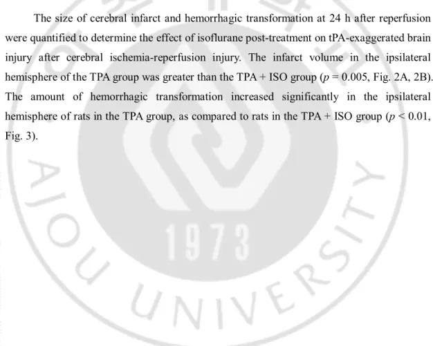
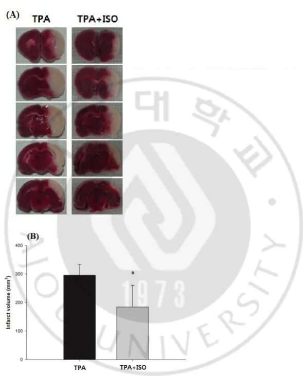
The size of cerebral infarct and hemorrhagic transformation at 24 h after reperfusion were quantified to determine the effect of isoflurane post-treatment on tPA-exaggerated brain injury after cerebral ischemia-reperfusion injury. The infarct volume in the ipsilateral hemisphere of the TPA group was greater than the TPA + ISO group (p = 0.005, Fig. 2A, 2B). The amount of hemorrhagic transformation increased significantly in the ipsilateral hemisphere of rats in the TPA group, as compared to rats in the TPA + ISO group (p < 0.01, Fig. 3).
8
Fig. 2.TTC (2,3,5-triphenyltetrazolium chloride) stained images of brain sections from an MCAO rats. (A) Representative TTC stained images of MCAO rat. (B) Quantification of
9
ischemia and 24 h of reperfusion. Animals in the TPA + ISO group received 1.5% isoflurane from the onset of reperfusion for one hour. MCAO, middle cerebral artery occlusion; TPA, control group; TPA + ISO, isoflurane post-treatment group; TTC, 2,3,5-triphenyltetrazolium chloride. Values shown are means ± SEM. *p value = 0.005 vs. TPA.
Fig. 3.The intracranial blood volume quantified by extracting hemoglobin from the intracranial hemisphere of rats.Animals in the TPA + ISO group received 1.5% isoflurane
from the onset of reperfusion for one hour. TPA, control group; TPA + ISO, isoflurane post-treatment group.*p value < 0.01 vs. TPA.
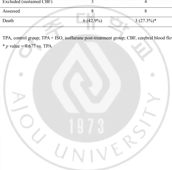
Three TPA rats and four TPA + ISO rats were excluded from assessment due to sustained cerebral blood flow following MCAO. Consistent with its beneficial effects on tPA therapy-induced intracranial hemorrhage in transient cerebral ischemia, isoflurane post-treatment reduced mortality in our rat model. Six rats (42.9 %) and three rats (27.3 %) died in the TPA and TPA + ISO groups, respectively (Table 1); however, statistical significance was lacking (p = 0.677).Physiologic parameters measured prior to ischemia, following ischemia, and 30 min following thrombolysis remained within normal range for both groups.
10
Table 1.Mortality after tPA therapy for transient cerebral ischemia.
TPA TPA + ISO
Total 17 15
Excluded (sustained CBF) 3 4
Assessed 8 8
Death 6 (42.9%) 3 (27.3%)*
TPA, control group; TPA + ISO, isoflurane post-treatment group; CBF, cerebral blood flow. * p value = 0.677 vs. TPA
11
Ⅳ
. DISCUSSION
Various studies have shown isoflurane as a neuroprotective anesthetic agent. (McMurtrey and Zuo, 2010; Lee et al, 2008; Khatibi et al, 2011) Although the neuroprotective features of pre- and post-treatment with isoflurane have been identified in various models of ischemia, the role of isoflurane in hemorrhage and ischemia-reperfusion is rather controversial. (McMurtrey and Zuo, 2010; Lee et al, 2008; Khatibi et al, 2011; Esposito et al, 2013; Li et al, 2014) In this study, post-treatment with isoflurane for 1 h not only decreased the cerebral infarct volume but also decreased hemorrhagic transformation in tPA-treated transient cerebral infarction in a rat model.
To our knowledge, no prior reports have acknowledged an association between post-treatment with isoflurane and its effects on tPA-exaggerated brain injury in a cerebral ischemia model. Such results could be of great significance because they suggest an intervention that may prevent the undesirable hemorrhagic side effects of tPA treatment.
Mechanisms of action for isoflurane post-treatment include cerebral protective effects in response to ischemia-reperfusion, cell necrosis, apoptosis, and altered signaling factors. (Lee et al 2008; Zhou et al, 2010) Vascular endothelial growth factor (VEGF) is a major regulator of normal and pathologic blood vessel growth. However, VEGF also has the unique property of inducing vascular leak. (Eliceiri et al, 1999; Weis et al, 2004) Matrix metalloproteinase-9 (MMP-9) is an enzymatic protein that degrade extracellular matrix, and may cause degradation of the BBB after cerebral ischemia-reperfusion. (Yang et al, 2007) MMP-9 can be activated by VEGF. Thus, MMP-9 activation may cause both breakdown of the BBB and intracranial hemorrhage after cerebral ischemia-reperfusion injury. Anesthetic pre- and post-treatment in ischemia-reperfusion models results in down-regulation of MMP-9 and VEGF expression. (Li et al, 2014; Lee et al, 2013) Thus, anesthetic agents could be used to reduce intracranial hemorrhage and the BBB disruption by lowering the levels of VEGF and MMP-9. Our study focused on hemorrhagic changes in the ischemia-reperfusion injured brain by quantifying the hemorrhagic content in brain tissues. Future, more extensive studies should focus on evaluating the plasma levels of MMP-9 and VEGF in ischemia-reperfusion models
12
over time. This will improve our understanding of signaling dynamics, instead of showing what happens at one point in time based on an end-point analysis of brain tissue.
The neuroprotective effect of both pre- and post-treatment with inhaled anesthetics has been shown previously. (Lee et al, 2008; Li et al, 2014) But, when it comes to clinical application, the role of post-treatment is likely of greater significance considering the nature of cerebral ischemia and the on-going efforts to improve neurological outcomes in stroke victims. Thus, the cerebral-protective effect of isoflurane demonstrated in our tPA-treated ischemia model may be more clinically relevant and provide important pre-clinical evidence in support of clinical trials of treatment for acute stroke.
The benefits of tPA for the treatment of cerebral stroke have been clouded by its potential hemorrhagic and neurotoxic side effects, as well as its short therapeutic time window. Our results from post-treatment with isoflurane in a cerebral ischemia-reperfusion model show that isoflurane can ameliorate the limitations of tPA. Thus, post-treatment with isoflurane may enhance the safety and efficacy, and lengthen the treatment window for tPA in stroke therapy.
There are several limitations to our study. First, as mentioned previously, MMP-9 and VEGF are regarded as relevant in the molecular pathobiology of intracranial hemorrhage. Thus, efforts to determine tissue levels for these factors will not only help to validate our findings, but will also allow us to understand the patterns of signaling. Second, efforts to assess cerebral hemorrhage, extravasation of red blood cells, and disruption of the BBB could also be adopted during experiments to obtain more in-depth results. Lastly, although the protective effect of isoflurane post-treatment within 24 h after the reperfusion could be seen here, further investigations are necessary to evaluate the long-term clinical outcomes or even potential adverse effect of the treatment.
13
Ⅴ
. CONCLUSION
In conclusion, our study showed that isoflurane post-treatment following tPA therapy for transient cerebral ischemia not only improved neurobehavioral outcomes, but also effectively decreased the infarct size and amount of hemorrhagic transformation.
14
REFERENCES
1. Belayev L, Alonso OF, Busto R, Zhao W, Ginsberg MD: Middle cerebral artery occlusion in the rat by intraluminal suture. Neurological and pathological evaluation of an improved model. Stroke 27: 1616-1622,1996
2. Choudhri TF, Hoh BL, Solomon RA, Connolly ES, Pinsky DJ: Use of a spectrophotometric hemoglobin assay to objectively quantify intracerebral hemorrhage in mice. Stroke28: 2296-2302, 1997
3. Eliceiri BP, Paul R, Schwartzberg PL, Hood JD, Leng J, Cheresh DA: Selective requirement for Src kinases during VEGF-induced angiogenesis and vascular permeability. Mol Cell 4: 915-924, 1999
4. Eissa A, Krass I, Bajorek BV: Optimizing the management of acute ischaemic stroke: a review of the utilizaion of intravenous recombinant tissue plasminogen activator (tPA). J Clin Pharm Ther 37: 620-629, 2012
5. Esposito E ME, Lo EH: Lower doses of isoflurane treatment has no beneficial effeccts in a rat model of intracerebral hemorrhage. BMC Neurosci 14: 129, 2013 6. Feigin VL, Forouzanfar MH, Krishnamurthi R, Mensah GA, Connor M, Bennett DA,
Moran AE, Sacco RL, Anderson L, Truelsen T, O’Donnell M, Venketasubramanian N, Barker Collo S, Lawes CM, Wang W, Shinohara Y, Witt E, Ezzati M, Naghavi M, Murray C: Global and regional burden of stroke during 1990-2010: findings from the Global Burden of Disease Study 2010. Lancet 383: 245-254, 2014
7. Hunter AJ, Hatcher J, Virley D, Nelson P, Irving E, Hadingham SJ, Parsons AA: Functional assessments in mice and rats after focal stroke. Neuropharmacology 39: 806-816, 2000
8. Khatibi NH, Ma Q, Rolland W, Ostrowski R, Fathali N, Martin R, Applegate R, Stier G, Tang J, Zhang JH: Isoflurane posttreatment reduces brain injury after an intracerebral hemorrhagic stroke in mice. Anesth Analg 113: 343-348, 2011
9. Lee JH, Cui HS, Shin SK, Kim JM, Kim SY, Koo B: Effect of propofol post-treatment on blood-brain barrier integrity and cerebral edema after transient cerebral
15
ischemia in rats. Neurochem Res 38: 2276-2286, 2013
10. Lee JJ, Li L, Jung H, Zuo Z: Postconditioning with isoflurane reduced ischemia-induced brain injury in rats. Anesthesiology 108: 1055-1062, 2008
11. Li G, Jia J, Fu J, Wang H, Ji K, Zang B: [The effects of preconditioning and postconditioning with isoflurane on focal cerebral ischemi/reperfusion injury in rats].
Zhonghua Wei Zhong Bing Ji Jiu Yi Xue 26: 431-435, 2014
12. Longa EZ, Weinstein PR, Carlson S, Cummins R: Reversible middle cerebral artery occlusion without craniectomy in rats. Stroke 20: 84-91, 1989
13. McMurtrey RJ, Zuo Z: Isoflurane preconditioning and postconditioning in rat hippocampal neurons. Brain Res 28: 184-190, 2010
14. Xing B, Chen H, Zhang M, Zhao D, Jiang R, Liu X, Zhang S: Ischemic post-conditioning protects brain and reduces inflammation in a rat model of focal cerebral ischemia/reperfusion. J Neurochem 105: 1737-1745, 2008
15. Wardlaw JM, Murray V, Berge E, Del Zoppo GJ: Thrombolysis for acute ischaemic stroke. Cochrane Database Syst Rev29: CD000213, 2014
16. Weis S, Shintani S, Weber A, Kirchmair R, Wood M, Cravens A, McSharry H, Iwakura A, Yoon Y, Himes N, Burstein D, Doukas J, Soll R, Losordo D, Cheresh D: Src blockade stabilizes a Flk/cadherin complex, reducing edema and tissue injury following myocardial infarction. J Clin Invest 113: 885-894, 2014
17. Yang Y, Estrada EY, Thompson JF, Liu W, Rosenberg GA: Matrix metalloproteinase-mediated disruption of tight junction proteins in cerebral vessels is reversed by synthetic matrix metalloproteinase inhibitor in focal ischemia in rat. J Cereb Blood
Flow Metab 27: 697-709, 2007
18. Yepes M, Roussel BD, Ali C, Vivien D: Tissue-type plasminogen activator in the ischemic brain: more than a thrombolytic. Trends Neurosci 32: 48-55, 2009
19. Zheng Y, Lan Y, Tang H, Zhu S: Propofol pretreatment attenuates aquaporin-4 over-expression and alleviates cerebral edema after transient focal brain ischemia reperfusion in rats. Anesth Analg 107: 2009-2016, 2008
20. Zhou Y, Lekic T, Fathali N, Ostrowski RP, Martin RD, Tang J, Zhang JH: Isoflurane posttreatment reduces neonatal hypoxic-ischemic brain injury in rats by the
16
sphingosine-1-phosphate/phosphatidylinositol-3-kinase/Akt pathway. Stroke 41: 1521-1527, 2010
21. Zhu H, Fan X, Yu Z, Liu J, Murata Y, Lu J, Zhao S, Hajjar KA, Lo EH, Wang X: Annexin A2 combined with low-dose tPA improves thrombolytic therapy in a rat model of focal embolic stroke. J Cereb Blood Flow Metab; 30: 1137-1146, 2010
17
- 국문요약 –
뇌경색 흰쥐 모델에서의 혈전 용해술로 인한 뇌손상에 대한
Isoflurane 후처치의 효과
아주대학교 대학원 의학과 (마취통증의학)
김 은 정
(지도교수: 김 진 수)
조직플라스미노겐활성제 (tissue-type plasminogen activator, tPA) 는 허혈성뇌졸중에 표준 치료 방법으로 여겨져 왔다. 하지만, tPA 의 좁은 치료 범위와 대뇌출혈을 증가시키는 단점은 환자 치료에 사용하는 데 있어서 주의를 요한다. 이러한 약제의 한계를 극복하기 위한 여러가지 연구들이 진행 중이며, 그 중 마취 약제는 허혈재관류 손상을 막을 수 있다고 알려져 있기에 마취 약제의신경계 보호작용에 대한 연구들이 진행되고 있다. 본 연구에서는 일시적인 뇌경색 치료를 위한 tPA 투여에 따른 뇌내 출혈 및 뇌 허혈에 대한 isoflurane 마취 약제 후처치가 가진 효과에 대해 알아보고자 하였다. 수컷 Sprague-Dawley 흰쥐를 대상으로 우측중대뇌동맥의 일시적 결찰을 통해 뇌내허혈을 유도한 후, 정맥내 tPA 를 주입하였다. tPA 주입을 마친뇌경색흰쥐 모델은
18 대조군과 isoflurane 후처치 군으로 무작위 배정되어 후자의 경우 재관류와 함께 1 시간 동안 1.5% isoflurane 을 후처치 하였다. 재관류 24 시간 후 시행한 신경행동 및 뇌조직 검사 결과, isoflurane 후처치 군은 대조군에 비해 우월한 행동 기능을 보였으며, 뇌경색 및 뇌내 출혈의 범위 역시 isoflurane 후처치 군에서 감소된 양상을 보였다. 핵심어: 조직플라스미노겐활성제, 두개내출혈, Isoflurane, 허혈성후처치