Association of Plasma Adipokines with Chronic Obstructive
Pulmonary Disease Severity and Progression
Yeon-Mok Oh1*, Byeong-Ho Jeong2*, Sook-Young Woo3, Su-Young Kim2, Hojoong Kim2, Ji-Hyun Lee4,
Seong Yong Lim5, Chin Kook Rhee6, Kwang Ha Yoo7, Jin Hwa Lee8, Hyoung Kyu Yoon9, Don D. Sin10, Sang-Do Lee1, Eun Kyung Kim4†, and Hye Yun Park2†; for the KOLD Study Group
1Department of Pulmonary and Critical Care Medicine, Clinical Research Center for Chronic Obstructive Airway Diseases, Asan Medical Center, University of Ulsan College of Medicine, Seoul;2Division of Pulmonary and Critical Care Medicine, Department of Medicine, Samsung Medical Center, Sungkyunkwan University School of Medicine, Seoul;3Biostatistics Team, Samsung Biomedical Research Institute, Seoul;4Department of Internal Medicine, Bundang CHA Medical Center, CHA University College of Medicine, Seongnam;5Department of Medicine, Kangbuk Samsung Hospital, Sungkyunkwan University School of Medicine, Seoul;6Department of Internal Medicine, Seoul St. Mary’s Hospital, Catholic University of Korea, Seoul;7Department of Internal Medicine, Konkuk University College of Medicine, Seoul;8Department of Internal Medicine, Ewha Womans University Mokdong Hospital, College of Medicine, Ewha Womans University, Seoul; and9Department of Internal Medicine, Yeouido St. Mary’s Hospital, Catholic University of Korea, Seoul, Korea; and10Respiratory Division, Department of Medicine, University of British Columbia, Vancouver, British Columbia, Canada ORCID IDs: 0000-0003-0116-4683 (Y.-M.O.); 0000-0002-3124-1718 (B.-H.J.); 0000-0001-9207-0433 (H.K.); 0000-0002-8287-5470 (J.-H.L.); 0000-0001-8098-3622 (S.Y.L.); 0000-0003-4533-7937 (C.K.R.); 0000-0003-0843-9862 (J.H.L.); 0000-0001-8189-4509 (S.-D.L.);
0000-0001-6239-1682 (E.K.K.); 0000-0002-5937-9671 (H.Y.P.).
Abstract
Rationale: Two adipokines, leptin and adiponectin, regulate metabolic and inflammatory systems reciprocally. The role of adiponectin in chronic obstructive pulmonary disease (COPD) has been studied. However, there are few data evaluating the relationship of plasma leptin with COPD severity or progression.
Objectives: The objective of this study was to evaluate the relationship of leptin, adiponectin, and the leptin/adiponectin ratio with COPD severity and progression according to COPD
phenotypes.
Methods: Plasma leptin and adiponectin levels were measured in 196 subjects with COPD selected from the Korean Obstructive Lung Disease cohort. Using a linear regression model and mixed linear regression, we determined the relationship of plasma leptin and adiponectin levels and the leptin/adiponectin ratio to COPD severity and progression over 3 years.
Measurements and Main Results: The concentration of adiponectin in plasma positively correlated with percent emphysema on initial computed tomography (CT) (adjustedP = 0.022), whereas plasma leptin concentrations and the leptin/adiponectin ratio exhibited a significant inverse correlation with initial FEV1(adjusted P = 0.013 for leptin and adjusted P = 0.041 for leptin/adiponectin ratio). Increased plasma leptin and leptin/adiponectin ratio were significantly associated with change in percent emphysema over 3 years (adjusted P = 0.037 for leptin and adjusted P = 0.029 for leptin/adiponectin ratio), whereas none of the adipokines demonstrated an association with FEV1decline over the 3-year period.
Conclusions: Plasma adiponectin and leptin vary according to COPD phenotypes. Plasma leptin and the leptin/adiponectin ratio, but not adiponectin, were significantly associated with changes in CT-assessed emphysema, suggesting a potential role as a biomarker in emphysema progression in patients with COPD.
Keywords: adiponectin; leptin; chronic obstructive pulmonary disease; emphysema
(Received in original form January 6, 2015; accepted in final form April 11, 2015 ) *These authors contributed equally to this work.
†These authors contributed equally to this work and should be considered as co-corresponding authors.
Supported by the Samsung Medical Center Foundation for Medical Research (SMO1140211).
Author Contributions: All authors contributed to and approved the final draft of the manuscript. Conception and design: Y.-M.O., H.Y.P. and E.K.K. Experiment and data acquisition: Y.-M.O., S.-Y.K., J.-H.L., S.Y.L., C.K.R, K.H.Y., J.H.L., H.K.Y., E.K.K. and S.-D.L. Analysis and interpretation: H.Y.P., B.-H.J., Y.-M.O., D.D.S. and S.-Y.W. Drafting the manuscript: H.Y.P., E.K.K., B.-H.J. and Y.-M.O. Critical revision of the manuscript: Y.-M.O., H.Y.P., E.K.K., B.-H.J., H.K., J.-H.L., S.Y.L., D.D.S. and S.-Y.W. Correspondence and requests for reprints should be addressed to Hye Yun Park, M.D., Ph.D., Division of Pulmonary and Critical Care Medicine, Department of Medicine, Samsung Medical Center, Sungkyunkwan University School of Medicine, 81 Irwon-ro, Gangnam-gu, Seoul 135-710, Korea. E-mail:
hyeyunpark@skku.edu;or Eun Kyung Kim, M.D., Ph.D., Department of Internal Medicine, Bundang CHA Medical Center, CHA University College of Medicine, Seongnam 463-712, Korea. E-mail: imekkim@cha.ac.kr
This article has an online supplement, which is accessible from this issue’s table of contents online at www.atsjournals.org Ann Am Thorac Soc Vol 12, No 7, pp 1005–1012, Jul 2015
Copyright© 2015 by the American Thoracic Society DOI: 10.1513/AnnalsATS.201501-005OC
Chronic obstructive pulmonary disease (COPD) is a major cause of mortality and morbidity worldwide, with an estimated 3 million annual deaths (1, 2). It is a chronic inflammatory lung disease characterized by accelerated decline in lung function and progressive parenchymal destruction leading to emphysema (3, 4). COPD is also associated with low-grade systemic inflammation, which contributes to the development of extrapulmonary complications and comorbidities such as cardiovascular disease, osteoporosis, depression, metabolic syndrome, and weight loss (5–7). Indeed, various systemic inflammatory markers, including C-reactive protein,fibrinogen, and leukocyte counts, have been associated with COPD severity, as well as with increased risk of
comorbidities in patients with COPD (7, 8). Adiponectin, a systemic biomarker associated with cardiovascular outcome in COPD (9), is a circulating hormone produced by adipose tissue, along with leptin and other cytokines (10, 11). The two hormones, adiponectin and leptin, have opposing effects on regulation of metabolic and inflammatory systems (11, 12). Adiponectin possesses antiinflammatory properties by reducing inflammatory mediators, whereas leptin plays an important role in up-regulating the inflammatory system. In cross-sectional studies of patients with stable COPD, however, circulating adiponectin levels were elevated in patients with COPD and correlated with systemic inflammation as assessed by serum tumor necrosis factor-a levels (13–15). Moreover, adiponectin has received attention as a biomarker reflecting disease activity such as dynamic hyperinflation and radiologically assessed emphysema (16, 17). Circulating leptin levels increase further during acute exacerbations and are significantly related to tumor necrosis factor-a and IL-6 levels on acute exacerbations (18, 19). Even in stable COPD, serum leptin levels are dysregulated (20). However, there are few data evaluating the relationship of circulating leptin levels with COPD severity according to COPD phenotypes. In addition, studies on the longitudinal relationship of two adipokines, leptin or adiponectin, with lung function and extent of emphysema assessed on chest computed tomography (CT) are limited. Therefore, the objective of this study was to investigate the association of adipokines with COPD severity, such as FEV1and the extent of emphysema at enrollment, and
then to explore the relationship of adipokines with COPD progression, using change of lung function and emphysema extent over 3 years.
Methods
Patients
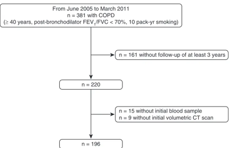
One hundred ninety-six subjects with COPD were from the Korean Obstructive Lung Disease (KOLD) cohort. Details of the KOLD Study have been published previously (21). Subjects were recruited from pulmonary clinics in 13 hospitals in the Republic of Korea from June 2005 to February 2011, and the inclusion criteria for this study were as follows: (1) those with COPD, as defined by a postbronchodilator FEV1/FVC less than 0.7 and more than 10 pack-years of smoking history; (2) those without history or radiographic evidence of tuberculosis, bronchiectasis, or other pulmonary disorders; (3) those with at least 3 years of follow-up; and (4) those with blood sample and chest high-resolution volumetric computed tomography (HRCT) at enrollment (Figure 1).
The KOLD cohort also includes nonsmoking patients with COPD (7% of overall patients with COPD). However, we restricted our analyses to patients with COPD having a more than 10-pack-year smoking history, as generally defined in large cohort or randomized controlled study for recruitment of patients with COPD. At yearly visits, subject smoking status was surveyed with a series of questions. Subjects were categorized as sustained quitters if they were nonsmokers at each annual visit for
3 years. Subjects who were smokers at each annual visit were defined as continued smokers and those whose smoking behavior varied were classified as intermittent quitters. The study protocol was approved by the institutional review boards of Asan Medical Center (Seoul, Korea; no. 2005-0010).
Adiponectin and Leptin Measurements
Blood samples, collected at enrollment when the patients were stable, were separated into their various components, aliquoted, transferred to the KOLD Data Coordinating Center on dry ice, and kept in–808C freezers until use. Plasma samples were thawed, and adiponectin and leptin concentrations were measured with a commercially available ELISA kit (B-Bridge International, Sunnyvale, CA) according to the manufacturer’s
instructions. The lower limits of detection of these assays were 0.0234 and 0.17 ng/ml for adiponectin and leptin, respectively.
Pulmonary Function Test
Spirometry was performed according to the recommendations of the American Thoracic Society, using a Vmax 22 (SensorMedics, Yorba Linda, CA) or a PFDX (MedGraphics, St. Paul, MN) (22). Absolute values of FVC and FEV1were obtained, and the
percentage predicted values (% predicted) for FEV1and FVC were calculated from equations obtained in a representative Korean sample (23). A positive
bronchodilator response was defined as an increase of at least 12% and 200 ml in FEV1 from baseline values (24).
n = 196 n = 220
n = 161 without follow-up of at least 3 years From June 2005 to March 2011
n = 381 with COPD
(≥ 40 years, post-bronchodilator FEV1/FVC < 70%, 10 pack-yr smoking)
n = 15 without initial blood sample n = 9 without initial volumetric CT scan
Figure 1. Study flow diagram. COPD = chronic obstructive pulmonary disease; CT = computed tomography.
Computed Tomography Data Acquisition and Analysis
Volumetric CT scans were performed on all subjects, using 16-channel, multidetector, CT scanners; because this was a multicenter study, the scanners were from three manufacturers. These included the Somatom Sensation 16 (Siemens Medical Solutions, Forchheim, Germany), the GE Lightspeed Ultra (General Electric
Healthcare, Milwaukee, WI), and the Philips Brilliance 16 (Philips Medical Systems, Best, The Netherlands). However, subjects used the same manufacturer of CT scanner over time. Subjects were scanned during suspended full inspiration and expiration in the supine position. CT parameters used in each CT scanner were as follows: 163 0.75 mm collimation, 100 eff. milliamperes-second (mAs), 140 peak kilovoltage (kVp) (Somatom Sensation 16); 163 0.625 mm, 200 mAs, 140 kVp, pitch 0.938, 0.5 seconds per rotation (GE Lightspeed); and 163 0.75 mm, 133 mAs, 140 kVp, pitch 1, 0.75 seconds per rotation (Philips 16). The acquired data were reconstructed according to a standard algorithm, with a thickness of 0.625–0.8 mm and an increment of 0.625– 0.8 mm. The CT machines were calibrated every week with an American Association of Physicists in Medicine (AAPM) standard phantom. Image data were stored in Digital Imaging and Communications in Medicine (DICOM) format; this is the international standard for interconnecting medical imaging devices on standard networks.
Using in-house software, images of the whole lung were extracted automatically and the attenuation coefficient of each pixel was measured and calculated. Emphysema was defined as a percentage of lung attenuation less than 950 Hounsfield units (HU) (25). The percent emphysema was determined for the total lung, as well as the upper lung (above the carina) and the lower lung (below the carina).
Statistical Analysis
To evaluate the relationship of plasma adiponectin, plasma leptin, and the ratio of leptin to adiponectin with (1) initial FEV1, (2) FEV1decline rate, (3) initial percent emphysema on HRCT, and (4) change in percent emphysema on HRCT, wefirst log-transformed adiponectin and leptin to achieve normal distribution and to mitigate the effects of outliers. Linear regression models were then used to account for the possible confounding effects of age, body
mass index (BMI), smoking status and pack-years of smoking at baseline, cardiovascular disease, positivity of bronchodilator response, initial percent emphysema, and use of inhaled
corticosteroid on the initial FEV1(L) and FEV1, % predicted. To evaluate
a relationship between adipokines and percent emphysema on initial HRCT scans, initial FEV1(L) was added to age, BMI, smoking status and pack-years of smoking at baseline, cardiovascular disease, and use of inhaled corticosteroid as covariates. Sex was excluded as a covariate because 97.4% of the population was male. Because HRCT was conducted at recruitment and after 3 years, the percentage change in emphysema was calculated as [(% emphysema at 3-yr follow-up)– (% emphysema at recruitment)]/3 years. To evaluate a relationship between adipokines and change of percent emphysema over 3 years, statistical adjustment were made for age, BMI, cardiovascular disease, initial percent emphysema, use of inhaled corticosteroid, and exacerbation and smoking status over 3 years.
Because some individuals, who initiated bronchodilators for COPD treatment at enrollment, experienced an increase in FEV1during thefirst 3 months, we calculated FEV1decline rate by using FEV1measured at 3 months as the baseline value. The FEV1decline rate over 3 years was analyzed by linear mixed effects model with a random intercept and slope, using FEV1values obtained at 3 months and at 1, 2, and 3 years. Statistical adjustments were made for age, BMI, cardiovascular disease, positivity of bronchodilator response, baseline FEV1(L), use of inhaled
corticosteroid, and exacerbation and smoking status over 3 years on FEV1decline, expressed in milliliters per year. Data are presented as median and interquartile range (IQR) for continuous variables and as frequency (percentage) for categorical variables. All tests were two-sided, and aP value less than 0.05 was considered significant. Data were analyzed with SPSS Statistics, version 21 (IBM, Armonk, NY) and R statistical software version 3.0.0.
Results
Clinical Characteristics of the Cohort Demographics and clinical characteristics are presented in Table 1. Of the 196
subjects, 191 (97.4%) were male and 5 (2.6%) were female, with a median age of 67 years. Median BMI was 23.3 kg/m2. Sixty-three (32.1%) patients were current smokers and 133 (67.9%) were ex-smokers with a median smoking history of 43 pack-years. The initial median FEV1was 1.39 L, corresponding to 47% predicted, and the extent of emphysema as determined by volume fraction of the lung less than –950 HU was 20.3%. All patients performed a bronchodilator test, and 69 (35.2%) exhibited a positive bronchodilator response. Median plasma adiponectin and leptin levels were 8,217 and 7.37 ng/ml, respectively.
Over 3 years of follow-up, sustained quitters, intermittent quitters, and continued smokers included 126 (64.3%), 30 (15.3%), and 40 (20.4%) patients, respectively. Of all the patients (total, 196), 107 (54.6%) patients reported a total of 214 exacerbations over 3 years (median, 0.3 events/person/yr [IQR, 0.0 to 0.7 events/person/yr]) and the median time tofirst exacerbation was 1.1 years (IQR, 0.4 to 1.8 yr). Only two patients did not use any inhaler during follow-up. The decline in FEV1 was–18.6 ml/year (95% confidence interval, –33.69 to –3.57) and median change in percent emphysema (n = 143 with HRCT scans after a 3-yr follow-up) was 0.29%/year (IQR,–0.65 to 1.69%/yr) for the total lung, 0.72%/year (IQR,–0.50 to 1.75%/yr) for the upper lung, and 0.28%/year (–0.68 to 1.66%/yr) for the lower lung.
Relationship of Plasma Adipokines to Parameters of COPD Severity
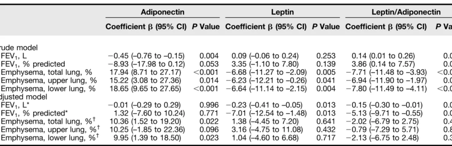
Plasma adiponectin levels were significantly associated with parameters of COPD severity (initial FEV1and percent emphysema of total, upper, and lower lung) (Table 2). This association was noted with respect to percent emphysema of the total lung (adjustedP = 0.022; Figure 2) and percent emphysema of the lower lung (adjusted P = 0.023) after covariate adjustment. Plasma leptin levels were significantly associated with initial percent emphysema (total, upper, and lower lung), but this association disappeared after adjusting for all covariates indicated previously. In contrast, plasma leptin levels exhibited a significant inverse correlation with initial FEV1(adjusted P = 0.013; Figure 2). Finally, the leptin/ adiponectin ratio was significantly associated with initial FEV1(L and % predicted) and initial percent emphysema (total, upper, and lower lung). After covariate adjustment, the
leptin/adiponectin ratio was inversely associated only with initial FEV1(adjusted P = 0.041 for FEV1[L] and adjustedP = 0.028 for FEV1, % predicted).
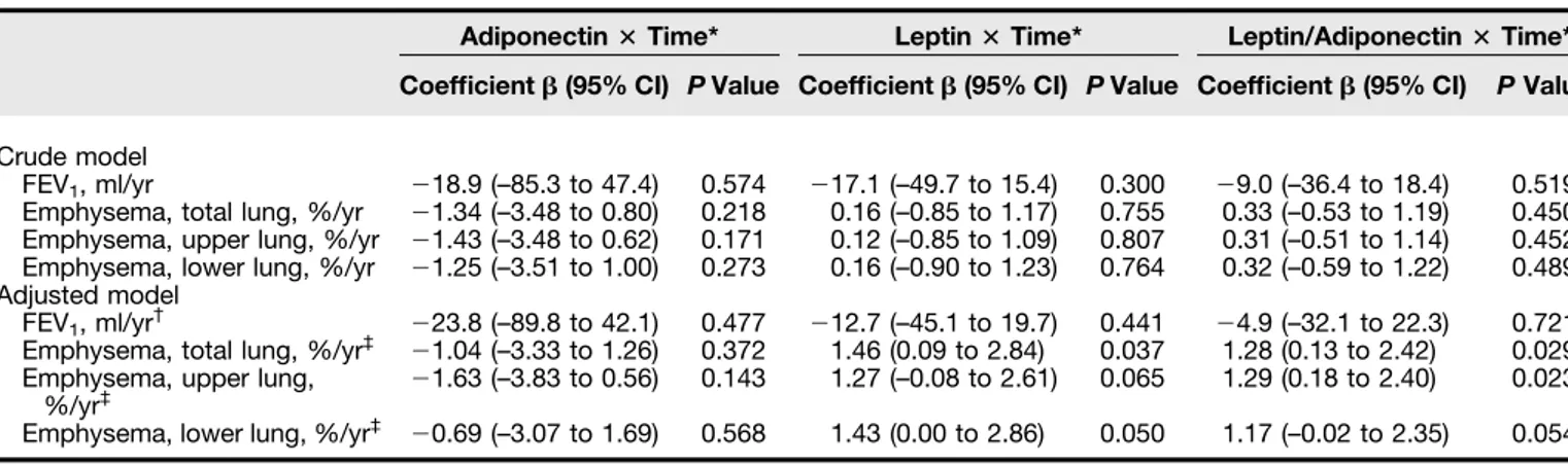
Relationship of Plasma Adipokines to Parameters of COPD Progression As shown in Table 3, there was no significant association between plasma adiponectin levels and parameters of COPD progression over time in both crude and adjusted states. However, there were significant positive correlations between plasma leptin levels and percent
emphysema progression of total lung and lower lung after covariate adjustment
(adjustedP = 0.037 for total lung [Figure 3] and adjustedP = 0.050 for lower lung). Moreover, there were significant positive correlations between the leptin/adiponectin ratio and percent emphysema progression of the total lung and upper lung after adjusting for covariates indicated previously (adjustedP = 0.029 for total lung and adjustedP = 0.023 for upper lung).
Discussion
The mainfindings of the present study were as follows: independent of other risk factors,
(1) plasma adiponectin concentrations are positively associated with initial percent emphysema; (2) plasma leptin concentrations and the leptin/adiponectin ratio are inversely associated with initial FEV1(L and % predicted); and (3) plasma leptin concentrations and the leptin/ adiponectin ratio were significantly related to CT-assessed emphysema progression over 3 years.
Adiponectin, a 30-kD protein secreted by adipocytes (i.e., adipokine), possesses antiinflammatory, antiatherosclerotic, and insulin-sensitizing properties (10, 26). COPDGene Study investigators have demonstrated an independent relationship between plasma adiponectin concentrations and percent emphysema in the lower lung in patients with COPD (16). We extended thisfinding by showing that plasma adiponectin concentrations are significantly associated with percent emphysema in both the total and lower lung by multivariable regression analysis. With respect to lung function, plasma adiponectin concentrations were inversely associated with initial FEV1, which was consistent with data from the COPDGene Study (16). However, this association disappeared after adjusting for covariates, including BMI.
Leptin, a 16-kD protein, is primarily a proinflammatory adipokine that affects both the innate and adaptive immune response (27). Leptin is also expressed in the human lung with adiponectin. Bruno and colleagues showed increased leptin expression in the bronchial mucosa of patients with COPD compared with normal subjects and smokers, which was related to airway inflammation and airflow obstruction (28). In an epidemiologic study, Sin and Man showed that circulating leptin levels was inversely related to FEV1among a representative sample of the U.S. nonobese adult population (29). We extend thesefindings by showing that higher plasma leptin concentrations have an independent association with lower FEV1in patients with stable COPD. However, there was no association between plasma leptin concentrations and emphysema extent in the present study.
There are several studies that investigated association between lung function decline and plasma adipokines (9, 30). To our knowledge, this is thefirst study to examine the relationship between plasma adipokines and change in
emphysema over a period of 3 years. Table 1. Demographics and clinical characteristics
Variable Value
Age, yr 67 (61–71)
Sex, male 191 (97.4)
Body mass index, kg/m2 23.3 (20.8–25.3) Smoking status at baseline
Current 63 (32.1) Ex-smoker 133 (67.9) Pack-years of smoking 43 (29–54) mMRC dyspnea scale 2 (1–2) Health status SGRQ total score 31 (21–49) Comorbidity Hypertension 59 (30.1) Diabetes mellitus 21 (10.7) Cardiovascular disease* 10 (5.1) Chronic liver disease 6 (3.1) Chronic kidney disease 2 (1.0)
Malignancy 1 (0.5)
Pulmonary function test
FEV1, L 1.39 (1.05–1.75)
FEV1, % predicted 47.0 (36.6–58.0)
FVC, L 3.22 (2.62–3.71)
FVC, % predicted 76.4 (64.5–87.8)
FEV1/FVC, % 45 (39–53)
Positive bronchodilator response 69 (35.2) Six-minute walk distance, m 441 (400–483) HRCT scans, parameters
Emphysema, total lung, % 20.3 (9.2–34.9) Emphysema, upper lung, % 20.3 (7.8–38.4) Emphysema, lower lung, % 19.1 (8.1–33.0) Medications No inhalers 84 (42.9) Use of ICS 83 (42.3) Use of LABA 80 (40.8) Use of LAMA 61 (31.1) Biomarkers Adiponectin, ng/ml 8,217 (5,473–11,273) Leptin, ng/ml 7.37 (3.36–12.11)
Definition of abbreviations: HRCT = high-resolution volumetric computed tomography; ICS = inhaled corticosteroid; LABA = long-actingb-agonist; LAMA = long-acting muscarinic antagonist; mMRC = modified Medical Research Council; SGRQ = St. George’s Respiratory Questionnaire.
Note: n = 196. Values are expressed as medians (interquartile range) or frequencies (%). *Cardiovascular disease included myocardial infarction, cerebrovascular disease, and peripheral arterial occlusive disease.
Unexpectedly, plasma adiponectin levels were not associated with emphysema progression. This lack of association gives a different view regarding the role of adiponectin as a contributor to emphysema development (16). On the basis of a cross-sectional study, it was not clear whether the
elevation in adiponectin is a causal determinant or reactive response in relation to emphysema. The role of adiponectin in emphysema development has been controversial in animal models (31–33). Adiponectin-deficient mice exhibit structural changes in the lung
(emphysema-like phenotype) and alveolar macrophage activation (31). In addition, the
emphysema-like phenotype was ameliorated by adiponectin supplementation, suggesting that adiponectin has a protective effect in the progression of structural lung parenchymal Table 2. Relationships of plasma adipokine concentrations with chronic obstructive pulmonary disease severity
Adiponectin Leptin Leptin/Adiponectin Coefficient b (95% CI) P Value Coefficient b (95% CI) P Value Coefficient b (95% CI) P Value Crude model
FEV1, L 20.45 (–0.76 to –0.15) 0.004 0.09 (–0.06 to 0.24) 0.253 0.14 (0.01 to 0.26) 0.033
FEV1, % predicted 28.93 (–17.98 to 0.12) 0.053 3.35 (–1.10 to 7.80) 0.139 3.86 (0.14 to 7.57) 0.042
Emphysema, total lung, % 17.94 (8.71 to 27.17) ,0.001 26.68 (–11.27 to –2.09) 0.005 27.71 (–11.48 to –3.93) ,0.001 Emphysema, upper lung, % 15.22 (3.08 to 27.36) 0.014 26.23 (–12.21 to –0.26) 0.041 26.94 (–11.90 to –1.97) 0.006 Emphysema, lower lung, % 18.65 (9.65 to 27.65) ,0.001 26.64 (–11.14 to –2.15) 0.004 27.80 (–11.49 to –4.11) ,0.001 Adjusted model
FEV1, L* 20.01 (–0.29 to 0.29) 0.996 20.23 (–0.41 to –0.05) 0.013 20.15 (–0.30 to –0.01) 0.041
FEV1, % predicted* 1.32 (–7.60 to 10.24) 0.771 27.01 (–12.54 to –1.48) 0.013 25.13 (–9.71 to –0.55) 0.028
Emphysema, total lung, %† 10.36 (1.52 to 19.20) 0.022 1.38 (–4.45 to 7.20) 0.641 22.02 (–6.79 to 2.75) 0.405 Emphysema, upper lung, %† 10.25 (–1.85 to 22.36) 0.096 3.16 (–4.75 to 11.08) 0.432 20.79 (–7.29 to 5.71) 0.811 Emphysema, lower lung, %† 9.95 (1.39 to 18.50) 0.023 1.04 (–4.60 to 6.68) 0.717 22.13 (–6.75 to 2.48) 0.364
Definition of abbreviation: CI = confidence interval.
Note: Coefficient = the mathematical weightings of the explanatory variables (the regression coefficient).
*Adjusted for age, body mass index, smoking status and pack-years of smoking at baseline, cardiovascular disease, positivity of bronchodilator response, initial percent emphysema, and use of inhaled corticosteroid.
†Adjusted for age, body mass index, smoking status and pack-years of smoking at baseline, cardiovascular disease, initial FEV
1(L), and use of inhaled
corticosteroid. 3.4 (2,512) 15 20 25 30 A 3.6 (3,981)
Adjusted coefficient = 10.36 (95% CI, 1.52 to 19.20), Adjusted P = 0.022
3.8 (6,310)
4.0 (10,000) Adiponectin (ng/ml)-log scale
Initial emphysema, total lung (%)
4.2 (15,849) 4.4 (25,119) 0.0 (1.0) 1.2 1.3 1.4 1.5 1.6 1.7 B 0.2 (1.6)
Adjusted coefficient = –0.23 (95% CI, –0.41 to –0.05), Adjusted P = 0.013
0.4 (2.5) 0.8 (6.3) 0.6 (4.0)
Leptin (ng/ml)-log scale
Initial FEV 1 (L) 1.2 (15.8) 1.0 (10.0) 1.4 (25.1)
Figure 2. Relationship of log-transformed adipokines with initial percent emphysema and FEV1. (A) Plasma adiponectin levels were significantly
associated with initial percent emphysema of total lung after adjustment for age, body mass index, smoking status and pack-years of smoking at baseline, cardiovascular disease, and use of inhaled corticosteroid and initial FEV1(L). (B) Plasma leptin levels were inversely associated with initial FEV1(L) after
adjustment for age, body mass index, smoking status and pack-years of smoking at baseline, cardiovascular disease, positivity of bronchodilator response, initial percent emphysema, and use of inhaled corticosteroid. CI = confidence interval.
changes (32). In contrast, Miller and colleagues showed that adiponectin-deficient mice were protected against tobacco-induced inflammation and emphysema (33). Therefore, the role of adiponectin in emphysema development remains uncertain.
Although the magnitude of this effect was relatively small, plasma leptin levels
showed a significant positive association with emphysema progression. Several animal studies have indicated a role for leptin in postnatal lung development (34, 35). Huang and colleagues demonstrated that leptin-deficient mice exhibited significantly decreased lung volume and lower alveolar surface area at 2 weeks of age relative to wild-type mice. Moreover,
these differences were amplified through 7 weeks of age. In another experiment, they showed that leptin replacement in leptin-deficient mice increased lung volume, which corresponded with enlarged alveolar size and surface area (35). Nevertheless, as there are limited data on leptin-related emphysema in human studies, uncertainty about whether leptin has a causal role in observed differences in emphysema outcomes in the present study remained.
Neither adiponectin and leptin nor the leptin/adiponectin ratio was related to lung function decline over 3 years. On the contrary, a study involving the Hokkaido COPD Cohort and COPD Quantification by Computed Tomography, Biomarkers, and Quality of Life (CBQ) study has shown that the ratio of leptin to adiponectin was associated with lung function decline (30). Compared with the reports based on the Hokkaido COPD Cohort and CBQ Study, the subjects with COPD in the present study had more severe airflow limitation, leading to slower lung function decline. This might result in a lack of significant association of adipokine levels with lung function decline. Another explanation would be the selection of adjustment factors. Although the rate of FEV1decline is affected by initial FEV1, bronchodilator reversibility, emphysema, and smoking status and exacerbations during follow-up (36, 37), the results of the previous study Table 3. Relationships of plasma adipokine concentrations with chronic obstructive pulmonary disease progression: annual change
Adiponectin3 Time* Leptin3 Time* Leptin/Adiponectin3 Time* Coefficient b (95% CI) P Value Coefficient b (95% CI) P Value Coefficient b (95% CI) P Value Crude model
FEV1, ml/yr 218.9 (–85.3 to 47.4) 0.574 217.1 (–49.7 to 15.4) 0.300 29.0 (–36.4 to 18.4) 0.519
Emphysema, total lung, %/yr 21.34 (–3.48 to 0.80) 0.218 0.16 (–0.85 to 1.17) 0.755 0.33 (–0.53 to 1.19) 0.450 Emphysema, upper lung, %/yr 21.43 (–3.48 to 0.62) 0.171 0.12 (–0.85 to 1.09) 0.807 0.31 (–0.51 to 1.14) 0.452 Emphysema, lower lung, %/yr 21.25 (–3.51 to 1.00) 0.273 0.16 (–0.90 to 1.23) 0.764 0.32 (–0.59 to 1.22) 0.489 Adjusted model
FEV1, ml/yr† 223.8 (–89.8 to 42.1) 0.477 212.7 (–45.1 to 19.7) 0.441 24.9 (–32.1 to 22.3) 0.721
Emphysema, total lung, %/yr‡ 21.04 (–3.33 to 1.26) 0.372 1.46 (0.09 to 2.84) 0.037 1.28 (0.13 to 2.42) 0.029 Emphysema, upper lung,
%/yr‡ 21.63 (–3.83 to 0.56)
0.143 1.27 (–0.08 to 2.61) 0.065 1.29 (0.18 to 2.40) 0.023 Emphysema, lower lung, %/yr‡ 20.69 (–3.07 to 1.69) 0.568 1.43 (0.00 to 2.86) 0.050 1.17 (–0.02 to 2.35) 0.054
Definition of abbreviation: CI = confidence interval.
*Interaction effect indicates whether the FEV1change over time differs depending on the level of adiponectin, the level of leptin, or the leptin/adiponectin
ratio.
†Adjusted for age, body mass index, cardiovascular disease, positivity of bronchodilator response, initial FEV
1(L), use of inhaled corticosteroid, and
exacerbation and smoking status during follow-up.
‡Adjusted for age, body mass index, cardiovascular disease, initial percent emphysema, use of inhaled corticosteroid, and exacerbation and smoking
status during follow-up.
Adjusted coefficient = 1.46 (95% CI, 0.09 to 2.84) Adjusted P = 0.037 0.0 (1.0) –2 0 –1 1 2 0.2 (1.6) 0.4 (2.5) 0.6 (4.0) 0.8 (6.3) 1.0 (10.0) Leptin (ng/ml)-log scale
Annual change of
emphysema, total lung (%)
1.2 (15.8)
1.4 (25.1)
Figure 3. Relationship between log-transformed leptin and emphysema progression. Plasma leptin levels were positively associated with annual change in percent emphysema, total lung after adjustment for age, body mass index, cardiovascular disease, initial percent emphysema, use of inhaled
were adjusted for age, sex, height, and body mass index only. Thus, it is unclear whether a fully adjusted model in that study would have shown similar results to those in the KOLD cohort.
It is important to note the limitations of the present study. First, this study used a relatively short follow-up duration with a small number of patients with COPD. Second, because this was a multicenter study cohort, three different types of CT scanner were used. However, to minimize variation, the imaging protocols were modified using similar reconstruction methods, resolution, and radiation dose. Considering that our baseline comparison data between adipokine and variables were consistent with previous studies (15, 16) (i.e., the association of adiponectin with BMI [P , 0.001], initial lung function and percent emphysema, the association between leptin and BMI [P , 0.001], and the inverse relationship between leptin and active smoking status [P , 0.001]), the selection bias from a small number of patients with COPD or different machines was likely minimal. Third, 143 of 196 patients (73%) who had an HRCT scan at the 3-year follow-up were included in the analysis related to emphysema progression. Comparison of baseline characteristics between patients with HRCT scans (n = 143) and those without HRCT scans (n = 53) at the 3-year follow-up (see Table E1 in the online supplement) revealed no significant differences in age, sex, BMI, smoking status, quality of life (St. George Respiratory Questionnaire score), FEV1, or plasma adipokine concentrations. However, patients with
HRCT scans showed significantly higher percent emphysema at the initial HRCT scan than did those without an HRCT scan at the 3-year follow-up. Given the inverse association between initial percent emphysema and emphysema progression in the present study (P = 0.023), inclusion of the 53 patients with relatively lower percent emphysema may portray a greater effect of the association between adipokine and COPD-related parameters. Fourth, compared with previous studies, the annual decline in FEV1(–19 ml/yr) in our study is relatively small (38). However, there is geographic variation in the annual rate of decline in FEV1, and Asian patients show a trend toward a reduced annual rate of decline in FEV1(–20 ml/yr) that is comparable to our study (39). In the Hokkaido COPD Cohort Study with Japanese patients, the annual decline in FEV1was–32 ml/year and most subjects in the cohort (71%) had mild to moderate COPD. Considering that the average decline in FEV1is faster in patients with mild to moderate COPD than in patients with severe or very severe COPD, the inclusion of mostly severe or very severe COPD (58%) in the present study may in part explain the relatively low decline in lung function over time. Fifth, 97% of the study population was male, due to the high prevalence of male smokers in Korea. Martinez and colleagues showed that emphysema is less extensive in females relative to males among patients with severe COPD (40). Thus, our results may be limited to a generalization of female patients. In addition, adiponectin and leptin concentrations were measured only
once from blood samples collected at the time of enrollment. Thus, how adiponectin and leptin levels change during COPD progression is unknown. Finally, because this study was conducted with non-population-based sampling, the results of the study might not be generalizable to the patients with COPD in a community.
In summary, thefindings from the present study suggest that each plasma adipokine possesses a different association with COPD severity according to COPD phenotype, and plasma leptin and the leptin/adiponectin ratio, but not adiponectin, are more reflective of emphysema progression. Additional human studies employing a larger sample size and long-term follow-up are needed to validate these preliminaryfindings.n
Author disclosures are available with the text of this article at www.atsjournals.org.
Acknowledgment: The authors thank the following members of the Korean Obstructive Lung Disease (KOLD) Study Group for the generous provision of data and samples: Tae-Hyung Kim, Tae Rim Shin, Sang Yeub Lee, Ho Il Yoon, Seung Soo Sheen, Joo Hun Park, Yong Bum Park, Changhwan Kim, Yong Il Hwang, Young Sam Kim, Ji Ye Jung, Yoon ki Hong, Seung Won Ra, Joon Beom Seo, Sang Min Lee, In A Jeong, Chang Hoon Lee, Sei Won Lee, Jae Seung Lee, Jin Won Huh, Ji Yong Moon, Hye Kyeong Park, Jin Woo Kim, Woo Jin Kim, Kang Hyeon Choi, Joo Ock Na, Doh Hyung Kim, Hye Sook Choi, Kwang Ha Lee, Myung Jae Park, and Sung Soon Lee. The KOLD Study has been supported by a grant from the Korean Health 21 R&D Project, Ministry of Health and Welfare, Republic of Korea (HI10C2020 and A102065).
References
1 Buist AS, McBurnie MA, Vollmer WM, Gillespie S, Burney P, Mannino DM, Menezes AM, Sullivan SD, Lee TA, Weiss KB, et al.; BOLD Collaborative Research Group. International variation in the prevalence of COPD (the BOLD Study): a population-based prevalence study. Lancet 2007;370:741–750.
2 Lozano R, Naghavi M, Foreman K, Lim S, Shibuya K, Aboyans V, Abraham J, Adair T, Aggarwal R, Ahn SY, et al. Global and regional mortality from 235 causes of death for 20 age groups in 1990 and 2010: a systematic analysis for the Global Burden of Disease Study 2010. Lancet 2012;380:2095–2128. [Published erratum appears in Lancet 381:628.]
3 Fletcher C, Peto R. The natural history of chronic airflow obstruction. BMJ 1977;1:1645–1648.
4 Global Initiative for Chronic Obstructive Lung Disease. Global strategy for the diagnosis, management and prevention of chronic obstructive pulmonary disease, updated 2014 [accessed 2015 May 28]; Available from: http://www.goldcopd.org/uploads/users/files/ GOLD_Report_2014_Jun11.pdf
5 Agust´ı A, Faner R. Systemic inflammation and comorbidities in chronic obstructive pulmonary disease. Proc Am Thorac Soc 2012;9:43–46.
6 MacNee W. Systemic inflammatory biomarkers and co-morbidities of chronic obstructive pulmonary disease. Ann Med 2013;45: 291–300.
7 Thomsen M, Dahl M, Lange P, Vestbo J, Nordestgaard BG. Inflammatory biomarkers and comorbidities in chronic obstructive pulmonary disease. Am J Respir Crit Care Med 2012;186: 982–988.
8 Vanfleteren LE, Spruit MA, Groenen M, Gaffron S, van Empel VP, Bruijnzeel PL, Rutten EP, Op’t Roodt J, Wouters EF, Franssen FM. Clusters of comorbidities based on validated objective
measurements and systemic inflammation in patients with chronic obstructive pulmonary disease. Am J Respir Crit Care Med 2013;187: 728–735.
9 Yoon HI, Li Y, Man SF, Tashkin D, Wise RA, Connett JE, Anthonisen NA, Churg A, Wright JL, Sin DD. The complex relationship of serum adiponectin to COPD outcomes COPD and adiponectin. Chest 2012; 142:893–899.
10 Fantuzzi G. Adipose tissue, adipokines, and inflammation. J Allergy Clin Immunol 2005;115:911–919, quiz 920.
11 La Cava A, Alviggi C, Matarese G. Unraveling the multiple roles of leptin in inflammation and autoimmunity. J Mol Med 2004;82:4–11. 12 Wolf AM, Wolf D, Rumpold H, Enrich B, Tilg H. Adiponectin induces the
anti-inflammatory cytokines IL-10 and IL-1RA in human leukocytes. Biochem Biophys Res Commun 2004;323:630–635.
13 Kirdar S, Serter M, Ceylan E, Sener AG, Kavak T, Karadağ F. Adiponectin as a biomarker of systemic inflammatory response in smoker patients with stable and exacerbation phases of chronic obstructive pulmonary disease. Scand J Clin Lab Invest 2009;69: 219–224.
14 Tomoda K, Yoshikawa M, Itoh T, Tamaki S, Fukuoka A, Komeda K, Kimura H. Elevated circulating plasma adiponectin in underweight patients with COPD. Chest 2007;132:135–140.
15 Chan KH, Yeung SC, Yao TJ, Ip MS, Cheung AH, Chan-Yeung MM, Mak JC; COPD Study Group of the Hong Kong Thoracic Society. Elevated plasma adiponectin levels in patients with chronic obstructive pulmonary disease. Int J Tuberc Lung Dis 2010;14:1193–1200. 16 Carolan BJ, Kim YI, Williams AA, Kechris K, Lutz S, Reisdorph N,
Bowler RP. The association of adiponectin with computed tomography phenotypes in chronic obstructive pulmonary disease. Am J Respir Crit Care Med 2013;188:561–566.
17 Leivo-Korpela S, Lehtim ¨aki L, Vuolteenaho K, Nieminen R, K ¨o ¨obi L, J ¨arvenp ¨a ¨a R, Kankaanranta H, Saarelainen S, Moilanen E. Adiponectin is associated with dynamic hyperinflation and a favourable response to inhaled glucocorticoids in patients with COPD. Respir Med 2014;108:122–128.
18 Krommidas G, Kostikas K, Papatheodorou G, Koutsokera A, Gourgoulianis KI, Roussos C, Koulouris NG, Loukides S. Plasma leptin and adiponectin in COPD exacerbations: associations with inflammatory biomarkers. Respir Med 2010;104:40–46.
19 Calikoglu M, Sahin G, Unlu A, Ozturk C, Tamer L, Ercan B, Kanik A, Atik U. Leptin and TNF-a levels in patients with chronic obstructive pulmonary disease and their relationship to nutritional parameters. Respiration 2004;71:45–50.
20 Gaki E, Kontogianni K, Papaioannou AI, Bakakos P, Gourgoulianis KI, Kostikas K, Alchanatis M, Papiris S, Loukides S. Associations between BODE Index and systemic inflammatory biomarkers in COPD. COPD 2011;8:408–413.
21 Park TS, Lee JS, Seo JB, Hong Y, Yoo JW, Kang BJ, Lee SW, Oh YM, Lee SD; KOLD Study Group. Study design and outcomes of Korean Obstructive Lung Disease (KOLD) Cohort Study. Tuberc Respir Dis (Seoul) 2014;76:169–174.
22 American Thoracic Society. Standardization of spirometry, 1994 update. Am J Respir Crit Care Med 1995;152:1107–1136. 23 Choi JK, Paek D, Lee JO. Normal predictive values of spirometry in
Korean population. Tuberc Respir Dis (Seoul) 2005;58:230–242. 24 Pellegrino R, Viegi G, Brusasco V, Crapo RO, Burgos F, Casaburi R,
Coates A, van der Grinten CP, Gustafsson P, Hankinson J, et al. Interpretative strategies for lung function tests. Eur Respir J 2005;26: 948–968.
25 Gevenois PA, de Maertelaer V, De Vuyst P, Zanen J, Yernault JC. Comparison of computed density and macroscopic morphometry in pulmonary emphysema. Am J Respir Crit Care Med 1995;152:653–657. 26 Ruan H, Lodish HF. Regulation of insulin sensitivity by adipose tissue–
derived hormones and inflammatory cytokines. Curr Opin Lipidol 2004;15:297–302.
27 Ali Assad N, Sood A. Leptin, adiponectin and pulmonary diseases. Biochimie 2012;94:2180–2189.
28 Bruno A, Chanez P, Chiappara G, Siena L, Giammanco S, Gjomarkaj M, Bonsignore G, Bousquet J, Vignola AM. Does leptin play a cytokine-like role within the airways of COPD patients? Eur Respir J 2005;26: 398–405.
29 Sin DD, Man SF. Impaired lung function and serum leptin in men and women with normal body weight: a population based study. Thorax 2003;58:695–698.
30 Suzuki M, Makita H, ¨Ostling J, Thomsen LH, Konno S, Nagai K, Shimizu K, Pedersen JH, Ashraf H, Bruijnzeel PL, et al.; Hokkaido COPD Cohort Study; Danish Lung Cancer Screening Trial Investigators. Lower leptin/adiponectin ratio and risk of rapid lung function decline in chronic obstructive pulmonary disease. Ann Am Thorac Soc 2014; 11:1511–1519.
31 Summer R, Little FF, Ouchi N, Takemura Y, Aprahamian T, Dwyer D, Fitzsimmons K, Suki B, Parameswaran H, Fine A, et al. Alveolar macrophage activation and an emphysema-like phenotype in adiponectin-deficient mice. Am J Physiol Lung Cell Mol Physiol 2008; 294:L1035–L1042.
32 Nakanishi K, Takeda Y, Tetsumoto S, Iwasaki T, Tsujino K, Kuhara H, Jin Y, Nagatomo I, Kida H, Goya S, et al. Involvement of endothelial apoptosis underlying chronic obstructive pulmonary disease–like phenotype in adiponectin-null mice: implications for therapy. Am J Respir Crit Care Med 2011;183:1164–1175.
33 Miller M, Pham A, Cho JY, Rosenthal P, Broide DH. Adiponectin-deficient mice are protected against tobacco-induced inflammation and increased emphysema. Am J Physiol Lung Cell Mol Physiol 2010;299:L834–L842.
34 Gnanalingham MG, Mostyn A, Gardner DS, Stephenson T, Symonds ME. Developmental regulation of the lung in preparation for life after birth: hormonal and nutritional manipulation of local glucocorticoid action and uncoupling protein-2. J Endocrinol 2006; 188:375–386.
35 Huang K, Rabold R, Abston E, Schofield B, Misra V, Galdzicka E, Lee H, Biswal S, Mitzner W, Tankersley CG. Effects of leptin deficiency on postnatal lung development in mice. J Appl Physiol 2008;105: 249–259.
36 Vestbo J, Edwards LD, Scanlon PD, Yates JC, Agusti A, Bakke P, Calverley PM, Celli B, Coxson HO, Crim C, et al.; ECLIPSE Investigators. Changes in forced expiratory volume in 1 second over time in COPD. N Engl J Med 2011;365:1184–1192.
37 Nishimura M, Makita H, Nagai K, Konno S, Nasuhara Y, Hasegawa M, Shimizu K, Betsuyaku T, Ito YM, Fuke S, et al.; Hokkaido COPD Cohort Study Investigators. Annual change in pulmonary function and clinical phenotype in chronic obstructive pulmonary disease. Am J Respir Crit Care Med 2012;185:44–52.
38 Tantucci C, Modina D. Lung function decline in COPD. Int J Chron Obstruct Pulmon Dis 2012;7:95–99.
39 Fukuchi Y, Fernandez L, Kuo HP, Mahayiddin A, Celli B, Decramer M, Kesten S, Liu D, Tashkin D. Efficacy of tiotropium in COPD patients from Asia: a subgroup analysis from the UPLIFT Trial. Respirology 2011;16:825–835.
40 Martinez FJ, Curtis JL, Sciurba F, Mumford J, Giardino ND, Weinmann G, Kazerooni E, Murray S, Criner GJ, Sin DD, et al.; National Emphysema Treatment Trial Research Group. Sex differences in severe pulmonary emphysema. Am J Respir Crit Care Med 2007;176: 243–252.