Seven Year-follow-up of a Brown Tumor in the Maxilla Associated
with Secondary Hyperparathyroidism in End-stage Renal Failure
Wonjin Kim1, Daham Kim1, Su Jin Lee1, Sung-Kil Lim1,2, Yumie Rhee1,2
1
Department of Internal Medicine, 2Endocrine Research Institute, Yonsei University College of Medicine, Seoul, Korea Hyperparathyroidism is a frequent complication of chronic kidney disease (CKD) as a result of prolonged hyperphosphatemia and hypocalcemia. Brown tumor is a rare bony complication of hyperparathyroidism as a result of increased osteoclastic activity and fibroblastic proliferation. Frequent sites of brown tumor are known as ribs, clavicles, mandible, and pelvic bone, but maxilla is very rare site. Twenty seven- year-old woman with stage V CKD on hemodialysis presented with maxillary mass which had gradually increased in size for 3 years. It was painless, but tooth derangement occurred. Initial laboratory findings revealed hypercalcemia (11.0 mg/dL), hyperphosphatemia (6.9 mg/dL), high creatinine (7.5 mg/dL), and high serum PTH (1729.9 pg/mL). The bone mineral density was significantly low (lumbar spine Z-score: -4.1, femur neck Z-score: -4.5). Radiologically, there were resorptive lesions in the maxilla. We performed total parathyroidectomy with transplanting half of her parathyroid gland on her right forearm. After surgery, serum PTH was markedly decreased to normal level. Immediate post-operative hypocalcemia developed without any change in serum Pi, then calcium gradually normalized. Seven years after the parathyroid surgery, she finally underwent renal transplantation that lead her calcium, phosphate and creatinine corrected to normal range, and the size of brown tumor has decreased further more. We report a case of long term follow up on a brown tumor in the maxilla which is infrequent site finally recovered.
Key Words: Brown tumor, Secondary hyperparathyroidism, Chronic kidney disease
Received: April 30, 2012 Revised: August 2, 2012 Accepted: August 20, 2012
Corresponding Author: Yumie Rhee, Department of Internal Medicine, Yonsei University College of Medicine, 50 Yonsei-ro, Seodaemun-gu, Seoul, 120-749, Korea
Tel: +82-02-2228-1973, Fax: +82-02-392-5548 E-mail: [email protected]
이차성 부갑상선 기능 항진증은 만성 신부전의 흔 한 합병증으로, 고인산혈증과 저칼슘혈증으로 특징 지어진다.1-3 갈색종(Brown tumor, osteitis fibrosa
cystica)은 파골 세포의 활성화 및 섬유세포의 증식 으로 인해 뼈에 발생하게 되는 부갑상선 기능 항진 증의 드문 합병증 가운데 하나로4, 이는 늑골, 쇄골, 하악골, 골반과 같은 부위에 주로 발생하고, 상악골 이나 경구개에서의 발생은 극히 드문 것으로 알려져 있다.5 갈색종은 이차성 부갑상선 기능항진증에서 약 1.5~1.75%, 일차성 부갑상선 기능 항진증에서 약 3~4% 발생하는 것으로 알려져 있다.6 이들 중 상악
Wonjin Kim, et al:Seven Year-follow-up of a Brown Tumor in the Maxilla Associated with Secondary Hyperparathyroidism in End-stage Renal Failure
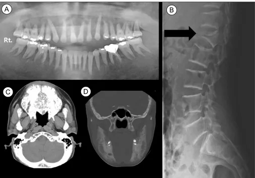
Fig. 1. Initial radiologic findings. (A) Panoramic view of maxilla and mandible: Multifocal bone demineralization is noted on both jaws. Generalized loss of lamina dura on whole dentition is also noted. (B) Lumbar-sacrum lateral x-ray: compression fracture of L1 (arrow). (C) and (D) Maxilla and mandible CT. (C) Axial soft tissue window setting image: Multifocal osteolytic lesions with bony expansion are noted on maxilla. (D) Coronal bone window setting image: Multifocal bone demineralization with bony expansion is noted on both mandiblar rami.
골에서 4.5~11.8% 정도로 발생하는 것으로 알려져 있다.7 1963년에 처음 국내에 보고되었고,5 최근까지 11개의 증례가 더 보고되었다. 골반 뼈 1예, 하악골 3예, 손목뼈 1예, 척추 뼈 1예, 갈비뼈 1예, 대퇴골 1 예, 손가락뼈 1예4,5,8-14가 있으며, 일차성 부갑상선 기능 항진증에 의해 상악골에 발생한 1예5와 이차성 부갑상선 기능 항진증에 의해 상악골에 발생한 갈색 종 1예15가 있다. 2008년부터 2011년까지 국외에서는 18개의 상악골 갈색종6,16-22이 보고되었고 이들은 대 부분 부갑상선 절제술을 통하여 치료되었다. 저자들 은 혈액투석을 받고 있는 만성 신부전 환자에서 발 생된 이차성 부갑상선 기능 항진증으로 인한 상악골 의 갈색종 1예를 7년간 장기 추적 검사하여 이에 대 해 보고하는 바이다.
증례 보고
환자: 27세, 여자 주소: 지속적인 크기 증가를 보이는 상악골의 종 물 및 하부 요통 현병력: 내원 3년 전부터 상악골의 종물이 점점 커 지기 시작하였으나 이에 대한 특별한 치료는 받지 않 았다. 환자는 입이 잘 다물어지지 않아 식사를 할 때 에 음식이 바깥으로 새어 나왔으며, 치열이 흐트러지 면서 잇몸에 간헐적인 출혈도 동반되었다. 또한 내원 1달 전부터 하부 요통 증상 발생하여 시행한 검사에 서 골다공증에 의한 압박 골절 발생하여 이에 대한 치료 위해 내분비내과로 협의 진료 의뢰되었다. 과거력: 1997년 고혈압으로 투약 시작하였고, 만 성 신부전 진단받고 혈액 투석 중이었다. 2003년 이 차성 부갑상선 기능 항진증 진단받았으나 치료 받고 있지 않았다. 가족력: 특이 사항 없었다. 진찰소견: 급성 병색을 보였으며, 혈압 170/90 mmHg, 맥박 95회/분, 호흡 20회/분, 체온 36.8oC이었 고 의식은 명료하였다. 하부 요통 있었으나 부종이 나 열감은 관찰되지 않았고, 상악골의 종괴는 치열 이상을 초래하였고 이로 인해 치아 사이가 벌어지면 서 잇몸이 파열되어 이로 인한 통증이 동반되었고, 또한 입을 잘 다물어지지 않았다(Fig. 1A, 5A). 그 외A B
Fig. 2. Parathyroid scan (MIBI). The inferior portion of bilateral thyroid lobe showed focal uptake of radioiodide and delayed washout, which indicates parathyroid lesion.
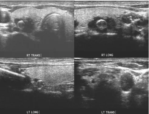
Fig. 3. Neck ultrasonography: dense calcified or non-calcified rim lesions, suspicious of parathyroid gland.
에는 신체 진찰 상 다른 이상 소견은 보이지 않았다. 검사 소견: 혈청 칼슘은 11.0 mg/dL (참고치: 8.5~ 10.5)로 증가되어 있었고, 인산염은 6.9 mg/dL (참고 치: 2.5~4.5)였으며, 부갑상선호르몬은 1,729.9 pg/mL (참고치: 10~65)였고, 크레아티닌은 7.5 mg/dL였다. 말초혈액검사에서 백혈구 4,300/uL (%), 혈색소
Wonjin Kim, et al:Seven Year-follow-up of a Brown Tumor in the Maxilla Associated with Secondary Hyperparathyroidism in End-stage Renal Failure
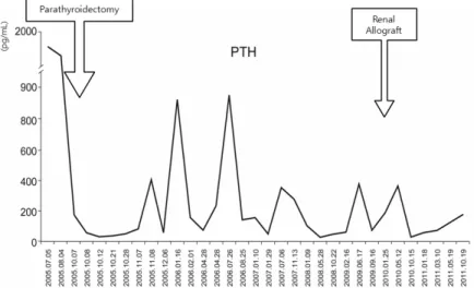
Fig. 4. Serial change of PTH.
Fig. 5. The features of brown tumor. (A) Before parathyroidectomy (B) 5-year postparathyroidectomy (C) 2-year post-renal transplantation. 9.0 g/dL, 혈소판 207,000 /uL였고, 생화학검사에서 아스파르테이트아미노전이효소(AST) 9 IU/L, 알라 닌아미노전이효소(ALT) 5 IU/L였으며, 알칼리인산 분해효소는 456 IU/L (참고치: 38~115), 총 빌리루 빈은 0.5 mg/dL였다. 혈청 총 콜레스테롤 149 mg/dL, BUN 33.2 mg/dL였다. 전해질 검사에서 Na+ 138 mmol/L, K+ 4.7 mmol/L, Cl- 98 mmol/L, total CO2 25
mmol/L였다. 25-수산화 비타민 D는 5.33 ng/mL (참 고치: 9.6~37.6)였다. 단순 흉부 엑스선 검사에서 경도의 심장비대가 있 었고, 단순 복부 엑스선 촬영에서 요로결석은 보이 지 않았다. 요추부 엑스선 검사상 요추 1번의 압박 성 골절이 있었다(Fig. 1B). 골밀도 검사상 요추부 Z-score -4.1, 대퇴부 -4.5로 심한 골다공증이 확인 되었고, 상악 파노라마 엑스선 검사상 양악골 치조 경선(lamina dura)의 전반적인 소실이 관찰되었다 (Fig. 1A). 당시 시행하였던 부갑상선 스캔(MIBI)에서 갑상 선 양쪽 하엽에서 부분적 조영 증강이 관찰되었고 (Fig. 2), 두경부 초음파 검사에서 양측 갑상선 하부 에 석회화 음영의 유무가 동반된 병변이 관찰되어 (Fig. 3), 부갑상선 선종을 의심할 수 있었다. 치료 및 경과: 고칼슘혈증 및 부갑상선 기능항진 증에 대해 부갑상선 전 절제술을 시행하였고 우측 상박에 부갑상선 자가이식을 시행하였다. 수술 후 조직 소견에서 부갑상선 비후가 관찰되었다. 수술 직후 환자에서 손발 저림 증상은 관찰되지 않았으 며, 수술 후 2일째 혈청 칼슘 6.7 mg/dL, 인산 2.6 mg/dL 소견 보여 염화칼슘(calcium chloride) 1,200 mg을 정주하였다. 혈청 부갑상선 호르몬은 수술 전 1,729.9 pg/mL에서 수술 후 5일째 19.22 pg/mL로 감 소되었다(Fig. 4). 이후 혈청 칼슘이 정상수치로 회복 되어 수술 후 22일째 퇴원하였다. 이후 외래에서 추 적 관찰하였으며, 상악골의 갈색종은 지속적으로 크
Fig. 6. Bone mineral density by dual x-ray absorptiometry, expressed by Z-score. 기 감소되어 입을 다물 수 있게 되었으며 통증 및 출혈도 없어졌다(Fig. 5b). 환자는 부갑상선 절제술 후 6년 뒤 신장 이식술 시행 받았고, 혈청 칼슘, 인 산 염, 크레아티닌 모두 정상 범위로 조절 중이다. 진단 당시 동반되었던 비타민 D 결핍증은 칼시트리 올 0.50 µg과 콜레칼시페롤 1000 IU, 탄산 칼슘 1,250 mg 복합제(DicamaxⓇ)를 복용하면서 정상 범위로 유 지 중이다. 부갑상선 절제술 후 매년 골밀도 검사를 시행하였고, 7년 후 시행한 골밀도 검사 상에서도 요추부 Z-score 0.7, 대퇴부 -0.2로 정상화되고 있으 며(Fig. 6), 갈색종의 크기도 더 감소되었다.
고 찰
부갑상선 기능 항진증은 혈청 내의 부갑상선 호르몬 농도 증가로 여러 질환을 유발하는 병으로 크게 원발성, 이차성, 삼차성, 이소성으로 분류한다. 원발성 부갑상 선 기능 항진증은 부갑상선 선종이나 드물게 부갑상선 암에 의하여 발생하고, 이차성 부갑상선 기능 항진증은 비타민 D 결핍증이나 만성 신부전 환자에서 장기간의 고인산혈증과 이에 따른 저칼슘혈증에 의하여 발생한 다. 오랜 기간 동안 지속적인 이차성 부갑상선 기능 항진증으로 부갑상선 세포 비후에서 자가 분비 (autonomous secretion)하는 선종으로 변하게 되면 삼차 성 부갑상선 기능 항진증이라 한다.9 만성 신부전으로 인한 이차성 부갑상선 기능 항진증 은 대부분 약물 및 식이조절, 비타민 D 보충, 인산의 섭취 조절 및 인산 결합제로 치료한다. 최근에는 칼슘유 사물질(calcimimetics)도 쓰는데 투석 중인 환자에서만 보험급여 처리가 되므로 현재로서는 제한적인 사용만 가능하다. 삼차성 부갑상선 기능 항진증으로 진행될 경우 부갑상선 전 또는 아전 절제술을 시행한다. 갈색종 즉, 낭성 섬유성 골염은 부갑상선 기능 항 진증의 후기에 나타나는 고립성 혹은 다발성 골병변 이다.5 대부분의 갈색종은 만성 신부전에 의한 이차 성 부갑상선 기능 항진증으로 인해 발생하게 된다.23 이차성 부갑상선 기능 항진증에서 갈색종의 발생률 은 약 1%에서 13%까지로 추정된다.24-26 과거에는 갈 색종의 원인이 대부분 일차성 부갑상선 기능 항진증 에서 발생하였으나, 최근에는 이차성 부갑상선 기능 항진증에서의 발생률이 증가하고 있다. 이것은 투석 을 받고 있는 말기 신부전 환자들의 생명 연장과 연 관이 있을 것으로 생각된다. 말기 신부전에서 갈색 종, 골연화증, 골경화증, 그리고 골다공증과 같이 여 러 가지 골격계 이상이 발생하게 된다.23 갈색종은 부갑상선 기능 항진증에 의한 광범위한 골흡수 과정의 결과로 알려져 있다. 증가된 부갑상 선호르몬으로 인해 파골세포의 작용이 증가가 되어Wonjin Kim, et al:Seven Year-follow-up of a Brown Tumor in the Maxilla Associated with Secondary Hyperparathyroidism in End-stage Renal Failure
Authors, Year
Sex/
Age Site of brown tumor
Hyperpara- thyroidism Calcium (mg/dL) Phosphate (mg/dL) Creatinine (mg/dL) Treatment Post- operative follow-up period Park et al., 201112
F/56 left maxilla Secondary 9.9 4.9 not
mentioned
Total parathyroidectomy & mass excision
12 months Kim et al.,
20103
F/32 mandible Secondary 10.4 5.0 not
mentioned
Total parathyroidectomy 6 months Mok et al.,
20109
M/44 left pelvic bone Primary 14.8 1.6 1.2 Total parathyroidectomy 12 months Chun et al.,
20094
F/39 hard palate Primary 14.2 not
mentioned
not mentioned
Left parathyroidectomy & mass excision
8 months Lee et al.,
200927
F/50 maxilla, mandible Primary not mentioned not mentioned not mentioned Parathyroidectomy not mentioned Park et al., 20087
F/35 lingual side of the mandible Secondary not mentioned not mentioned not mentioned
Excision of the mandible lesion
not mentioned Park et al.,
20088
F/70 right wrist Primary 11.0 1.9 not
mentioned
Total parathyroidectomy 12 months Choi et al., 20062 M/34 spine: L5 compression fracture Primary 12.8 1.0 not mentioned
Right parathyroidectomy not mentioned Mok et al.,
20031
M/80 right 10th rib Primary 16.7 2.9 2.7 Total parathyroidectomy not mentioned Chon et al.,
200310
M/52 femur shaft, phalanges
Primary 11.3 1.8 0.3 Parathyroidectomy not
mentioned Lee et al.,
199911
M/18 right 5th phalanx Primary 12.1 2.2 0.6 Total parathyroidectomy 8 months
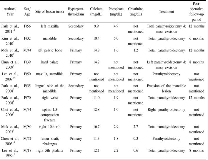
Table 1. Case reports of brown tumor in Korea
골흡수가 진행된다. 활성화된 다핵 파골세포들이 뼈 안에 미세골절을 생성하게 되어 출혈 및 그 주위로 혈철소를 포함하는 대식세포들이 둘러싸게 된다. 이 것을 신호로 섬유모세포들이 섬유주 표면과 골수에 모이게 되고 섬유주의 주변으로 섬유화를 촉진하게 된다. 갈색종은 이러한 골흡수와 섬유화의 진행으로 육안으로 보이는 낭종이 발생한 것이다.23,26 이것은 대부분 서서히 자라고 통증을 동반하는 종괴의 형태 로 나타난다. 주로 늑골, 쇄골, 하악골, 골반에 발생 하고, 상악골이나 경구개에서의 발생은 극히 드물게 발생한다.5 본 증례에서도 내원 3년 전부터 상악골 종괴의 크기가 서서히 증가하였고 종괴 자체는 통증 을 유발하지는 않았지만 주위 압박으로 인해 잇몸이 찢어지면서 통증 및 출혈을 동반하였다. 부갑상선 기능 항진증에 의한 갈색종에 대한 일반 적인 치료 방법은 부갑상선 절제술로 알려져 있다. 그러나 부갑상선 절제술 후의 갈색종 치료 방법에 대해서는 이견이 있다. Scott 등27은 부갑상선 호르몬 의 증가로 인해 갈색종은 서서히 소실되지만 골 파 괴를 동반한 광범위한 낭성변화가 있는 갈색종의 경 우는 수술이 필요하다고 보고하였고, Daniel 등28은 갈색종의 자연소실이 더디거나 오히려 크기가 커지 는 경우, 기능적 장애를 초래하는 경우에 국소 소파 술이나 외과적 적출술을 시행해야 한다고 보고하였 다. 반면 부갑상선 절제술 후 갈색종이 정상적인 골 로 전환되면서 골병변이 소실된다는 주장도 있는데, Knevezic 등29은 부갑상선 절제술 후 젊은 나이일 경 우에는 골 병변의 소실이 몇 달 사이에도 일어날 수
기능 항진증에 의해 갈색종이 발생하였으며, 3명은 이차성 부갑상선 기능 항진증에 의해 발생하 였다.4,10,15 일차성 부갑상선 기능 항진증이 있는 환자 들에서 혈청 칼슘은 11.0~16.7 mg/dL로 증가되어 있 었고, 이차성 부갑상선 기능 항진증 환자들은 9.9~ 10.4 mg/dL로 정상범위 내에 있었다. 대부분의 환자 에서 부갑상선 절제술을 시행하였고, 1예에서만 갈 색종 적출술을 시행하였다.10 증례들에서는 1년 정도 의 단기간 추적관찰 후에 보고를 하였다. 본 증례는 부갑상선 절제술 시행 후 7년간의 장기 추적 관찰 중이며, 수술적인 치료 없이 갈색종의 종 괴는 지속적으로 감소하고 있다. 발생 초기에 있었 던 저작 시 불편감이나 통증 및 출혈은 없고, 현재 칼시트리올 0.25 mcg을 복용하면서 매년 시행하는 골밀도 검사에서 골밀도가 정상화되었다.
요 약
저자들은 만성 신부전으로 인한 부갑상선 기능 항 진증의 드문 합병증인 상악골의 갈색종을 진단하였 고, 부갑상선 절제술 시행 후 갈색종의 지속적인 크 기 감소가 관찰되었다. 이차성 부갑상선 기능 항진 증으로 인한 갈색종을 수술적인 치료 없이 7년간의 장기간 추적 관찰을 통하여 크기의 감소와 통증의 소실 등의 호전을 경험하였기에 보고하는 바이다.참 고 문 헌
1. Kronenberg HM. Hormones and disorders of mineral metabolism. 11th ed. Philadelphia: Saunders; 2008. 2. Fraser WD. Hyperparathyroidism. Lancet 2009;374:
145-58.
3. JT P. Harrison's principles of internal medicine. 17th ed: McGraw-Hill Professional; 2008.
with Primary Hyperparathyroidism. Korean J Otorhinolaryngol-Head Neck Surg AID 2009;52: 612-5.
6. Di Daniele N, Condo S, Ferrannini M, Bertoli M, Rovella V, Di Renzo L, et al. Brown tumour in a patient with secondary hyperparathyroidism resis-tant to medical therapy: case report on successful treatment after subtotal parathyroidectomy. Int J Endocrinol 2009;2009:827652.
7. Triantafillidou K, Zouloumis L, Karakinaris G, Kalimeras E, Iordanidis F. Brown tumors of the jaws associated with primary or secondary hyper-parathyroidism. A clinical study and review of the literature. Am J Otolaryngol 2006;27:281-6. 8. Mok JO. A Case of Brown Tumor with Severe
Hypercalcemia Caused by Parathyroid Adenoma. J Korean Soc Endocrinol 2003;18:221-6.
9. Choi YW, Ok CS. Brown Tumor of The Spine with Compression Fracture: A Case Report. J Korean Radiol Soc 2006;54:33-7.
10. Park JW, Choi BR, Gang TI, Huh KH, Yi WJ, Choi SC. Mandibular brown tumor in renal osteo-dystrophy. Korean J Oral Maxillofac Radiol 2008; 38:229-31.
11. Park H, Kang GH, Kim SG, Kim JJ, Baek NN, Kim DM, et al. Brown Tumor of the Ulna and Radius: An Unusual Presentation of Primary Hyperparathyroidism. J Korean Endocr Soc AID - 10.3803/jkes.2008.23.5.347 [doi] 2008;23:347-51. 12. Mok JY, Kim HY, Ter HC, Kim SO, Kim DK,
Han JS, et al. A Case of Primary Hyperparathy-roidism with Rapid Regression of a Brown Tumor after Parathyroidectomy. J Korean Endocr Soc AID - 10.3803/jkes.2010.25.1.50 [doi] 2010;25:50-5.
Wonjin Kim, et al:Seven Year-follow-up of a Brown Tumor in the Maxilla Associated with Secondary Hyperparathyroidism in End-stage Renal Failure
13. Chon S, Kim YH, Park JY, Ko KP, Park CY, Kim DY, et al. A Case of Cystic Parathyroid Adenoma Presenting as Severe Bony Lesion. J Korean Soc Endocrinol 2003;18:214-20.
14. Lee SK, Moon SD, Kim HS, Park EJ, Ahn SJ, Han JH, et al. A case of mediastinal parathyroid adenoma presenting as fracture of brown tumor. The korean Journal of Medicine 1999;56:113-8. 15. Park DW, Lee CG, Lee JY, Kim HK. A case of
brown tumor of the maxilla associated with secon-dary hyperparathyroidism. Korean J otorhinolaryngol Head Neck Surg 2011;54:304-7.
16. LB. Fatma, S. Barbouch, BH. Fethi, BA. Imen, K. Karima, H. Imed, et al. Brown tumors in patients with chronic renal failure and secondary hyper-parathyroidism: report of 12 cases. Saudi J Kidney Dis Transpl 2010;4:772-7.
17. Nabi Z, Algailani M, Abdelsalam M, Asaad L, Albaqumi M. Regression of brown tumor of the maxilla in a patient with secondary hyperparathy-roidism after a parathyroidectomy. Hemodial Int 2010;14:247-9.
18. Marlene Corr Pinto, Scheila Maria Gabeta Sass, Cl dia Paragua Pupo Sampaio, Danielle Salvatti Campos. Brown tumor in a patient with hyper-parathyroidism secondary to chronic renal failure. Braz J Otorhinolaryngol 2010;76:404.
19. Sutbeyaz Y, Yoruk O, Bilen H, Gursan N. Primary hyperparathyroidism presenting as a palatal and mandibular brown tumor. J Craniofac Surg 2009; 20:2101-4.
20. Firat. Selvi, S. Cakarer, R. Tanakol, SD. Guler, C. Keskin. Brown tumour of the maxilla and mandible: a rare complication of tertiary hyperparathyroidism. Dentomaxillofacial radiology 2009;38:53-8.
21. Proimos E, Chimona TS, Tamiolakis D, Tzanakakis MG, Papadakis CE. Brown tumor of the maxillary sinus in a patient with primary
hyperparathy-roidism: a case report. J Med Case Reports 2009; 3:7495.
22. Resendiz-Colosia JA, Rodriguez-Cuevas SA, Flores-Diaz R, Juan MH, Gallegos-Hernandez JF, Barroso-Bravo S, et al. Evolution of maxillofacial brown tumors after parathyroidectomy in primary hyperparathyroidism. Head Neck 2008;30:1497-504. 23. Fargen KM, Lin CS, Jeung JA, Yachnis AT, Jacob
RP, Velat GJ. Vertebral Brown Tumors Causing Neurologic Compromise. World Neurosurg 2011. 24. Griffiths HJ, Ennis JT, Bailey G. Skeletal changes
following renal transplantation. Radiology 1974; 113:621-6.
25. Kaya RA, Cavusoglu H, Tanik C, Kahyaoglu O, Dilbaz S, Tuncer C, et al. Spinal cord compression caused by a brown tumor at the cervicothoracic junction. Spine J 2007;7:728-32.
26. Fineman I, Johnson JP, Di-Patre PL, Sandhu H. Chronic renal failure causing brown tumors and myelopathy. Case report and review of patho-physiology and treatment. J Neurosurg 1999;90: 242-6.
27. Scott SN, Graham SM, Sato Y, Robinson RA. Brown tumor of the palate in a patient with primary hyperparathyroidism. Ann Otol Rhinol Laryngol 1999;108:91-4.
28. Daniels JS. Primary hyperparathyroidism presenting as a palatal brown tumor. Oral Surg Oral Med Oral Pathol Oral Radiol Endod 2004;98:409-13.
29. Knezevic G, Uglesic V, Kobler P, Svajhler T, Bagatin M. Primary hyperparathyroidism: evalua-tion of different treatments of jaw lesions based on case reports. Br J Oral Maxillofac Surg 1991;29: 185-8.
30. Lee JK, Cho SD, Leem DH. A case report: brown tumor of the maxilla and mandible in association with primary hyperparathyroidism. J Korean Assoc Maxillofac Plat Reconstr Surg 2009;31:61-6.