865
Partial Nephrectomy for Complex Renal Tumors
Kyung Hwa Choi, Cheol Kyu Oh, Wooju Jeong, Enrique Ian S.Lorenzo, Woong Kyu Han, Koon Ho Rha
From the Department of Urology, Urological Science Institute, Yonsei University College of Medicine, Seoul, Korea
Purpose: Robot-assisted laparoscopic partial nephrectomy (RLPN) is gaining acceptance as an alternative to open partial nephrectomy and laparoscopic partial nephrectomy for small renal masses. However, it still remains a technically challenging procedure even for experienced laparo-scopists. Endophytic tumors or renal hilar tumors pose an additional challenge.
Materials and Methods: We reviewed the medical records of 11 patients (mean age: 49.3 years; range: 31-67 years) who underwent RLPN for small, complex renal masses including hilar tumors and endophytic tumors.
RLPN was performed with the Da VinciⓇ
surgical system (Intuitive Surgical, Sunnyvale, USA) with three robot arms and intraoperative ultrasonography (Tile-proⓇ
System).
Results: RLPN was performed successfully without complications in all cases. The mean tumor size was 3.2 cm (range, 1.1-8.0 cm). The mean operative time was 177 minutes (range, 150-260 minutes), and the mean warm ischemia time was 32 minutes (range, 25-41 minutes). The mean estimated blood loss was 177 ml (range, 50-350 ml), and the mean hospital stay was 4 days (range, 3-7 days). Pathology found four patients with clear cell type renal cell carcinoma, one with multilocular multicystic renal cell carcinoma, two with papillary type, one with chromophobe type, and three with angiomyolipoma.
Conclusions: RLPN is a feasible and safe surgery for complex renal tumors. In our experiences, RLPN could be a nephron-sparing surgical option for patients with compromised renal function and it could be an alternative to open partial nephrectomy and laparoscopic partial nephrectomy for a select group of patients. (Korean J Urol 2009;50:865-869)
Key Words: Robotics, Nephrectomy, Kidney neoplasms
Korean Journal of Urology Vol. 50 No. 9: 865-869, September 2009
DOI: 10.4111/kju.2009.50.9.865
Received:June 17, 2009 Accepted:August 27, 2009 Correspondence to: Koon Ho Rha
Department of Urology, Urological Science Institute, Yonsei University College of Medicine, 134, Sinchon-dong, Seodaemun- gu, Seoul 120-752, Korea TEL: 02-2228-2318 FAX: 02-312-2538 E-mail: [email protected]
Ⓒ The Korean Urological Association, 2009
INTRODUCTION
Nephron-sparing surgery is now considered the standard of care for small renal tumors (<4 cm) [1]. Partial nephrectomy was conducted initially for patients with a solitary kidney, with bilateral renal masses, or with compromised renal function. It has since been used for patients with normal contralateral kidneys with the development of surgical techniques and technologies.
With the development of laparoscopy, laparoscopic partial
nephrectomy (LPN) has been offered as an alternative to open surgery for small renal masses. It has been increasingly performed due to advantages such as less perioperative and postoperative morbidity, faster recovery, and better cosmetic results. Its safety, efficacy, and oncologic outcomes did not differ significantly from those of open surgery [1,2]. However, endophytic or renal hilar tumors are challenging for laparo-scopy in terms of operative angles and mechanical handling; hence, they are most often taken care of though converted open surgery [3]. Another option is the use of laparoscopic radical nephrectomy. Issues such as adequacy of resection, vascular
Table 1. Patients' demographic data
No. of patients 11 Mean age, years (range) 49.36 (31-67) Sex
Male 5
Female 6
Mean preoperative Cra, mg/dl (range) 1.01 (0.7-2.3) Side of involvement
Right 3
Left 8
a
: preoperative serum creatinine
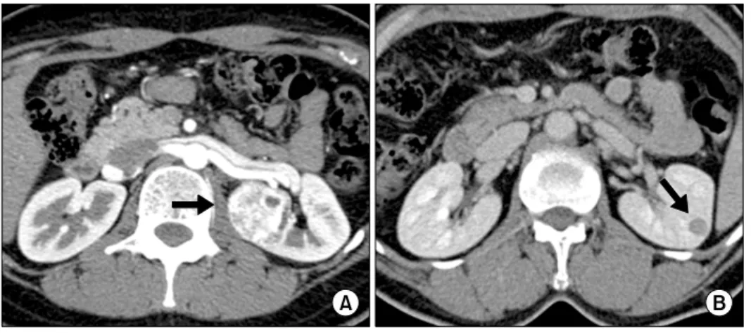
Fig. 1. (A) Large posterior renal
hilar tumor demonstrating proximity to renal hilar vessels and abutting the renal pelvis. CT demonstrates left 3.5 cm solid upper pole renal mass. (B) Small lateral endophytic tumor. CT demonstrates left 2 cm solid midportion renal mass.
control, watertight closure, and ischemia time are some of the challenges to the urologist.
Recently, after the introduction of surgical robots including the Da VinciⓇ surgical system (Intuitive Surgical, Sunnyvale, USA), robot-assisted kidney procedures such as pyeloplasty and radical nephrectomy have been successfully performed by using robotic technology. Robot-assisted laparoscopic partial nephrectomy (RLPN) has also been used more with the development of surgical skills. Compared with laparoscopic surgery with two-dimensional images and a long learning curve, robot-assisted surgery has many advantages such as the multiple joints with seven degrees of freedom, high-definition three-dimensional imaging, hand-held function, and elimination of tremor. These factors enable resection of a tumor located in a position often times too extremely difficult for LPN [4]. Robotics application thus makes nephron-sparing surgery feasible for complex tumors. We present our initial outcomes in 11 successfully conducted RLPN cases.
MATERIALS AND METHODS 1. Subjects
From September 2006 to March 2009, RLPN was performed in a total of 52 cases by a single surgeon. Eleven of these were for complex renal tumors (endophytic or hilar) (Fig. 1). Table 1 shows the patients’ demographic data. In imaging studies taken before the operation, all of the tumors were considered to be confined to the kidney. All operations were conducted transperitoneally with the Da VinciⓇ surgical system. One patient was diagnosed with chronic renal failure. The serum creatinine level was 2.3 mg/dl. All other patients had normal renal function of both kidneys.
2. Surgical technique
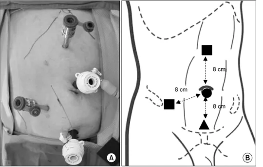
1) Position and port placement: The patient was positioned with some modifications to the conventional lumbotomy position [4]. Pneumoperitoneum was established by using the Veress needle just above the umbilicus. A 12 mm camera port was placed in its place. Three 8 mm robot instrument ports were placed as was done by Park et al (Fig. 2) [4]. An optional 5 mm assistant port was placed subxiphoid when deemed necessary.
2) Ultrasound and tumor exposure: Bowel and kidney mobilization was done in the usual manner [4]. By using the robotic scissors and bipolar forceps, the renal vein and renal artery were exposed to complete mobilization of the kidney. Gerota’s fascia was dissected on the area of the tumor. For endophytic tumors, their precise location and depth were evaluated with preoperative computed tomography images and intra-operative laparoscopic ultrasound (Tile-proⓇ System). Margins were then marked with monopolar cautery.
3) Robot-assisted tumor excision: Tumor resection was conducted under warm ischemia time. The renal artery and
Table 2. The operative parameters for 11 patients undergoing robotic partial nephrectomy
Warm Estimated Mean Tumor Operative Hospital
Tumor ischemic blood change
No. size time stay Pathology
location time loss of Cra (cm) (min) (days)
(min) (ml) (ml/dl)
1 H+E 1.7 160 25 300 4 0.1 Angiomyolipoma
2 H 2.8 150 29 100 5 0.5 Clear cell RCC, Fuhrman 2 3 H+E 2.7 167 26 50 4 0.1 Clear cell RCC, Fuhrman 2 4 H+E 3.0 168 41 100 7 0.2 Cystic clear cell RCC, Fuhrman 2 5 E 1.1 172 32 200 4 0.4 Angiomyolipoma
6 H 3.7 187 37 350 3 0.5 Papillary RCC, Type I, Furhman 2 7 H 8.0 218 38 200 3 0.4 Papillary RCC, Type I, Furhman 1 8 H 3.0 163 32 200 6 −0.1 Angiomyolipoma
9 H+E 3.7 260 35 200 4 0.1 Clear cell RCC, Fuhrman 2 10 H 4.0 245 30 200 3 0.4 Multilocular cystic RCC 11 E 1.5 190 30 50 4 0.1 Chromophobe, RCC, Fuhrman 3 Mean 3.2 177 32 177 4 0.25 RCC 8, angiomyolipoma 3 H: hilar, E: endophytic, RCC: renal cell carcinoma, a: increased serum creatinine (Cr) at discharge
Fig. 2. (A) Port site placement for
left robotic partial nephrectomy. (B) Schematic of port site placement demonstrating a 12 mm periumbili-cal camera port (round), two 8 mm robotic instrument ports (square), and a 12 mm assistant port.
renal vein were clamped individually with two laparoscopic bulldog clamps using the assistant port. The tumor was then resected along the previously marked margins. Margins of resection were then sent for frozen section biopsy. Any minor bleeding observed was coagulated with the bipolar forceps. The specimen was placed in an entrapment bag and kept temporarily out of the operating field.
4) Renorrhaphy: Hemostasis was obtained by electrocautery, suturing, and application of a hemostatic agent. Both robotic arms were installed with needle drivers and the renal
parenchyma defect was sutured after repairing the collecting system with VicrylⓇ 2-0. The area of the repair was over-layered with SurgicelⓇ and tissue glue was applied over. The kidney was replaced in its anatomical position and the bulldog clamps were carefully pulled out. The kidney and the sur-rounding tissues were inspected for bleeding, which was controlled as needed. The specimen packed in the entrapment bag was delivered through the camera port site. All trocars were then removed under direct visualization after inserting the drain.
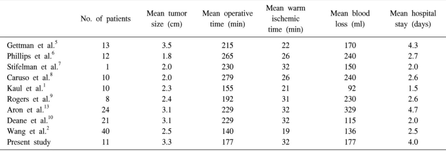
Table 3. Comparison of contemporary reports on robotic partial nephrectomy
Mean warm
Mean tumor Mean operative Mean blood Mean hospital No. of patients ischemic
size (cm) time (min) loss (ml) stay (days) time (min) Gettman et al.5 13 3.5 215 22 170 4.3 Phillips et al.6 12 1.8 265 26 240 2.7 Stifelman et al.7 1 2.0 230 32 150 2.0 Caruso et al.8 10 2.0 279 26 240 2.6 Kaul et al.1 10 2.3 155 21 92 1.5 Rogers et al.9 8 2.4 192 31 230 2.6 Aron et al.13 24 3.1 229 32 329 4.7 Deane et al.10 21 3.1 229 32 115 2.0 Wang et al.2 40 2.5 140 19 136 2.5 Present study 11 3.3 177 32 177 4.0 RESULTS
The operative parameters are summarized in Table 2. There were no incidences of intraoperative complications such as conversion to total nephrectomy or to open surgery nor injury to vascular structures or adjacent organs. None of the patients required blood transfusion. No postoperative complication developed. Comparing the serum creatinine before and after the surgery (before discharge), it increased by an average of 0.28 mg/dl in 10 patients. It decreased by 0.1 mg/dl in one patient. Pathologic examination revealed eight patients with renal cell carcinoma and three with angiomyolipoma. The final pathology report on 3 of the 11 patients showed the tumor abutting the surgical margin. The frozen section margins, however, were free of cancer. Of these 3 patients, one had angiomyolipoma, one papillary, and one clear cell type renal cell carcinoma. The average follow-up period was 6.5 months (range, 1 to 12 months). Only one patient was lost in follow-up. The latest follow-up found all subjects with normal creatinine levels except the patient with chronic renal failure. In this patient, the serum creatinine level increased from 2.3 mg/dl to 2.8 mg/dl postoperatively, but the level was stabilized at 2.5 mg/dl after 9 months. There were no remarkable findings in chest X-rays and abdominal computed tomography scans taken for all patients during the follow-up period.
DISCUSSION
With the introduction of robotic technology, laparoscopic
partial nephrectomy has been performed more often with robot assistance [1,2,4-10]. The surgical technique is based on conventional laparoscopy and has been proven to have surgical and oncologic outcomes similar to those of open partial nephrectomy [11,12]. Early studies on RLPN have also shown that its outcomes are no different from conventional laparoscopic and open techniques [1,5,13]. However, LPN is technically difficult especially with issues of vascular control and watertight closure of the collecting system and capsule under the limitation of warm ischemia time. It is for these reasons that renal hilar or endophytic tumors are an additional challenge to a minimally invasive surgeon. Often times, these tumors are treated via laparoscopic radical nephrectomy or open partial nephrectomy. Our institution has conducted a good number of RLPNs aside from other robotic cases and hence has developed more skill and confidence in RLPN than with LPN [4]. Because of this, the authors attempted RLPN in complex renal tumors.
The advantages of the Da VinciⓇ
surgical system (Intuitive Surgical, Sunnyvale, USA) as provided by the EndoWristⓇ and the three-dimensional, high-definition imaging allow a minimally invasive surgeon to reach difficult areas as well as conduct precise and elaborate dissection and suturing. This may eventually broaden indications for nephron-sparing surgery. With the addition of the Tile-proⓇ system, assessment of the depth of an endophytic tumor and determination of the proximity of the tumor to the hilar blood vessels with color Doppler can be efficiently done intraoperatively. This aids the surgeon in doing a more accurate resection.
were comparable with those of current reports (Table 3). Although the cases were complex, warm ischemia time and estimated blood loss were similar to those of RLPN for general tumors. Operative time was also similar to other reports indicating no delay associated with the difficulty in resection and repair of complex renal tumors. There were also no intraoperative or postoperative complications observed. For mean hospital stay, the lowest number of days was 1.5 [1] and our institution’s experience was 4 days. The seemingly prolonged stay in our institution may be due to factors such as the health care system in Korea and patients’ preference. Although no emergency situations were encountered in this study, it is also important that a competent and experienced bedside assistant is present who should be able to respond immediately when a situation requiring conversion to open surgery emerges. This is because the primary surgeon is in an unsterile condition at the console [7].
The use of robotic technology entails a higher cost, which has led to most renal tumors being treated via conventional laparoscopy or open surgery. However, RLPN may be offered in cases of complex renal tumors, especially for patients for whom nephron-sparing surgery is the only option.
CONCLUSIONS
RLPN is feasible and safe for complex renal tumors such as hilar and endophytic types. Because these types of surgeries implicate a high degree of difficulty, proper patient selection and adequate robotic surgery skills are advocated for a safe and successful surgery. Long-term studies should also be conducted to verify surgical and oncologic outcomes in the long run.
REFERENCES
1. Kaul S, Laungani R, Sarle R, Stricker H, Peabody J, Littleton R, et al. da Vinci-assisted robotic partial nephrectomy:
technique and results at a mean of 15 months of follow-up. Eur Urol 2007;51:186-91.
2. Wang AJ, Bhayani SB. Robotic partial nephrectomy versus laparoscopic partial nephrectomy for renal cell carcinoma: single-surgeon analysis of >100 consecutive procedures. Urology 2009;73:306-10.
3. Kim KY, Kim DK, Woo SH, Kim ET, Lee SB. Laparoscopic partial nephrectomy: an useful method of decision making for determining the approach and surgical method based on the systematic classification of tumor location. Korean J Urol 2008;49:1067-73.
4. Park SY, Kim HJ, Seo JW, Cho KS, Ham WS, Rha KH. Robot-assisted laparoscopic partial nephrectomy. Korean J Urol 2008;49:387-91.
5. Gettman MT, Blute ML, Chow GK, Neururer R, Bartsch G, Peschel R. Robotic-assisted laparoscopic partial nephrectomy: technique and initial clinical experience with DaVinci robotic system. Urology 2004;64:914-8.
6. Phillips CK, Taneja SS, Stifelman MD. Robot-assisted laparo-scopic partial nephrectomy: the NYU technique. J Endourol 2005;19:441-5.
7. Stifelman MD, Caruso RP, Nieder AM, Taneja SS. Robot- assisted laparoscopic partial nephrectomy. JSLS 2005;9:83-6. 8. Caruso RP, Phillips CK, Kau E, Taneja SS, Stifelman MD.
Robot assisted laparoscopic partial nephrectomy: initial experi-ence. J Urol 2006;176:36-9.
9. Rogers CG, Metwalli A, Blatt AM, Bratslavsky G, Menon M, Linehan WM, et al. Robotic partial nephrectomy for renal hilar tumors: a multi-institutional analysis. J Urol 2008;180:2353-6. 10. Deane LA, Lee HJ, Box GN, Melamud O, Yee DS, Abraham JB, et al. Robotic versus standard laparoscopic partial/wedge nephrectomy: a comparison of intraoperative and perioperative results from a single institution. J Endourol 2008;22:947-52. 11. Allaf ME, Bhayani SB, Rogers C, Varkarakis I, Link RE,
Inagaki T, et al. Laparoscopic partial nephrectomy: evaluation of long-term oncological outcome. J Urol 2004;172:871-3. 12. Lane BR, Gill IS. 5-year outcomes of laparoscopic partial
nephrectomy. J Urol 2007;177:70-4.
13. Aron M, Koenig P, Kaouk JH, Nguyen MM, Desai MM, Gill IS. Robotic and laparoscopic partial nephrectomy: a matched- pair comparison from a high-volume centre. BJU Int 2008; 102:86-92.