plSSN: 1976-8257 eISSN: 2234-2753 Toxicol. Res. Vol. 35, No. 4, pp. 403-410 (2019)
https://doi.org/10.5487/TR.2019.35.4.403 Open Access
403
Theracurmin (Highly Bioavailable Curcumin) Prevents High Fat
Diet-Induced Hepatic Steatosis Development in Mice
Jin Won Yang1
, Hee Kyung Yeo2
, Jee Hye Yun2
and Jung Un Lee3
1College of Pharmacy, Woosuk University, Wanju, Korea 2
HANDOK Inc., Seoul, Korea
3ChemOn Inc., Suwon, Korea
Abstract
Curcumin, a hydrophobic polyphenol isolated from the Curcuma longa L. plant, has many pharmacological prop-erties, including antioxidant, anti-inflammatory, and chemo-preventive activities. Curcumin has been shown to have potential in preventing nonalcoholic fatty liver disease (NAFLD). However, the low bioavailability of cumin has proven to be a major limiting factor in its clinical adoption. Theracurmin, a highly bioavailable cur-cumin that utilizes micronized technology showed improved biological absorbability in vivo. The aim of this study was to investigate the role of theracurmin in modulating hepatic lipid metabolism in vivo. A fatty liver mouse model was produced by feeding mice a high fat diet (HFD; 60% fat) for 12 weeks. We found that treatment for 12 weeks with theracurmin significantly lowered plasma triacylglycerol (TG) levels and reduced HFD-induced liver fat accumulation. Theracurmin treatment lowered hepatic TG and total cholesterol (T-CHO) levels in HFD-fed mice compared to controls. In addition, theracurmin administration significantly reduced lipid peroxidation and cellular damage caused by reactive oxygen species in HFD-fed mice. Overall, these results suggest that theracur-min has the ability to control lipid metabolism and can potentially serve as an effective therapeutic remedy for the prevention of fatty liver.
Key words: Theracurmin, Curcumin, Nonalcoholic fatty liver disease (NAFLD), High fat diet (HFD), Fatty liver, Steatosis
INTRODUCTION
Non-alcoholic fatty liver disease (NAFLD) has been rec-ognized as a common liver disease worldwide. The term represents a broad spectrum of liver damage ranging from simple steatosis to nonalcoholic steatohepatitis (NASH), progressive fibrosis, and cirrhosis (1,2). NAFLD is strongly associated with metabolic syndrome-associated conditions such as obesity, dyslipidemia, diabetes, hypertension, and insulin resistance (3,4). The detailed pathogenesis of
NAFLD is not completely known, but excessive fatty acid [triacylglycerol (TG)] and cholesterol (CHO) accumula-tion in the liver has been linked to the development of NASH, cirrhosis, and cancer (5,6). Although current ther-apeutic approaches have focused on the treatment of the underlying risk factors for these metabolic conditions, no standard strategy has yet been approved for NAFLD ther-apy (2,6).
Curcumin, a natural yellow polyphenol that exists in herbal remedies and the dietary spice turmeric, has been
Correspondence to: Jin Won Yang, College of Pharmacy, Woo-suk University, 443 Samnyero, Samnye-eup, Wanju-gun, Jeon-buk 55338, Korea
E-mail: [email protected]
This is an Open-Access article distributed under the terms of the Creative Commons Attribution Non-Commercial License (http:// creativecommons.org/licenses/by-nc/3.0) which permits unre-stricted non-commercial use, distribution, and reproduction in any medium, provided the original work is properly cited.
List of Abbreviations: ACAT, acyl-CoA:cholesterol acyltransfer-ase; GSH, glutathione; HDL, high-density lipoprotein; H&E, hema-toxylin and eosin; HFD, high fat diet; HMG-CoA, 3-hydroxy-3-methylglutaryl-CoA; LDL, low-density lipoprotein; MDA, malondi-aldehyde; NAFLD, Nonalcoholic fatty liver disease; NASH, nonal-coholic steatohepatitis; ND, normal diet; Nrf2, nuclear factor erythroid-2-related factor-2; ROS, reactive oxygen species; SREBPs, sterol regulatory elementbinding proteins; TBA, thiobarbituric acid; T-CHO, Total cholesterol; TG, triacylglycerol.
shown to possess antioxidant, anti-inflammatory, antimi-crobial, and chemopreventive activities, and has been demonstrated to prevent obesity and diabetes in animal models (7,8). Curcumin also exerted beneficial effects against hypercholesterolemia and dyslipidemia in rodent animal models, as well as in two randomized double-blind NAFLD clinical trials (9,10).
Although curcumin has been shown to be protective against dyslipidemia and NAFLD, its therapeutic out-comes and clinical use is limited by low oral bioavailability owing to its very low intestinal absorption and hydropho-bic properties, leading to poor solubility (11-13). Many studies investigating curcumin delivery systems, includ-ing submicron suspensions, phosphatidylcholine complexes, and solid lipid nanoparticles, have been performed with the aim of improving curcumin oral bioavailability (14-16). Among these, thearcurmin, a highly bioavailable cur-cumin developed using micronized-technology, has sig-nificantly increased bioavailability and water solubility relative to curcumin. In rat and human studies, theracur-min absorption was 30-fold higher than that of commer-cially available curcumin (14). Moreover, the maximum curcumin plasma concentration increased over 50-fold when theracurmin was used instead of curcumin powder (14).
Theracurmin has been reported to be effective against a variety of pathological conditions including cardiovascu-lar disease, esophageal cancer, inflammatory bowel dis-ease, and osteoarthritis (11,14,17). However, no basic or clinical studies regarding the efficacy of theracurmin against NAFLD, including hepatic steatosis, have been performed. In this study, we evaluated the preventive effect of theracurmin on hepatic steatosis in mice fed with a high fat diet (HFD). We uncovered that theracurmin treatment prevented the accumulation of TG and total cholesterol (T-CHO), as well as the lipid peroxidation, normally observed in the livers of HFD-fed mice. Our findings suggested that theracurmin is protective against NAFLD through lipid metabolism and oxidative stress modulation, and has ther-apeutic potential.
MATERIALS AND METHODS
Animals and treatment. Animal experiments were
performed in accordance with the requirements of the Animal Care and Ethics Committees of Gyeonggi bio cen-ter (2017-08-0001). C57BL/6N mice at 4 weeks of age were maintained in a standard condition (23 ± 3oC, 55 ± 15% humidity with a 12-hr light/dark cycle), pathogen-free environment and had access to a sterile standard rodent chow diet and water ad libitum. After a one week adaptive period, Male C57BL/6N mice at 5 weeks of age were started on either a normal diet (ND) or HFD 60% w/w for 12 weeks. Vehicle (normal saline), theracurmin (500, 1,000, and 2,000 mg/kg, as a curcumin 150, 300, 600 mg/
kg), or silymarin (25 mg/kg) were orally administered to mice seven times per week during 12 weeks of the diet feeding.
Histopathological analysis. The left lateral lobe of the liver was sliced and tissue slices were fixed in 10% buff-ered-neutral formalin, embedded in paraffin. The liver slices were used to generate 3-4µm sections in a cryostat. Tissue sections were stained with H&E and Oil red O staining.
After that the histological profiles of individual cross trimmed hepatic tissues were light microscopically observed (Model Eclipse 80i, Nikon, Tokyo, Japan). To observe more detail histopathological changes, the steatohepatitis regions (under OR staining) and mean hepatocyte diame-ters (under HE staining) were calculated using an auto-mated image analysis process (iSolution FL ver 9.1, IMT i-solution Inc., Vancouver, Quebec, Canada) on the restricted view fields. Steatohepatitis regions, the percentage of fatty deposited regions in hepatic parenchyma, were calculated as percentages of lipid deposited regions between restricted histological view field of liver (Mean hepatic steatosis regions - %/mm2 of hepatic parenchyma) under cryostat and oil red staining. Mean diameters of hepatocytes were also calculated in restricted view fields on a computer monitor under paraffin embedding and HE staining using an automated image analysis process.
Measurement of hepatic TG and total cholesterol
contents. TG (Catalog#K622-100, Biovision, San
Fran-cisco, CA, USA) and T-CHO (Catalog#K603, Biovision) contents were measured using commercial kits.
Biochemical parameters. Serum was collected after
centrifugation at 3,000 rpm for 10 min. Serum TG log#OSR61118, Beckman coulter, CA, USA), T-CHO (Cata-log#OSR6116, Beckman coulter), LDL-C (Catalog#6183, Beckman coulter), and HDL-C (Catalog#OSR6187, Beck-man coulter), were analyzed using commercial kits from Chemistry Analyzer (AU680, Beckman coulter).
Measurement of hepatic malondialdehyde and
glu-tathione contents. Malondialdehyde (MDA) levels was
determined by the thiobarbituric acid (TBA) method (Cat-alog#STA-330, Cellbiolabs, San Francisco, CA, USA). TBA reaction was performed according to manufactory guidance. Glutathione (GSH) were analyzed using com-mercial kits (Catalog#ADI-900-160, ENZO Life Science, Vileurbanne, France).
Data analysis. Statistical analyses were performed
using SPSS statistics 22 for medical science. LSD test was used to examine the significant inter-group differences. Statistical significance was accepted at either p < 0.05 or p < 0.01.
plSSN: 1976-8257 eISSN: 2234-2753
RESULTS
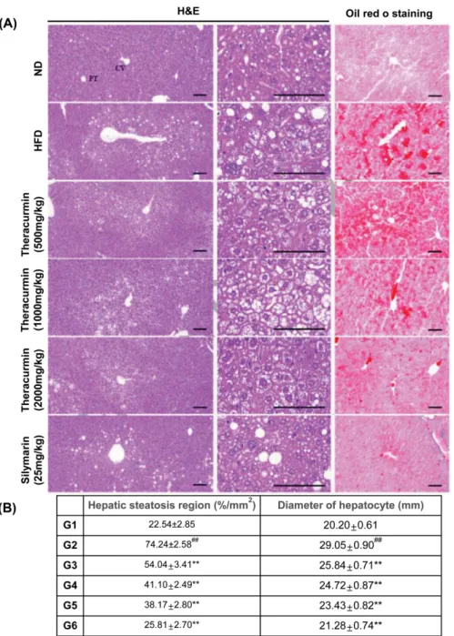
Theracurmin inhibits HFD-induced hepatic steatosis.
To examine the effect of theracurmin on liver fat
accumu-lation, mice were fed with a HFD (60% fat) for 12 weeks and then treated with vehicle, theracurmin, or silymarin (25 mg/kg; reference control). Since the main histological fea-ture of hepatic steatosis is liver fat accumulation, hepatic
Fig. 1. Effects of theracurmin on hepatic lipid accumulation in mice fed with HFD. (A) Liver sections were stained with H&E or
oil-Red O staining. The mice were fed either a ND or HFD for 12 weeks. theracurmin (500, 1,000, and 2,000 mg/kg) or silymarin (25 mg/ kg) (reference control) was administered to the mice at the same time. H&E staining. The livers of the mice were stained with H&E after a treatment with 500, 1,000, and 2,000 mg/kg theracurmin for 12 weeks. CV, Central vein; PT, Portal triad area; Scale bars = 100µm. Oil Red O staining. Each photo represents their groups after staining with Oil Red O in the liver. Scale bars = 100 µm. (B) Histomorphometric analysis. Measurement of hepatic steatosis region (%/mm2
of hepatic parenchyma) and diameter of hepatocyte (mm/hepatocyte). Data were expressed as mean ± SEM statistically analyzed by LSD-test methods. Significant versus normal control,
##
p < 0.01; significant versus HFD-fed group, **p < 0.01 (n = 10). G1: ND (normal saline), n = 10; G2: HFD + vehicle (normal saline), n = 10; G3: HFD + theracurmin (500 mg/kg/day), n = 10; G4: HFD + theracurmin (1,000 mg/kg/day), n = 10; G5: HFD + theracurmin (2,000 mg/kg/day), n = 10; G6: HFD + reference control (silymarin 25 mg/kg/day), n = 10.
fat deposition was measured. Histopathological analysis using hematoxylin and eosin (H&E) and Oil Red O tissue staining found that HFD fed mice exhibited increased hepatocyte fat accumulation (Fig. 1A). Theracurmin treat-ment at doses of 500, 1,000, and 2,000 mg/kg signifi-cantly reduced these pathological changes in the tissue, as did oral administration of silymarin (Fig. 1B).
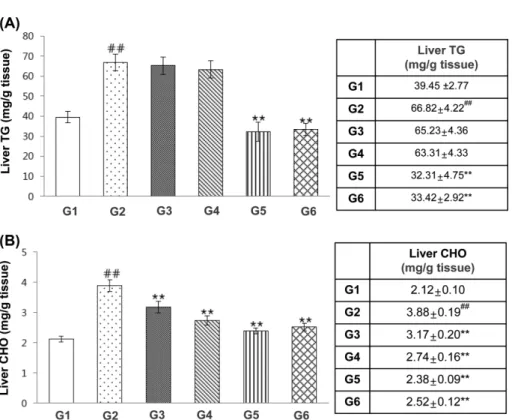
Theracurmin improves the accumulation hepatic TG
and T-CHO in HFD-fed mice. TG and T-CHO
accumu-lation in hepatocyte cytoplasm is the hallmark of NAFLD (1). Theracurmin treatment (2,000 mg/kg) for 12 weeks reduced HFD-induced hepatic TG content increases, as did oral administration of silymarin (Fig. 2A). In addi-tion, hepatic CHO levels were significantly decreased in theracurmin-treated mice relative to ND controls [ND: 2.12 ± 0.10 mg/dL; HFD, 3.88 ± 0.19 mg/dL; HFD + Theracurmin (500 mg/kg), 3.17 ± 0.20 mg/dL; HFD + Theracurmin (1,000 mg/kg), 2.74 ± 0.16 mg/dL; HFD + Theracurmin (2,000 mg/kg), 2.38 ± 0.09 mg/dL] (Fig. 2B). Accordingly, HFD treatment for 12 weeks induced increases in hepatocyte and hepatic steatosis region diam-eter increases, phenomena notably attenuated by adminis-trations of theracurmin at doses of 500, 1,000, and 2,000 mg/kg (Fig. 1B).
Theracurmin reduces plasma TG but does not affect
body weight in HFD-fed mice. Next, we examined the
effect of theracurmin on serum lipid levels. Theracurmin treatment significantly reduced HFD-induced plasma TG level increases but did not affect plasma total CHO, low-density lipoprotein (LDL), and high-low-density lipoprotein (HDL) levels (Fig. 3). However, the administration of theracurmin did not affect body weight gain or food intake amounts (Supplementary Fig. 1).
Theracurmin inhibits HFD-induced lipid peroxidation.
Lipid peroxidation and cellular damage by reactive oxy-gen species is characterized by membrane lipid break-down and the production of lipid peroxides (18). As observed in Fig. 4A, liver MDA production, a marker of lipid peroxidation, was elevated in HFD-treated mice rela-tive to normal diet-fed controls. This elevation of hepatic MDA was significantly attenuated by theracurmin admin-istration at a dosage of 2,000 mg/kg. Theracurmin treat-ment (1,000 and 2,000 mg/kg doses) also increased hepatic GSH content versus untreated HFD-fed mice (Fig. 4B).
DISCUSSION
Curcumin has been shown to reduce hepatic steatosis,
Fig. 2. Effects of theracurmin on the accumulation of hepatic TG and CHO in HFD-fed mice. (A, B) Measurement of accumulation
of TG and CHO in the liver from mice of each group. Data were expressed as mean ± SEM statistically analyzed by LSD-test meth-ods. Significant versus normal control, ##
p < 0.01; significant versus HFD-fed group, **p < 0.01 (n = 10). G1: ND (normal saline), n = 10; G2: HFD + vehicle (normal saline), n = 10; G3: HFD + theracurmin (500 mg/kg/day), n = 10; G4: HFD + theracurmin (1,000 mg/kg/day), n = 10; G5: HFD + theracurmin (2,000 mg/kg/day), n = 10; G6: HFD + reference control (silymarin 25 mg/kg/day), n = 10.
plSSN: 1976-8257 eISSN: 2234-2753
inflammation, insulin resistance, diabetes, and atheroscle-rosis by regulating hepatic lipid metabolism and plasma lipid homeostasis (19,20). However, the low oral bioavail-ability of curcumin limits its clinical adoption (11). To overcome this, theracumin, a submicron crystal solid dis-persion of curcumin, was formulated to enhance
cur-cumin bioavailability through enhanced water solubility and absorption (7,11).
We demonstrate herein that theracurmin administration exerted anti-steatotic activity in the livers of mice fed with high fat diets. In the current study, theracurmin adminis-tration led to significant decreases in both hepatic TG and
Fig. 3. Effects of theracurmin on the serum TG, CHO, LDL, and HDL in HFD-fed mice. (A-D) serum TG levels (A), serum CHO levels
(B), serum LDL levels (C), serum HDL levels (D) in mice fed a normal diet or high-fat-diet for 12 weeks. Data were expressed as mean ± SEM statistically analyzed by Q-test and LSD-test methods. Significant versus normal control, ##
p < 0.01; significant versus HFD-fed group, **p < 0.01 (n = 10). G1: ND (normal saline), n = 10; G2: HFD + vehicle (normal saline), n = 10; G3: HFD + theracurmin (500 mg/kg/day), n = 10; G4: HFD + theracurmin (1,000 mg/kg/day), n = 10; G5: HFD + theracurmin (2,000 mg/kg/day), n = 10; G6: HFD + reference control (silymarin 25 mg/kg/day), n = 10.
total CHO levels in mice fed a HFD for 12 weeks. Exces-sive fatty acids, derived from diet or lipolysis, results in hepatocyte lipid droplet accumulation, a representative feature of NAFLD (21). Consequently, specific lipotoxic lip-ids, including ceramide, diacylglycerols, and lysophospha-tidyl choline species, from these droplets induce hepatocellular injury in NASH (2,21). In addition, an imbalance between intrahepatic CHO and the removal of CHO from hepato-cytes leads to CHO accumulation in the liver (22,23). This extensive dysregulation of hepatic CHO homeostasis has been shown to accentuate hepatocellular injury and liver inflammation in NAFLD development. Thus, preventing TG and CHO accumulation in the liver may prove promis-ing for NAFLD treatment.
Curcumin has been reported to alleviate HFD-induced obesity in mice through the inhibition of sterol regulatory element-binding proteins (SREBPs) such as SREBP-1 and SREBP-2. SREBPs are key transcription factors that mod-ulate the expression of genes related to lipid synthesis (24). Specifically, SREBP-1c activation results in lipid-medi-ated lipotoxicity that contributes to metabolic syndrome-associated conditions including obesity, diabetes mellitus, hepato-steatosis, dyslipidemia, inflammation, and fibrosis in various organs (24,25). SREBP-2 is a crucial transcrip-tion factor involved in the regulatranscrip-tion of CHO metabolism
(26). We also confirmed that theracurmin treatment atten-uated the increase in SREBP-1 induced by HFD (Supple-mentary Fig. 2). Curcumin has also been shown to reduce CHO accumulation via inhibition of 3-hydroxy-3-methyl-glutaryl-CoA (HMG-CoA) reductase, the rate-limiting enzyme of CHO synthesis, and CoA:cholesterol acyl-transferase (ACAT), the main enzyme responsible for the intracellular esterification of CHO. It was also observed that theracurmin administration attenuated the increase HFD-associated increase in hepatic T-CHO levels (Fig. 2B). Although theracurmin treatment did not affect serum T-CHO in mice fed a HFD for 12 weeks, a low dosage of theracurmin (200 mg/kg or 400 mg/kg) resulted in a sup-pression of serum T-CHO levels in mice fed an HFD for 8 weeks (data not shown). Thus, it is plausible that theracur-min may regulate the extensive dysregulation of hepatic CHO homeostasis necessary for the development of hepatic steatosis found in HFD-fed mice. At the present time, the expression of hepatic HMG-CoA reductase and ACAT remains unexplored and will be a focus of further study.
Lipid accumulation as part of NAFLD progression results in increased vulnerability to oxidative stress, leading to an increase in inflammation, endoplasmic reticulum stress, mitochondrial dysfunction, and an inability of hepatocytes to synthesize endogenous antioxidants (27). Oxidative stress
Fig. 4. Effects of theracurmin on oxidative stress in HFD-fed mice. Measurement of hepatic MDA and GSH contents in the liver
from mice of each group. Data were expressed as mean ± SEM statistically analyzed by Q-test and LSD-test methods. Significant versus normal control, ##
p < 0.01; significant versus HFD-fed group, *p < 0.05; **p < 0.01 (n = 10). G1: ND (normal saline), n = 10; G2: HFD + vehicle (normal saline), n = 10; G3: HFD + theracurmin (500 mg/kg/day), n = 10; G4: HFD + theracurmin (1,000 mg/kg/day), n = 10; G5: HFD + theracurmin (2,000 mg/kg/day), n = 10; G6: HFD + reference control (silymarin 25 mg/kg/day), n = 10.
plSSN: 1976-8257 eISSN: 2234-2753
is caused by an imbalance between the formation of reac-tive nitrogen species and antioxidant defenses (28). Although the main processes for producing oxidizing species is related to the production of hydrogen peroxide in peroxi-somes and oxidative metabolism in mitochondria, lipid peroxidation, the major consequence of oxidative stress, produces extremely reactive aldehyde components such as 4-hydroxy-2-nonenal and MDA, leading to intracellular damage in the liver (29,30). Curcumin also has been shown to alleviate reactive oxygen species (ROS)-induced lipid peroxidation in mitochondria (31). These findings are con-sistent with our previous results, which saw theracurmin treatment contributing to a decrease in lipid peroxide levels and the induction of glutathione, both phenomena playing crucial roles in the detoxification and antioxidant systems involved in hepatic steatosis. Curcumin has been reported to induce the expression of antioxidant enzymes by up-regulating nuclear factor erythroid-2-related factor-2 (Nrf2) (32,33). In addition, curcumin contributed to a decrease in ROS production through the activation of Nrf2 in the mus-cles of HFD-fed mice (34). Due to this link between anti-oxidant enzyme expression and lipid peroxidation control, further studies should be conducted to address whether the decrease in lipid peroxidation observed after theracurmin treatment is related antioxidant enzyme levels or Nrf2 activation.
In conclusion, theracurmin appeared to play a crucial role in the prevention of hepatic steatosis by mediating the inhibition of TG/T-CHO biosynthesis and lipid peroxida-tion in the liver, suggesting that theracurmin can poten-tially be a new candidate as a therapeutic option for the treatment of fatty liver.
ACKNOWLEDGMENTS
This work was financially supported by Handok Inc.
CONFLICT OF INTEREST
The Authors who have taken part in this study declared that they do not have anything to disclose regarding fund-ing or conflict of interest with respect to this manuscript. Hee Hye Yun is former employee of HANDOK, Inc.
Received December 26, 2018; Revised February 27, 2019; Accepted March 8, 2019
REFERENCES
1. Browning, J.D. and Horton, J.D. (2004) Molecular media-tors of hepatic steatosis and liver injury. J. Clin. Invest., 114, 147-152.
2. Friedman, S.L., Neuschwander-Tetri, B.A., Rinella, M. and Sanyal, A.J. (2018) Mechanisms of NAFLD development
and therapeutic strategies. Nat. Med., 24, 908-922.
3. Birkenfeld, A.L. and Shulman, G.I. (2014) Nonalcoholic fatty liver disease, hepatic insulin resistance, and type 2 dia-betes. Hepatology, 59, 713-723.
4. Marra, F., Gastaldelli, A., Svegliati Baroni, G., Tell, G. and Tiribelli, C. (2008) Molecular basis and mechanisms of pro-gression of non-alcoholic steatohepatitis. Trends Mol. Med., 14, 72-81.
5. Kotronen, A. and Yki-Jarvinen, H. (2008) Fatty liver: a novel component of the metabolic syndrome. Arterioscler. Thromb. Vasc. Biol., 28, 27-38.
6. Rinella, M.E. (2015) Nonalcoholic fatty liver disease: a sys-tematic review. JAMA, 313, 2263-2273.
7. Sunagawa, Y., Hirano, S., Katanasaka, Y., Miyazaki, Y., Funamoto, M., Okamura, N., Hojo, Y., Suzuki, H., Doi, O., Yokoji, T., Morimoto, E., Takahashi, T., Ozawa, H., Imai-zumi, A., Ueno, M., Kakeya, H., Shimatsu, A., Wada, H., Hasegawa, K. and Morimoto, T. (2015) Colloidal submi-cron-particle curcumin exhibits high absorption efficiency-a double-blind, 3-way crossover study. J. Nutr. Sci. Vitami-nol., 61, 37-44.
8. Ohno, M., Nishida, A., Sugitani, Y., Nishino, K., Inatomi, O., Sugimoto, M., Kawahara, M. and Andoh, A. (2017) Nanoparticle curcumin ameliorates experimental colitis via modulation of gut microbiota and induction of regulatory T cells. PLoS ONE, 12, e0185999.
9. Farzaei, M.H., Zobeiri, M., Parvizi, F., El-Senduny, F.F., Marmouzi, I., Coy-Barrera, E., Naseri, R., Nabavi, S.M., Rahimi, R. and Abdollahi, M. (2018) Curcumin in liver dis-eases: a systematic review of the cellular mechanisms of oxidative stress and clinical perspective. Nutrients, 10, E855. 10. Maria, M., Eleni, P., George, V., Eftychia, T. and Constanti-nos, G. (2018) Effects of curcumin consumption on human chronic diseases: A narrative review of the most recent clini-cal data. Phytother. Res., 32, 957-975.
11. Imaizumi, A. (2015) Highly bioavailable curcumin (Thera-curmin): its development and clinical application. Pharma-Nutrition, 3, 123-130.
12. Ding, L., Li, J., Song, B., Xiao, X., Zhang, B., Qi, M., Huang, W., Yang, L. and Wang, Z. (2016) Curcumin res-cues high fat diet-induced obesity and insulin sensitivity in mice through regulating SREBP pathway. Toxicol. Appl. Pharmacol., 304, 99-109.
13. Shao, W., Yu, Z., Chiang, Y., Yang, Y., Chai, T., Foltz, W., Lu, H., Fantus, I.G. and Jin, T. (2012) Curcumin prevents high fat diet induced insulin resistance and obesity via atten-uating lipogenesis in liver and inflammatory pathway in adi-pocytes. PLoS ONE, 7, e28784.
14. Sasaki, H., Sunagawa, Y., Takahashi, K., Imaizumi, A., Fukuda, H., Hashimoto, T., Wada, H., Katanasaka, Y., Kakeya, H., Fujita, M., Hasegawa, K. and Morimoto, T. (2011) Innovative preparation of curcumin for improved oral bioavailability. Biol. Pharm. Bull., 34, 660-665.
15. Cuomo, J., Appendino, G., Dern, A.S., Schneider, E., McK-innon, T.P., Brown, M.J., Togni, S. and Dixon, B.M. (2011) Comparative absorption of a standardized curcuminoid mix-ture and its lecithin formulation. J. Nat. Prod., 74, 664-669. 16. Gota, V.S., Maru, G.B., Soni, T.G., Gandhi, T.R., Kochar, N.
solid lipid curcumin particle formulation in osteosarcoma patients and healthy volunteers. J. Agric. Food Chem., 58, 2095-2099.
17. Sunagawa, Y., Wada, H., Suzuki, H., Sasaki, H., Imaizumi, A., Fukuda, H., Hashimoto, T., Katanasaka, Y., Shimatsu, A., Kimura, T., Kakeya, H., Fujita, M., Hasegawa, K. and Morimoto, T. (2012) A novel drug delivery system of oral curcumin markedly improves efficacy of treatment for heart failure after myocardial infarction in rats. Biol. Pharm. Bull., 35, 139-144.
18. Cichoz-Lach, H. and Michalak, A. (2014) Oxidative stress as a crucial factor in liver diseases. World J. Gastroenterol., 20, 8082-8091.
19. Oner-Iyidogan, Y., Kocak, H., Seyidhanoglu, M., Gurdol, F., Gulcubuk, A., Yildirim, F., Cevik, A. and Uysal, M. (2013) Curcumin prevents liver fat accumulation and serum fetuin-A increase in rats fed a high-fat diet. J. Physiol. Biochem., 69, 677-686.
20. Liu, Y., Cheng, F., Luo, Y., Zhan, Z., Hu, P., Ren, H., Tang, H. and Peng, M. (2017) PEGylated curcumin derivative attenuates hepatic steatosis via CREB/PPAR-gamma/CD36 pathway. BioMed Res. Int., 2017, 8234507.
21. Greenberg, A.S., Coleman, R.A., Kraemer, F.B., McMana-man, J.L., Obin, M.S., Puri, V., Yan, Q.W., Miyoshi, H. and Mashek, D.G. (2011) The role of lipid droplets in metabolic disease in rodents and humans. J. Clin. Invest., 121, 2102-2110.
22. Ioannou, G.N. (2016) The role of cholesterol in the patho-genesis of NASH. Trends Endocrinol. Metab., 27, 84-95. 23. Walenbergh, S.M. and Shiri-Sverdlov, R. (2015)
Choles-terol is a significant risk factor for non-alcoholic steatohepa-titis. Expert Rev. Gastroenterol. Hepatol., 9, 1343-1346. 24. Shimano, H. and Sato, R. (2017) SREBP-regulated lipid
metabolism: convergent physiology - divergent pathophysi-ology. Nat. Rev. Endocrinol., 13, 710-730.
25. Yang, J.W., Kim, H.S., Im, J.H., Kim, J.W., Jun, D.W., Lim, S.C., Lee, K., Choi, J.M., Kim, S.K. and Kang, K.W. (2016) GPR119: a promising target for nonalcoholic fatty liver dis-ease. FASEB J., 30, 324-335.
26. Moore, K.J., Rayner, K.J., Suarez, Y. and Fernandez-Her-nando, C. (2011) The role of microRNAs in cholesterol efflux and hepatic lipid metabolism. Annu. Rev. Nutr., 31, 49-63.
27. Tariq, Z., Green, C.J. and Hodson, L. (2014) Are oxidative stress mechanisms the common denominator in the progres-sion from hepatic steatosis towards non-alcoholic steatohep-atitis (NASH)? Liver Int., 34, e180-e190.
28. Rolo, A.P., Teodoro, J.S. and Palmeira, C.M. (2012) Role of oxidative stress in the pathogenesis of nonalcoholic steato-hepatitis. Free Radic. Biol. Med., 52, 59-69.
29. Spahis, S., Delvin, E., Borys, J.M. and Levy, E. (2017) oxi-dative stress as a critical factor in nonalcoholic fatty liver disease pathogenesis. Antioxid. Redox Signal., 26, 519-541. 30. Negre-Salvayre, A., Auge, N., Ayala, V., Basaga, H., Boada,
J., Brenke, R., Chapple, S., Cohen, G., Feher, J., Grune, T., Lengyel, G., Mann, G.E., Pamplona, R., Poli, G., Portero-Otin, M., Riahi, Y., Salvayre, R., Sasson, S., Serrano, J., Shamni, O., Siems, W., Siow, R.C.M., Wiswedel, I., Zarkovic, K. and Zarkovic, N. (2010) Pathological aspects of lipid peroxida-tion. Free Radic. Res., 44, 1125-1171.
31. Wei, Q.Y., Chen, W.F., Zhou, B., Yang, L. and Liu, Z.L. (2006) Inhibition of lipid peroxidation and protein oxidation in rat liver mitochondria by curcumin and its analogues. Bio-chim. Biophys. Acta, 1760, 70-77.
32. Scapagnini, G., Vasto, S., Abraham, N.G., Caruso, C., Zella, D. and Fabio, G. (2011) Modulation of Nrf2/ARE pathway by food polyphenols: a nutritional neuroprotective strategy for cognitive and neurodegenerative disorders. Mol. Neuro-biol., 44, 192-201.
33. Yang, C., Zhang, X., Fan, H. and Liu, Y. (2009) Curcumin upregulates transcription factor Nrf2, HO-1 expression and protects rat brains against focal ischemia. Brain Res., 1282, 133-141.
34. He, H.-J., Wang, G.-Y., Gao, Y., Ling, W.-H., Yu, Z.-W. and Jin, T.-R. (2012) Curcumin attenuates Nrf2 signaling defect, oxidative stress in muscle and glucose intolerance in high fat diet-fed mice. World J. Diabetes, 3, 94.