Improved prognostic stratification using NCCN- and
GELTAMO-international prognostic index in patients with diffuse large
B-cell lymphoma
Junshik Hong1, Seok Jin Kim2, Myung Hee Chang3, Jeong-A Kim4, Jae-Yong Kwak5, Jin Seok Kim6, Dok Hyun Yoon7, Won Sik Lee8, Young Rok Do9, Hye Jin Kang10, Hyeon-Seok Eom11, Yong Park12, Jong-Ho Won13, Yeung-Chul Mun14, Hyo Jung Kim15, Jung Hye Kwon16, Jee Hyun Kong17, Sung Yong Oh18, Sunah Lee19, Sung Hwa Bae20, Deok-Hwan Yang21, Hyun Jung Jun22, Ho Sup Lee23, Hwan Jung Yun24, Soon Il Lee25, Min Kyoung Kim26, Jun Ho Yi27, Jae Hoon Lee28, Won Seog Kim2 and Cheolwon Suh7
1Department of Internal Medicine, Seoul National University College of Medicine, Seoul, South Korea
2Department of Medicine, Samsung Medical Center, Sungkyunkwan University School of Medicine, Seoul, South Korea 3Department of Hematology-Oncology, National Health Insurance Service Ilsan Hospital, Goyang, South Korea 4Department of Internal Medicine, St. Vincent’s Hospital, The Catholic University of Korea, Seoul, South Korea 5Department of Internal Medicine, Chonbuk National University Medical School & Hospital, Jeonju, South Korea 6Department of Internal Medicine, Yonsei University College of Medicine, Seoul, South Korea
7Department of Oncology, Asan Medical Center, University of Ulsan College of Medicine, Seoul, South Korea
8Department of Internal Medicine, Inje University College of Medicine, Inje University Busan Paik Hospital, Busan, South Korea 9Division of Hematology-Oncology, Department of Medicine, Dongsan Medical Center, Keimyung University School of
Medicine, Daegu, South Korea
10Department of Internal Medicine, Korea Cancer Center Hospital, Korea Institute of Radiological and Medical Sciences,
Seoul, South Korea
11Hematology-Oncology Clinic, National Cancer Center, Goyang, South Korea
12Department of Internal Medicine, Korea University Anam Hospital, College of Medicine, Seoul, South Korea 13Department of Internal Medicine, Soon Chun Hyang University, Seoul, South Korea
14Department of Internal Medicine, Ewha Womans University, Seoul, South Korea
15Department of Internal Medicine, Hallym University Sacred Heart Hospital, Hallym University College of Medicine,
Chuncheon, South Korea
16Department of Internal Medicine, Kangdong Sacred Heart Hospital, Seoul, South Korea
17Division of Hematology-Oncology, Department of Medicine, Wonju Severance Christian Hospital, Yonsei University College
of Medicine, Wonju, South Korea
18Department of Internal Medicine, Dong-A University Hospital, Busan, South Korea 19Department of Internal Medicine, Daegu Fatima Hospital, Daegu, South Korea
20Department of Internal Medicine, Catholic University of Daegu School of Medicine, Daegu, South Korea 21Department of Hemato-Oncology, Chonnam National University Hwasun Hospital, Jeollanamdo, South Korea 22Department of Internal Medicine, Seoul Medical Center, Seoul, South Korea
23Department of Internal Medicine, Kosin University Gospel Hospital, Busan, South Korea
24Department of Hemato-Oncology, Chungnam National University Hospital, Daejeon, South Korea 25Department of Internal Medicine, Dankook University College of Medicine, Cheonan, South Korea 26Department of Medicine, Yeungnam University College of Medicine, Daegu, South Korea
27Department of Internal Medicine, Chung Ang University, Seoul, South Korea
28Department of Internal Medicine, Gachon University College of Medicine, Incheon, South Korea
Correspondence to: Seok Jin Kim, email: [email protected]
Keywords: non-hodgkin lymphoma, diffuse large B-cell lymphoma, international prognostic index, prognosis, overall survival Received: July 18, 2017 Accepted: August 27, 2017 Published: September 18, 2017
www.impactjournals.com/oncotarget/ Oncotarget, 2017, Vol. 8, (No. 54), pp: 92171-92182
Copyright: Hong et al. This is an open-access article distributed under the terms of the Creative Commons Attribution License 3.0
(CC BY 3.0), which permits unrestricted use, distribution, and reproduction in any medium, provided the original author and source are credited.
ABSTRACT
The National Comprehensive Cancer Network (NCCN)-International Prognostic Index (IPI) and GELTAMO (Grupo Español de Linfomas/Trasplante Autólogo de Médula Ósea)-IPI were developed to enable better risk prediction of patients with diffuse large B-cell lymphoma (DLBCL). The present study compared the effectiveness of risk prediction between IPI, NCCN-IPI, and GELTAMO-IPI in patients with DLBCL particularly in terms of determining high-risk patients. Among 439 patients who were enrolled to a prospective DLBCL cohort treated with R-CHOP immunochemotherapy, risk groups were classified according to the three IPIs and the prognostic significance of individual IPI factors and IPI models were analyzed and compared. All three IPI effectively separated the analyzed patients into four risk groups according to overall survival (OS). Estimated 5-year OS of patients classified as high-risk according to the IPI was 45.7%, suggesting that the IPI is limited in the selection of patients who are expected to have a poor outcome. In contrast, the 5-year OS of patients stratified as high-risk according to NCCN- and GELTAMO-IPI was 31.4% and 21.9%, respectively. The results indicate that NCCN- and GELTAMO-IPI are better than the IPI in predicting patients with poor prognosis, suggesting the superiority of enhanced, next-generation IPIs for DLBCL.
INTRODUCTION
The International Prognostic Index (IPI) has been widely adapted in clinical practice since its introduction almost 25 years ago for patients with aggressive non-Hodgkin lymphoma (NHL) [1]. The IPI is clinically useful because it is reproducible, allows convenient scoring and categorizes patients. Several modified versions of IPI according to the subtypes of NHL have been described [2– 4]. The modifications and the original IPI that comprises five factors, has been used in patients with aggressive NHL, including DLBCL [1]. The addition of rituximab to chemotherapy has improved the outcome of patients with DLBCL, and necessitated a re-evaluation of the role of the IPI. It was concluded that the IPI remains a valid prognostic indicator for patients with DLBCL in the rituximab era [5].
Despite maintaining its overall prognostic value, criticisms of the IPI are that it cannot effectively separate patients who are expected to have a poor outcome in the rituximab era [6]: contrast to pre-rituximab era, 5-year overall survival (OS) of the IPI-defined high-risk group was significantly improved, approaching 40 to 50% [7–10], suggesting that even patients classified into the poorest risk group according to the IPI have up to a 50% chance of cure. Sehn et al. reported a convergence of Kaplan-Meier curves among high-intermediate (HI) and high-risk categories defined by the IPI, and suggested a Revised-IPI (R-IPI) for better prediction of survival [7]. However, in the R-IPI, the 4-year OS of patients with
high-risk category was 55%, and patients expected to have dismal prognosis were not distinguished [7].
In 2014, Zhou et al. proposed the National Comprehensive Cancer Network (NCCN)-IPI [8], which applied enhanced stratifications and scoring of age and serum lactate dehydrogenase (LDH) ratio to the upper limit of normal (ULN). In addition, they included the involvement of major extranodal organs [bone marrow, central nervous system (CNS), liver/gastrointestinal tract, and lung] as a factor of the NCCN-IPI instead of conventional definition of “involvement of >1 extranodal sites” according to the IPI. In their study using the NCCN study cohort comprising 1,650 individuals from seven NCCN centers, and the British Columbia Cancer Agency (BCCA) validation cohort (n = 1,138), the 5-year OS of NCCN-IPI-defined high-risk patients was 33% in the NCCN cohort and 38% in the BCCA cohort, suggesting the improved selection of high-risk group compared to the IPI [8]. Recently, the Grupo Español de Linfomas/Trasplante Autólogo de Médula Ósea (GELTAMO)-IPI Project Investigators proposed a new IPI incorporating the elevation of beta-2 microglobulin (B2MG) above ULN and enhanced scoring system but different from NCCN-IPI (Table 1). They reported that the GELTAMO-IPI yielded a better discrimination of high-risk DLBCL patients compared to the NCCN-IPI (5-year OS 39% vs. 49%) [9].
The purpose of the present study was to validate and compare the effectiveness of the risk assessment between IPI, NCCN-IPI, and GELTAMO-IPI among patients with DLBCL treated with rituximab-CHOP
(R-CHOP) immunochemotherapy, particularly in terms of determining high-risk patients.
RESULTS
Patient characteristics and classification
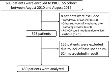
Among 603 patients who enrolled in the PROCESS study, 164 patients were excluded [8 patients did not satisfy inclusion criteria and 156 patients lacked data of baseline serum beta-2-microglobulin (B2MG)] and the remaining 439 patients who had complete clinical, radiologic, and laboratory data enabling their classification according to the three IPI schemes were included in the current study (Figure 1).
The baseline characteristics of the analyzed patients were summarized in Table 2. Overall characteristics of the 439 patients did not deviate from
those of all the patients from the PROCESS cohort. During the median follow-up duration of 55.0 months (95% CI 53.1 - 57.0), 133 patients (30.3%) underwent progression-free survival (PFS) events and 120 patients (27.3%) died. Five-year PFS and OS rates were 66.8% and 70.6%, respectively.
According to the IPI, the proportion of low-risk group was the highest (43%). In the NCCN-IPI, the number of low-intermediate (LI)-risk group was the highest (45%). In the GELTAMO-IPI, most patients were classified into LI-risk group (61%). Patterns of Distributio of patients according to the three IPI were overall similar with those of original NCCN and GELTAMO studies (Figure 2) [8, 9]. The NCCN- and GELTAMO-IPI classified a relatively smaller proportion of patients into the high-risk group (8.9% in NCCN-IPI and 6.8% in GELTAMO-IPI, respectively), compared to the IPI (18.2% in the high-risk group).
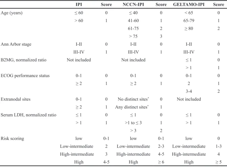
Table 1: Comparison of factors and scoring of IPI, NCCN-IPI, and GELTAMO-IPI
IPI Score NCCN-IPI Score GELTAMO-IPI Score
Age (years) ≤ 60 0 ≤ 40 0 < 65 0
> 60 1 41-60 1 65-79 1
61-75 2 ≥ 80 2
> 75 3
Ann Arbor stage I-II 0 I-II 0 I-II 0
III-IV 1 III-IV 1 III-IV 1
B2MG, normalized ratio Not included Not included ≤ 1 0
> 1 1
ECOG performance status 0-1 0 0-1 0 0-1 0
≥ 2 1 ≥ 2 1 2 1
3-4 2
Extranodal sites 0-1 0 No distinct sites* 0 Not included ≥ 2 1 Any distinct sites* 1
Serum LDH, normalized ratio ≤ 1 0 ≤ 1 0 ≤ 1 0
> 1 1 >1 to ≤ 3 1 > 1 1
> 3 2
Risk scoring low 0-1 low 0-1 low 0
Low-intermediate 2 Low-intermediate 2-3 Low-intermediate 1-3 High-intermediate 3 High-intermediate 4-5 High-intermediate 4
High 4-5 High ≥ 6 High ≥ 5
*Distinct extrnodal sites defined by NCCN-IPI: central nervous system, bone marrow, liver/gastrointestinal tract, or lung IPI, International Prognostic Index; NCCN, National Comprehensive Cancer Network; GELTAMO, Grupo Español de Linfomas/Trasplante Autólogo de Médula Ósea; B2MG, beta-2 microglobulin; ECOG, Eastern Cooperative Oncology Group; LDH, lactate dehydrogenase.
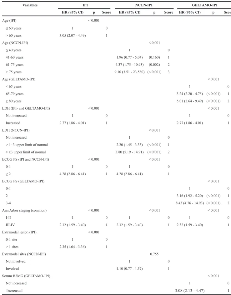
Prognostic significance of individual IPI factor in the IPI, NCCN-IPI, and GELTAMO-IPI
All five factors of the IPI showed a significant difference of OS with hazard ratios (HRs) between 2.27 to 4.10 (Table 3). Enhanced stratification of age (in the and GELTAMO-IPI), serum LDH (in the NCCN-IPI), and performance status (PS; in the GELTAMO-IPI) resulted in more effective risk stratification, except in groups between ≤ 40 vs. 41-60 years of age in the NCCN-IPI (p = 0.175). Involvement of extranodal sites designated by the NCCN-IPI failed to show prognostic significance (p = 0.755). Patients with an increased serum B2MG level showed significantly inferior OS compared to those with not increased B2MG. Ann Arbor staging lost its prognostic significance in the multivariate analyses performed in all three IPIs. Otherwise, most factors maintained an independent prognostic significance (Table 4).
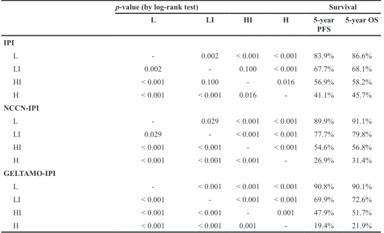
Stratification of patients according to the IPI, NCCN-IPI, and GELTAMO-IPI
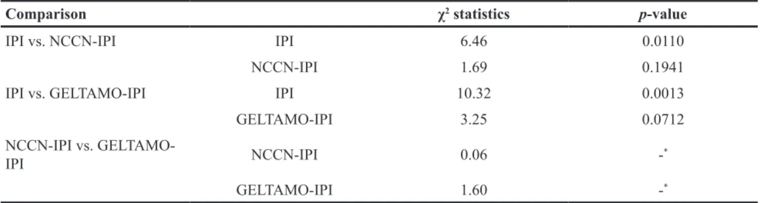
All three IPI schemes effectively separated the analyzed patients into four risk groups according to OS (Table 5 and Figure 3). Estimated 5-year OS of patients classified as high-risk group according to IPI was 45.7%, suggesting that the IPI is limited in the selection of patients who are expected to have poor outcome. In contrast, the 5-year OS of patients stratified as high-risk according to NCCN- and GELTAMO-IPI were 31.4%, and 21.9%, respectively (Table 5). In the reclassification calibration statistic analysis, NCCN- and GELTAMO-IPI showed superior risk prediction (separating patients into high-risk vs. non-high-risk) compared to the IPI (Table 6). Comparison between NCCN- and GELTAMO-IPI was not statistically feasible as patient numbers of high-risk group
by either of two IPIs were small and 23 patients were classified into high-risk by both NCCN- and GELTAMO-IPI.
DISCUSSION
In the present study, the NCCN- and GELTAMO-IPI, the revised versions of the IPI that feature enhanced scoring systems (and the addition of serum B2MG in case of GELTAMO-IPI), showed improved prognostic power to detect patients with dismal prognosis compared to the IPI.
The population we analyzed reflects a real-world clinical practice of DLBCL patients because they were accrued from 27 medical centers of a nation-wide distribution, and our prospective cohort had no specific interventions relevant to patient selection or additional investigative therapy. Our patients had a median age of 60 years (57 years in the NCCN-cohort and 63 years in the BCCA-cohort of the NCCN-IPI study and 60 years in the GELTAMO-IPI study, respectively). Forty eight percent of the patients were > 60 years of age, 57% were males, 51% were LDH >1x ULN, 50% were Ann Arbor stage III or IV, and 12% of the analyzed patients were PS >1, showing that these characteristics had not significantly deviated from the populations in the original studies of the NCCN- and GELTAMO-IPI.
In the present study, involvement of the NCCN-designated extranodal sites had no prognostic significance. The prognostic implication of gastrointestinal tract, one of the designated involved sites, is controversial. Studies have suggested poor survival [11], no association [10], and even favorable outcomes [12]. In a Japanese retrospective study of 1,221 patients, the involvement of the small intestine was an IPI-independent poor prognostic factor, whereas involvement of stomach or colon was not [13]. In addition, the involvement of extranodal sites other than
Table 2: Patient characteristics
Analyzed patients n = 439
n %
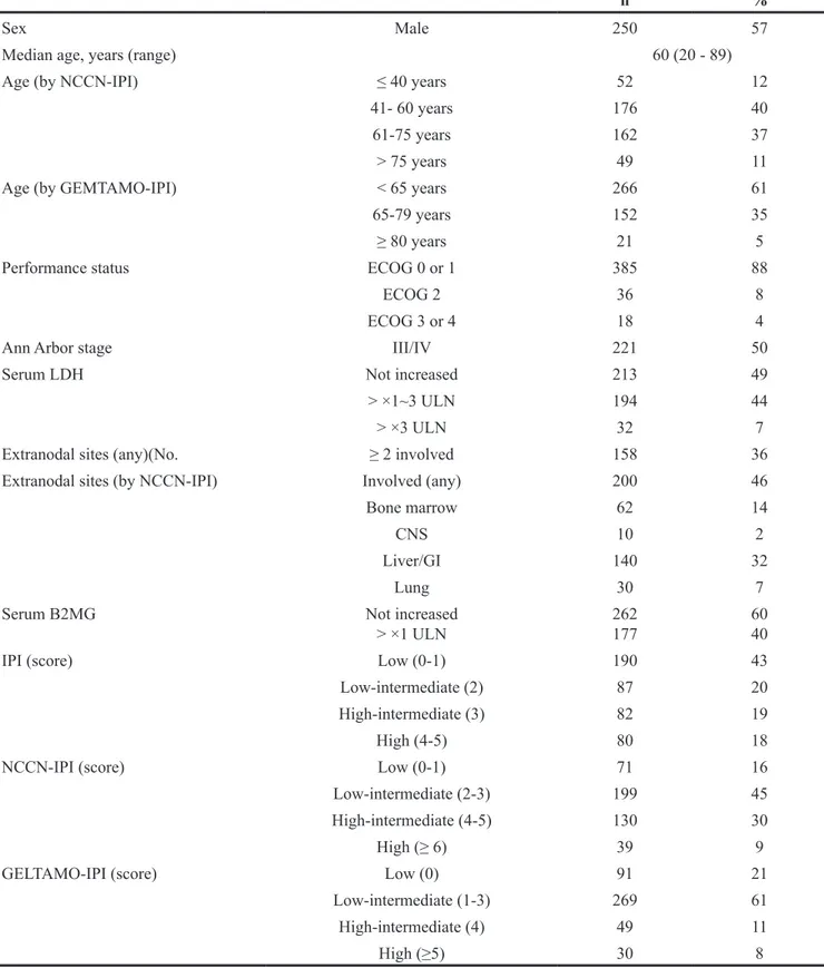
Sex Male 250 57
Median age, years (range) 60 (20 - 89)
Age (by NCCN-IPI) ≤ 40 years 52 12
41- 60 years 176 40
61-75 years 162 37
> 75 years 49 11
Age (by GEMTAMO-IPI) < 65 years 266 61
65-79 years 152 35
≥ 80 years 21 5
Performance status ECOG 0 or 1 385 88
ECOG 2 36 8
ECOG 3 or 4 18 4
Ann Arbor stage III/IV 221 50
Serum LDH Not increased 213 49
> ×1~3 ULN 194 44
> ×3 ULN 32 7
Extranodal sites (any)(No. ≥ 2 involved 158 36
Extranodal sites (by NCCN-IPI) Involved (any) 200 46
Bone marrow 62 14
CNS 10 2
Liver/GI 140 32
Lung 30 7
Serum B2MG Not increased
> ×1 ULN 262177 6040
IPI (score) Low (0-1) 190 43
Low-intermediate (2) 87 20
High-intermediate (3) 82 19
High (4-5) 80 18
NCCN-IPI (score) Low (0-1) 71 16
Low-intermediate (2-3) 199 45
High-intermediate (4-5) 130 30
High (≥ 6) 39 9
GELTAMO-IPI (score) Low (0) 91 21
Low-intermediate (1-3) 269 61
High-intermediate (4) 49 11
High (≥5) 30 8
ECOG, Eastern Cooperative Oncology Group; LDH, Lactate dehydrogenase; IPI, International Prognostic Index; NCCN, National Comprehensive Cancer Network; B2MG, beta-2 microglobulin; GELTAMO, Grupo Español de Linfomas/ Trasplante Autólogo de Médula Ósea.
Table 3: Univariate Cox regression analysis for impacts of variables from 3 IPIs on overall survival
Variables IPI NCCN-IPI GELTAMO-IPI
HR (95% CI) p Score HR (95% CI) p Score HR (95% CI) p Score
Age (IPI) < 0.001 ≤ 60 years 1 0 > 60 years 3.05 (2.07 - 4.49) 1 Age (NCCN-IPI) < 0.001 ≤ 40 years 1 0 41-60 years 1.96 (0.77 - 5.04) (0.160) 1 61-75 years 4.37 (1.75 - 10.93) (0.002) 2 > 75 years 9.10 (3.51 - 23.580) (< 0.001) 3 Age (GELTAMO-IPI) < 0.001 < 65 years 1 0 65-79 years 3.24 (2.20 - 4.75) (< 0.001) 1 ≥ 80 years 5.01 (2.64 - 9.49) (< 0.001) 2
LDH (IPI- and GELTAMO-IPI) < 0.001 < 0.001
Not increased 1 0 1 0
Increased 2.77 (1.86 - 4.01) 1 2.77 (1.86 - 4.01) 1
LDH (NCCN-IPI) < 0.001
Not increased 1 0
> 1~3 upper limit of normal 2.20 (1.45 - 3.33) (< 0.001) 1 > x3 upper limit of normal 8.80 (5.19 - 14.91) (< 0.001) 2 ECOG PS (IPI and NCCN-IPI) < 0.001 < 0.001
0-1 1 0 1 0 ≥ 2 4.28 (2.86 - 6.41) 1 4.28 (2.86 - 6.41) 1 ECOG PS (GELTAMO-IPI) < 0.001 0-1 1 0 2 3.16 (1.92 - 5.20) (< 0.001) 1 3-4 8.43 (4.76 - 14.93) (< 0.001) 2
Ann Arbor staging (common) < 0.001 < 0.001 < 0.001
I-II 1 0 1 0 1 0
III-IV 2.32 (1.59 - 3.40) 1 2.32 (1.59 - 3.40) 1 2.32 (1.59 - 3.40) 1 Extranodal lesion (IPI) < 0.001
0-1 site 1 0
> 1 sites 2.35 (1.64 - 3.36) 1
Extranodal sites (NCCN-IPI) 0.755
Not involved 1 0
Involved 1.10 (0.77 - 1.57) 1
Serum B2MG (GELTAMO-IPI) < 0.001
Not increased 1 0
Increased 3.08 (2.13 - 4.47) 1
NCCN, National Comprehensive Cancer Network; GELTAMO, Grupo Español de Linfomas/Trasplante Autólogo de Médula Ósea; IPI, International Prognostic Index; LDH, lactate dehydrogenase; ECOG, Eastern Cooperative Oncology Group; B2MG, beta-2 microglobulin.
Figure 2: Patient distributions according to risk group in respective three IPIs.
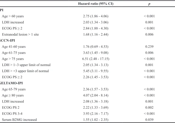
Table 4: Results of multivariate analysis of IPI factors in respective 3 IPIs on overall survival Hazard ratio (95% CI) p
IPI
Age > 60 years 2.75 (1.86 - 4.06) < 0.001
LDH increased 2.03 (1.34 - 3.06) 0.001
ECOG PS ≥ 2 2.84 (1.88 - 4.30) < 0.001
Extranodal lesion > 1 site 1.68 (1.16 - 2.44) 0.006 NCCN-IPI
Age 41-60 years 1.76 (0.69 - 4.53) 0.239
Age 61-75 years 3.63 (1.45 - 9.08) 0.006
Age > 75 years 6.51 (2.48 - 17.15) < 0.001
LDH > 1~3 upper limit of normal 2.05 (1.34 - 3.13) 0.001 LDH > ×3 upper limit of normal 5.45 (3.11 - 9.55) < 0.001
ECOG PS ≥ 2 2.26 (1.45 - 3.53) < 0.001 GELTAMO-IPI Age 65-79 years 2.36 (1.57 - 3.53) < 0.001 Age ≥ 80 years 4.07 (2.04 - 8.14) < 0.001 LDH increased 2.08 (1.36 - 3.18) 0.001 ECOG PS 2 2.22 (1.33 - 3.69) 0.002 ECOG PS 3-4 3.93 (2.16 - 7.17) < 0.001 Serum B2MG increased 1.55 (1.02 - 2.35) 0.039
IPI, International Prognostic Index; LDH, lactate dehydrogenase; ECOG, Eastern Cooperative Oncology Group; NCCN, National Comprehensive Cancer Network; GELTAMO, Grupo Español de Linfomas/Trasplante Autólogo de Médula Ósea; B2MG, beta-2 microglobulin.
the NCCN-defined lesions has been suggested as a poor prognostic indicator, including the genitourinary tract [14] and female reproductive organ [10]. In the validation of the IPI by the GELTAMO group, the NCCN-designated extranodal disease lost its prognostic value in multivariate analysis [9], and therefore it was not included in GELTAMO-IPI. A large-scale retrospective analysis (n = 25,992) using the Surveillance, Epidemiology, and End Results (SEER) database from 2004 to 2009 reported that
sites of extranodal involvement are more prognostic than the number of involved sites [11]. However, a Danish-Canadian study reported that involvement of three or more extranodal sites is independently associated with dismal outcomes [10]. Considering the above results, the prognostic impact of extranodal sites in terms of its number or anatomic location is still an area of debate.
In our study, advanced Ann Arbor staging lost its prognostic significance in multivariate analyses Table 5: Results of risk stratification according to IPI, NCCN-IPI, and GELTAMO-IPI
p-value (by log-rank test) Survival
L LI HI H 5-year PFS 5-year OS IPI L - 0.002 < 0.001 < 0.001 83.9% 86.6% LI 0.002 - 0.100 < 0.001 67.7% 68.1% HI < 0.001 0.100 - 0.016 56.9% 58.2% H < 0.001 < 0.001 0.016 - 41.1% 45.7% NCCN-IPI L - 0.029 < 0.001 < 0.001 89.9% 91.1% LI 0.029 - < 0.001 < 0.001 77.7% 79.8% HI < 0.001 < 0.001 - < 0.001 54.6% 56.8% H < 0.001 < 0.001 < 0.001 - 26.9% 31.4% GELTAMO-IPI L - < 0.001 < 0.001 < 0.001 90.8% 90.1% LI < 0.001 - < 0.001 < 0.001 69.9% 72.6% HI < 0.001 < 0.001 - 0.001 47.9% 51.7% H < 0.001 < 0.001 0.001 - 19.4% 21.9%
IPI, International Prognostic Index; NCCN, National Comprehensive Cancer Network; GELTAMO, Grupo Español de Linfomas/Trasplante Autólogo de Médula Ósea.
performed in each IPI. The prognostic significance of Ann Arbor staging (I/II vs. III/IV) has been reproduced in several studies using large cohorts [8, 9, 13]. However, several lines of evidence suggest that the prognostic role of Ann Arbor staging is at least more limited than other IPI factors in the rituximab era. Ziepert et al. performed a meta-analysis of three large clinical trials [5] and reported that for patients who received rituximab-containing immunochemotherapy, Ann Arbor staging was not prognostic of OS in the MInT trial (p = 0.5217), MegaCHOEP study (p = 0.107), and RICOVER-60 trial (p = 0.061). Moreover, application of positron emission tomography/computed tomography (PET/ CT) in response evaluation may affect the mitigation of prognostic significance of Ann Arbor staging. In the Danish-Canadian study conducted by El-Galaly et al., patients were staged and restaged by PET/CT. The authors reported no significant difference of prognosis among patients with stage I, II, and III, with only stage IV patients displaying an inferior OS. The 3-year OS were 89% [95% confidence interval (CI), 83-95%], 76% (95% CI, 62-90%), 82% (95% CI, 70-94%), and 62% (95% CI, 54-70%) for stage I, II, III, and IV disease, respectively [10]. The authors stated that the increased sensitivity of PET/CT may have upstaged a part of patients, particularly by detecting extranodal sites that would not be found by conventional CT [10]. Our study also integrated PET/ CT for response evaluation. It is noteworthy that as the modality of response evaluation shift from CT to PET/ CT, stage migration may occur, which may attenuate the prognostic significance of Ann Arbor staging.
Recently published studies reproduced the overall satisfactory prognostic stratification of DLBCL patients according to the NCCN-IPI in 100 to 443 DLBCL patients [10, 15–19]. However, in the aforementioned Danish-Canadian study, the NCCN-IPI was suboptimal to identify the high-risk group, showing that 3-year OS of patients with high-risk group was 48% [10]. Therefore, some modification of the NCCN-IPI, such
as integrating other clinical or laboratory factors into the index, was tried to further improve the separation of patients expecting dismal outcomes. The GELTAMO-IPI was developed after a validation study of the NCCN-IPI using 2,156 patients with DLBCL from the archives of 20 hospitals in the GELTAMO network in Spain [9]. In the development of GELTAMO-IPI, enhanced scorings were used in age and PS and involvement of extranodal sites were excluded. Notably, serum B2MG was included as an IPI factor. B2MG is a component of the major histocompatibility complex class I molecule, and it is present on all nucleated cells [20]. Elevated serum B2MG has been used as a prognostic indicator in the International Staging System of multiple myeloma [21] and the Follicular Lymphoma International Prognostic Index-2 of follicular lymphoma [22], and its potential role as a prognostic biomarker was reported in many subtypes of mature lymphoid malignancies [23–26] and lymphoma-associated hemophagocytic lymphohistiocytosis [27]. The mechanism of the relationship of elevated serum B2MG to poor prognosis has been suggested, with B2MG proposed to be an indicator of heavy tumor burden with high cellular turnover rate [28]. However, this remains unclear considering that the elevation of B2MG was independent to serum LDH or Ann Arbor staging in previous studies [9, 29] as well as the present study. Further investigations are required for this issue.
In the present study, we did not integrate any biologic prognostic markers recently defined or suggested by the advance of genomics, molecular biology, or immunology in the field of DLBCL. Cell of origin [30], stromal gene signature or its protein expression [31–33], double hit [34], or co-expression of MYC and BCL2/BCL6 (double expresser) [35] were not analyzed. However, the present aim is to validate and compare IPIs, and the above integrations are beyond the scope of the study. It is limitation of our study that we could not compare the efficiency of selecting high-risk group between NCCN- and GELTAMO-IPI.
Table 6: Results of reclassification calibration statistics
Comparison χ2 statistics p-value
IPI vs. NCCN-IPI IPI 6.46 0.0110
NCCN-IPI 1.69 0.1941
IPI vs. GELTAMO-IPI IPI 10.32 0.0013
GELTAMO-IPI 3.25 0.0712
NCCN-IPI vs.
GELTAMO-IPI NCCN-IPI 0.06 -*
GELTAMO-IPI 1.60 -*
*It was impossible to calculate p-value because two cells have more than 20 subjects in the reclassification table. IPI, International Prognostic Index; NCCN, National Comprehensive Cancer Network; GELTAMO, Grupo Español de Linfomas/Trasplante Autólogo de Médula Ósea.
In conclusion, our study shows that NCCN- and GELTAMO-IPI have a significant advantage in predicting patients with poor prognosis, with 5-year OS rate of approximately 20 to 30%, by using basic clinical information and blood tests that are inexpensive and have a rapid turnaround time. Therefore, when selecting high-risk patients, it would be more reasonable to use NCCN- or GELTAMO-IPI rather than the IPI in clinical practice.
MATERIALS AND METHODS
PatientsAnalyses were conducted with patients enrolled in the PROCESS (Prospective Cohort Study with Risk-Adapted Central Nervous System Evaluation in DLBCL) study from 27 hospitals belonging to the Consortium for Improving Survival of Lymphoma (CISL) in Korea. The original purpose, inclusion and exclusion criteria, and detailed information on the study conduct were as previously described [36]. Briefly, to evaluate risk factors of CNS relapse in patients with DLBCL, adult patients with newly diagnosed DLBCL planning to receive three weekly R-CHOP as a primary treatment were included. Patients with primary CNS DLBCL were excluded. Baseline evaluation of CNS involvement of DLBCL was recommended in any symptomatic patients or with features indicating a high risk for CNS involvement. However, the evaluation was not obligatory and there were no other specific interventions for the treatment of DLBCL. An interim and final response evaluation was conducted using PET/CT. The study started on August 2010 and completed patient enrollment on August 2012. Follow-up data regarding disease status and survival was updated every 6 months, with the latest update performed in February 2017. This study was approved by the institutional review boards of the participating institutions.
IPI, NCCN-IPI, and GELTAMO IPI
Risk groups were classified according to the scores calculated as described in the IPI, NCCN-IPI, and GELTAMO IPI, respectively. For the analysis with serum LDH and B2MG levels, normalized values (ratio to the ULN of each participated institution) were calculated and used.
Statistical analysis
PFS was time from the date of diagnosis to the date of disease progression, relapse, or last follow-up, or death from any cause. OS was defined time from diagnosis to death from any cause. Patient survival was analyzed using the Kaplan-Meier method and compared by log-rank test. Multivariate analyses by backward conditional Cox regression model were conducted with variables that had p < 0.1 in univariate analysis. Values were two-sided
and the significance of statistics was accepted at the level of p < 0.05. To compare the ability to predict high-risk patients between two IPIs, the risk category of each IPI was modified into either a high-risk or a non-high-risk group (patients with low-risk + LI risk + HI risk] defined by respective IPI. A reclassification calibration statistic was used, in which an IPI with smaller statistic (χ2) and larger p-value are considered to have better risk prediction than its counterpart [37].
Author contributions
Conception and study design: JH, SJK; Acquisition of data: MHC, J-AK, J-YK, JSK, DHY, WSL, YRD, HJ Kang, H-SE, YP, J-HW, Y-CM, HJ Kim, JH Kwon, JH Kong, SYO, SL, SHB, D-HY, HJJ, YSK, HJY, SIL, MKK, EKP, JHL, WSK, and CS; Analysis of data and interpretation; JH, SJK, MHC, J-AK, J-YK, JSK, DHY, WSL, YRD, HJ Kang, H-SE, YP, J-HW, Y-CM, HJ Kim, JH Kwon, JH Kong, SYO, SL, SHB, D-HY, HJJ, YSK, HJY, SIL, MKK, EKP, JHL, WSK, and CS; Writing the manuscript: JH; Manuscript review: JH, SJ K, MHC, J-AK, J-YK, JSK, DHY, WSL, YRD, HJ Kang, H-SE, YP, J-HW, Y-CM, HJ Kim, JH Kwon, JH Kong, SYO, SL, SHB, D-HY, HJJ, YSK, HJY, SIL, MKK, EKP, JHL, WSK, and CS.
ACKNOWLEDGMENTS
We appreciate Medical Research Collaborating Center, Seoul National University Hospital, Seoul, Korea for statistical consultation.
CONFLICTS OF INTEREST
The authors declare no conflicts of interest.
FUNDING
There was no specific funding for this study.
REFERENCES
1. International Non-Hodgkin’s Lymphoma Prognostic Factors Project. A predictive model for aggressive non-Hodgkin’s lymphoma. N Engl J Med. 1993; 329: 987-94. https://doi. org/10.1056/NEJM199309303291402.
2. Solal-Celigny P, Roy P, Colombat P, White J, Armitage JO, Arranz-Saez R, Au WY, Bellei M, Brice P, Caballero D, Coiffier B, Conde-Garcia E, Doyen C, et al. Follicular lymphoma international prognostic index. Blood. 2004; 104: 1258-65. https://doi.org/10.1182/blood-2003-12-4434. 3. Gutierrez-Garcia G, Garcia-Herrera A, Cardesa T,
Martinez A, Villamor N, Ghita G, Martinez-Trillos A, Colomo L, Setoain X, Rodriguez S, Gine E, Campo E,
Lopez-Guillermo A. Comparison of four prognostic scores in peripheral T-cell lymphoma. Ann Oncol. 2011; 22: 397-404. https://doi.org/10.1093/annonc/mdq359.
4. Geisler CH, Kolstad A, Laurell A, Raty R, Jerkeman M, Eriksson M, Nordstrom M, Kimby E, Boesen AM, Nilsson-Ehle H, Kuittinen O, Lauritzsen GF, Ralfkiaer E, et al. The Mantle Cell Lymphoma International Prognostic Index (MIPI) is superior to the International Prognostic Index (IPI) in predicting survival following intensive first-line immunochemotherapy and autologous stem cell transplantation (ASCT). Blood. 2010; 115: 1530-3. https:// doi.org/10.1182/blood-2009-08-236570.
5. Ziepert M, Hasenclever D, Kuhnt E, Glass B, Schmitz N, Pfreundschuh M, Loeffler M. Standard International prognostic index remains a valid predictor of outcome for patients with aggressive CD20+ B-cell lymphoma in the rituximab era. J Clin Oncol. 2010; 28: 2373-80. https://doi. org/10.1200/JCO.2009.26.2493.
6. Bari A, Marcheselli L, Sacchi S, Marcheselli R, Pozzi S, Ferri P, Balleari E, Musto P, Neri S, Aloe Spiriti MA, Cox MC. Prognostic models for diffuse large B-cell lymphoma in the rituximab era: a never-ending story. Ann Oncol. 2010; 21: 1486-91. https://doi.org/10.1093/annonc/mdp531. 7. Sehn LH, Berry B, Chhanabhai M, Fitzgerald C, Gill K,
Hoskins P, Klasa R, Savage KJ, Shenkier T, Sutherland J, Gascoyne RD, Connors JM. The revised International Prognostic Index (R-IPI) is a better predictor of outcome than the standard IPI for patients with diffuse large B-cell lymphoma treated with R-CHOP. Blood. 2007; 109: 1857-61. https://doi.org/10.1182/blood-2006-08-038257. 8. Zhou Z, Sehn LH, Rademaker AW, Gordon LI, Lacasce
AS, Crosby-Thompson A, Vanderplas A, Zelenetz AD, Abel GA, Rodriguez MA, Nademanee A, Kaminski MS, Czuczman MS, et al. An enhanced International Prognostic Index (NCCN-IPI) for patients with diffuse large B-cell lymphoma treated in the rituximab era. Blood. 2014; 123: 837-42. https://doi.org/10.1182/blood-2013-09-524108. 9. Montalban C, Diaz-Lopez A, Dlouhy I, Rovira J,
Lopez-Guillermo A, Alonso S, Martin A, Sancho JM, Garcia O, Sanchez JM, Rodriguez M, Novelli S, Salar A, et al. Validation of the NCCN-IPI for diffuse large B-cell lymphoma (DLBCL): the addition of beta2 -microglobulin yields a more accurate GELTAMO-IPI. Br J Haematol. 2017; 176: 918-28. https://doi.org/10.1111/bjh.14489. 10. El-Galaly TC, Villa D, Alzahrani M, Hansen JW, Sehn
LH, Wilson D, de Nully Brown P, Loft A, Iyer V, Johnsen HE, Savage KJ, Connors JM, Hutchings M. Outcome prediction by extranodal involvement, IPI, R-IPI, and NCCN-IPI in the PET/CT and rituximab era: a Danish-Canadian study of 443 patients with diffuse-large B-cell lymphoma. Am J Hematol. 2015; 90: 1041-6. https://doi. org/10.1002/ajh.24169.
11. Castillo JJ, Winer ES, Olszewski AJ. Sites of extranodal involvement are prognostic in patients with diffuse large B-cell lymphoma in the rituximab era: an analysis of the
Surveillance, Epidemiology and End Results database. Am J Hematol. 2014; 89: 310-4. https://doi.org/10.1002/ ajh.23638.
12. Lopez-Guillermo A, Colomo L, Jimenez M, Bosch F, Villamor N, Arenillas L, Muntanola A, Montoto S, Gine E, Colomer D, Bea S, Campo E, Montserrat E. Diffuse large B-cell lymphoma: clinical and biological characterization and outcome according to the nodal or extranodal primary origin. J Clin Oncol. 2005; 23: 2797-804. https://doi. org/10.1200/JCO.2005.07.155.
13. Takahashi H, Tomita N, Yokoyama M, Tsunoda S, Yano T, Murayama K, Hashimoto C, Tamura K, Sato K, Ishigatsubo Y. Prognostic impact of extranodal involvement in diffuse large B-cell lymphoma in the rituximab era. Cancer. 2012; 118: 4166-72. https://doi.org/10.1002/cncr.27381.
14. Lu CS, Chen JH, Huang TC, Wu YY, Chang PY, Dai MS, Chen YC, Ho CL. Diffuse large B-cell lymphoma: sites of extranodal involvement are a stronger prognostic indicator than number of extranodal sites in the rituximab era. Leuk Lymphoma. 2015; 56: 2047-55. https://doi.org/10.3109/10 428194.2014.982636.
15. Hong J, Lee S, Chun G, Jung JY, Park J, Ahn JY, Cho EK, Shin DB, Lee JH. Baseline renal function as a prognostic indicator in patients with newly diagnosed diffuse large B-cell lymphoma. Blood Res. 2016; 51: 113-21. https://doi. org/10.5045/br.2016.51.2.113.
16. Spiegel JY, Cheung MC, Guirguis HR, Buckstein R. Validation of the NCCN-IPI in both de novo and transformed diffuse large B cell lymphoma. Leuk Lymphoma. 2017; 58: 214-7. https://doi.org/10.1080/1042 8194.2016.1179295.
17. Huang CE, Chen YY, Lu CH, Chen PT, Lee KD, Chen CC. Validation of an enhanced International Prognostic Index (NCCN-IPI) in an Asian cohort of patients with diffuse large B cell lymphoma. Ann Hematol. 2015; 94: 1063-5. https://doi.org/10.1007/s00277-014-2293-8.
18. Ochi Y, Kazuma Y, Hiramoto N, Ono Y, Yoshioka S, Yonetani N, Matsushita A, Imai Y, Hashimoto H, Ishikawa T. Utility of a simple prognostic stratification based on platelet counts and serum albumin levels in elderly patients with diffuse large B cell lymphoma. Ann Hematol. 2017; 96: 1-8. https://doi.org/10.1007/ s00277-016-2819-3.
19. Bishton MJ, Hughes S, Richardson F, James E, Bessell E, Sovani V, Ganatra R, Haynes AP, McMillan AK, Fox CP. Delineating outcomes of patients with diffuse large b cell lymphoma using the national comprehensive cancer network-international prognostic index and positron emission tomography-defined remission status; a population-based analysis. Br J Haematol. 2016; 172: 246-54. https://doi.org/10.1111/bjh.13831.
20. Huang WC, Havel JJ, Zhau HE, Qian WP, Lue HW, Chu CY, Nomura T, Chung LW. Beta2-microglobulin signaling blockade inhibited androgen receptor axis and caused apoptosis in human prostate cancer cells. Clin Cancer
Res. 2008; 14: 5341-7. https://doi.org/10.1158/1078-0432. CCR-08-0793.
21. Greipp PR, San Miguel J, Durie BG, Crowley JJ, Barlogie B, Blade J, Boccadoro M, Child JA, Avet-Loiseau H, Kyle RA, Lahuerta JJ, Ludwig H, Morgan G, et al. International staging system for multiple myeloma. J Clin Oncol. 2005; 23: 3412-20. https://doi.org/10.1200/JCO.2005.04.242. 22. Federico M, Bellei M, Marcheselli L, Luminari S,
Lopez-Guillermo A, Vitolo U, Pro B, Pileri S, Pulsoni A, Soubeyran P, Cortelazzo S, Martinelli G, Martelli M, et al. Follicular lymphoma international prognostic index 2: a new prognostic index for follicular lymphoma developed by the international follicular lymphoma prognostic factor project. J Clin Oncol. 2009; 27: 4555-62. https://doi. org/10.1200/JCO.2008.21.3991.
23. Rodriguez J, Conde E, Gutierrez A, Lahuerta JJ, Arranz R, Sureda A, Zuazu J, Fernandez de Sevilla A, Bendandi M, Solano C, Leon A, Varela MR, Caballero MD, et al. The adjusted International Prognostic Index and beta-2-microglobulin predict the outcome after autologous stem cell transplantation in relapsing/refractory peripheral T-cell lymphoma. Haematologica. 2007; 92: 1067-74.
24. Vassilakopoulos TP, Nadali G, Angelopoulou MK, Siakantaris MP, Dimopoulou MN, Kontopidou FN, Karkantaris C, Kokoris SI, Kyrtsonis MC, Tsaftaridis P, Pizzolo G, Pangalis GA. The prognostic significance of beta(2)-microglobulin in patients with Hodgkin’s lymphoma. Haematologica. 2002; 87: 701-8; discussion 8. 25. Yoo C, Yoon DH, Kim S, Huh J, Park CS, Park CJ, Lee
SW, Suh C. Serum beta-2 microglobulin as a prognostic biomarker in patients with mantle cell lymphoma. Hematol Oncol. 2016; 34: 22-7. https://doi.org/10.1002/hon.2188. 26. Yoo C, Yoon DH, Yoon S, Kim S, Huh J, Park CJ, Lee
SW, Suh C. Prognostic impact of beta(2)-microglobulin in patients with non-gastric mucosa-associated lymphoid tissue lymphoma. Leuk Lymphoma. 2015; 56: 688-93. https://doi.org/10.3109/10428194.2014.917640.
27. Jiang T, Ding X, Lu W. The prognostic significance of Beta2 microglobulin in patients with hemophagocytic lymphohistiocytosis. Dis Markers. 2016; 2016: 1523959. https://doi.org/10.1155/2016/1523959.
28. Shi C, Zhu Y, Su Y, Chung LW, Cheng T. Beta2-microglobulin: emerging as a promising cancer therapeutic target. Drug Discov Today. 2009; 14: 25-30. https://doi. org/10.1016/j.drudis.2008.11.001.
29. Seo S, Hong JY, Yoon S, Yoo C, Park JH, Lee JB, Park CS, Huh J, Lee Y, Kim KW, Ryu JS, Kim SJ, Kim WS, et al. Prognostic significance of serum beta-2 microglobulin in patients with diffuse large B-cell lymphoma in the rituximab era. Oncotarget. 2016; 7: 76934-43. https://doi. org/10.18632/oncotarget.12734.
30. Rosenwald A, Wright G, Chan WC, Connors JM, Campo E, Fisher RI, Gascoyne RD, Muller-Hermelink HK, Smeland EB, Giltnane JM, Hurt EM, Zhao H, Averett L, et al. The use of molecular profiling to predict survival after chemotherapy for diffuse large-B-cell lymphoma. N Engl J Med. 2002; 346: 1937-47. https://doi.org/10.1056/ NEJMoa012914.
31. Lenz G, Wright G, Dave SS, Xiao W, Powell J, Zhao H, Xu W, Tan B, Goldschmidt N, Iqbal J, Vose J, Bast M, Fu K, et al. Stromal gene signatures in large-B-cell lymphomas. N Engl J Med. 2008; 359: 2313-23. https://doi.org/10.1056/ NEJMoa0802885.
32. Meyer PN, Fu K, Greiner T, Smith L, Delabie J, Gascoyne R, Ott G, Rosenwald A, Braziel R, Campo E, Vose J, Lenz G, Staudt L, et al. The stromal cell marker SPARC predicts for survival in patients with diffuse large B-cell lymphoma treated with rituximab. Am J Clin Pathol. 2011; 135: 54-61. https://doi.org/10.1309/AJCPJX4BJV9NLQHY.
33. Cardesa-Salzmann TM, Colomo L, Gutierrez G, Chan WC, Weisenburger D, Climent F, Gonzalez-Barca E, Mercadal S, Arenillas L, Serrano S, Tubbs R, Delabie J, Gascoyne RD, et al. High microvessel density determines a poor outcome in patients with diffuse large B-cell lymphoma treated with rituximab plus chemotherapy. Haematologica. 2011; 96: 996-1001. https://doi.org/10.3324/haematol.2010.037408. 34. Johnson NA, Slack GW, Savage KJ, Connors JM,
Ben-Neriah S, Rogic S, Scott DW, Tan KL, Steidl C, Sehn LH, Chan WC, Iqbal J, Meyer PN, et al. Concurrent expression of MYC and BCL2 in diffuse large B-cell lymphoma treated with rituximab plus cyclophosphamide, doxorubicin, vincristine, and prednisone. J Clin Oncol. 2012; 30: 3452-9. https://doi.org/10.1200/JCO.2011.41.0985.
35. Hu S, Xu-Monette ZY, Tzankov A, Green T, Wu L, Balasubramanyam A, Liu WM, Visco C, Li Y, Miranda RN, Montes-Moreno S, Dybkaer K, Chiu A, et al. MYC/BCL2 protein coexpression contributes to the inferior survival of activated B-cell subtype of diffuse large B-cell lymphoma and demonstrates high-risk gene expression signatures: a report from The International DLBCL Rituximab-CHOP Consortium Program. Blood. 2013; 121: 4021-31; quiz 250. https://doi.org/10.1182/blood-2012-10-460063.
36. Kim SJ, Hong JS, Chang MH, Kim JA, Kwak JY, Kim JS, Yoon DH, Lee WS, Do YR, Kang HJ, Eom HS, Park Y, Won JH, et al. Highly elevated serum lactate dehydrogenase is associated with central nervous system relapse in patients with diffuse large B-cell lymphoma: results of a multicenter prospective cohort study. Oncotarget. 2016; 7: 72033-43. https://doi.org/10.18632/oncotarget.12459.
37. Cook NR, Ridker PM. Advances in measuring the effect of individual predictors of cardiovascular risk: the role of reclassification measures. Ann Intern Med. 2009; 150: 795-802.