Ultrasound guided serratus planes block (SPB) which is performed on the interfascial layer between serratus anterior and latissimus dorsi muscle has recently been described as a regional anesthetic technique to provide analgesia to the anterolateral chest wall [1]. It provide analgesia via blockade of the lateral cutaneous branches of the thoracic intercostal nerves that pierce serratus anterior muscles [2]. SPB is technically simple and can be safely performed with ultrasound. Ultrasound guided SPB was introduced for regional anesthesia [1]. Few reports demonstrate the efficacy of SPB for unilateral chest wall pain [3]. Ultrasound guided SPB has some advantage in patients with wide range of chest wall pain, bleeding tendency and cardiovascular disease. In this case report, we report two cases applying ultrasound guided SPB as an alternative technique to avoid side effect of established interventional treatment such as paravertebral block, thoracic epidural block and multi-level intercostal block.
CASE REPORT
1. Case 1
An 162 cm, 50 kg, 83-year-old man was referred to us from department of thoracic surgery for complain of left chest pain. The
Clinical Application of Ultrasound-Guided Serratus Anterior Plane
Block: Two Cases
Sai Ju Seo, Do-Hyeong Kim, Duck Mi Yoon, Kyung Bong Yoon
Department of Anesthesiology and Pain Medicine, Anesthesia and Pain Research Institute, Yonsei University College of Medicine, Seoul, Korea
Ultrasound guided serratus plane block (SPB) which is performed on the interfascial plane between serratus ansterior and latissimus dorsi muscle has recently been described as a regional anesthetic technique to provide analgesia to the anterolateral chest wall. It provides analgesia of hemithorax via blockade of the lateral cutaneous branches of the thoracic intercostal nerves that pierce serratus anterior. In this case report, we applied ultrasound guided SPB to two patients. One was a patient who complained wide range of anterolateral chest pain due to chest wall pain syndrome, and had low platelet due to alcoholic liver cirrhosis. The other was a patient who complained lateral chest wall pain due to herpes zoster, and couldn't discontinue antiplatelet therapy due to recent percutaneous transluminal coronary angioplasty. In both cases, ultrasound guided SPB has some advantages than established thoracic regional block including multilevel intercostal nerve block, thoracic paravertebral block and epidural block. It provides wide range of analgesia with single injection. It is safer and easier to perform by using ultrasound. It could provide analgesia without hemodynamic instability. This technique could be a good alternative to established regional block in patient with wide range of unilateral chest wall pain, cardiovascular disease and bleeding tendency.
Key Words: block, nerve, chronic pain, thoracic wall.
Accepted: December 22, 2014
IJP
International Journal of PainCorrespondence to: Kyung Bong Yoon, Department of Anesthesiology and Pain Medicine, Anesthesia and Pain Research Institute, Yonsei University College of Medicine, 50-1, Yonsei-ro, Seodaemun-gu, Seoul 120-752, Korea. Tel: +82-2-2228-5770, Fax: +82-2-2228-7897, E-mail: kbyoon@ yuhs.ac
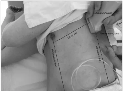
pain site was from T3 to T7 on left anterolateral hemithorax. The pain started two months ago and aggravated within two weeks. He described his pain as stabbing and moving pattern. The pain was aggravated in motionless status and relieved with manual massage on pain site. He had sleep disturbance due to pain and reported his pain score on an 11-point numeric rating scale (NRS, 0 = no pain, 10 = worst pain) as 7 and 8. To evaluate the possibility of heart or lung disease as a pain generator, various examinations including coronary angio-computed tomography (CT), transthoracic echocardiography and chest CT were performed. There was no remarkable finding which can explain the origin of pain. The result of laboratory test including troponin-T and creatine kinase was within normal range. In chest CT, old rib fracture in left anterior arc was found. He stated that the old rib fracture was 20-years-old. On physical examination, the pain was produced by palpation in multiple rib levels of left hemithorax above and below 5th rib level. The patient had already received oral medications including aceclofenac 200 mg, gabapentin 900 mg, tramadol 120 mg and acetaminophen 900 mg a day but these did not provide any relief in pain. Wide range of pain area and his low platelet count as 70,000/μl due to his alcoholic liver cirrhosis history made us consider simpler and safer regional block technique. We decided to perform the ultrasound guided SPB as a diagnostic and therapeutic treatment block. The procedure was performed with ultrasound machine (SonoSite, Bothell, WA, USA) and 13-6 MHz linear probe (SonoSite, Bothell, WA, USA). The patient was placed in lateral decubitus position with the painful side upward. The both arms of the patient were stretched forward side by side. We counted the ribs in the mid-clavicular line using ultrasound, until we identified the fifth rib. After confirming the fifth rib, the ultrasound probe was moved laterally along the rib and placed over mid-axillary line vertically (Fig. 1). On the surface plane, the subcutaneous tissue was identified, in the intermediated plane, the latissimus dorsi and serratus anterior muscle were identified, and in the deep plane, the fifth rib, sixth rib, the pleura and the lungs were identified by ultrasound (Fig. 2A). The serratus plane which is a plane between serratus anterior and lattisimus dorsi can be confirmed easily. As an extra reference point, we used the thoracodorsal artery; this aids in the identification of the plane superficial to the serratus muscle (Fig. 2B). The 25-gauge 50 mm needle (Junglim industry, Jinchungun, Chungbuk, Korea) was introduced following the lower edge of the probe under real time ultrasound using in plane technique form a caudal to a cephalad direction, directing the needle tip between fifth rib and sixth rib and positioning the needle tip between serratus anterior and lattisimus dorsi muscle. Anesthetic boluses of 3 ml were introduced to perform hydrodissection of the interfacial plane (Fig. 2C). After confirmation needle tip on serratus plane, total 20 ml (0.4 ml/kg) of 0.25% ropivacaine was
Fig. 1. Probe was placed over mid-axillary line vertically and needle was introduced from a caudal to a cephalad direction using in-plane technique.
injected. After the procedure, we compressed the injection site gently for five minute for hemostasis. Thirty minute after injection, we demonstrated dermatomal hypoesthesia from T3 to T7 in clavicular line, T2 to T8 in axillary line and T2 to T9 in mid-scapular line. One hour after injection, he reported his pain score as 2 on NRS. He reported pain score as 3 to 4 on NRS with oral medication later on.
2. Case 2
A 165 cm, 61 kg, 67-year-old man referred to us from department of dermatology for chest wall pain due to herpes zoster lasted for three weeks. The pain area was lateral chest wall in the left T4 dermatome. He described his pain as stabbing and complained sleep disturbance due to pain. He reported his pain score on NRS as 8. In physical examination, allodynia to touch and hypoesthesia was found. The patient had already received oral pain medications including pregabalin 300 mg, ibuprofen 600 mg, codein 30 mg and acetaminophen 750 mg a day. We performed paravertebral block on left T4 level with 0.5% lidocaine 10 ml. After paravertebral block, he reported his pain score decreased to 3 on NRS. The block was performed with 2 week intervals, total 3 times and the pain score was between 1 and 2 on NRS with pregabalin 150 mg later on. After three months, he revisited our pain clinic due to recurrence of pain on lateral chest wall. His pain score was 9 on NRS. The pain was lasted 1 week and was not relieved by pregabalin 150 mg and tramadol 200 mg. Due to his recent history of coronary angioplasty just 4 weeks ago, he was on dual antiplatelet therapy including aspirin and clopidogrel. The result of laboratory test including platelet count and coagulation function test was within normal range. But he could not discontinue anti-coagulants. Considering the history of cardiovascular disease and the bleeding tendency due to anti-coagulation, we decided to perform ultrasound-guided SPB. The procedure was carried out in using the same technique as in case 1. At the 4th rib level, total 25 ml (0.4 ml/kg) of 0.25% ropivacaine was injected on the serratus plane.
Thirty minute after injection, the patient demonstrated dermatomal hypoesthesia from T2 to T7 in mid-clavicular line, T2 to T9 in mid-axillary line and T2 to T9 in mid-scapular line. One hour after injection, he reported his pain score as 2 on NRS. His blood pressure and heart rate were monitored every 10 minutes and no hemodynamic deterioration was founded during one hour. He reported pain score as 3 on NRS with pregabalin 150 mg later on.
DISSCUSSION
We used ultrasound-guided SPB in two cases. In first case, the patient reported unexplained chest pain. After exclusion of cardiac
A B C
Fig. 2. Sonographic images during serratus plane block. (A) Short axis view, dotted line shows serratus plane. (B) Thoracodorsal artery in serratus plane. (C) Hydrodissection of serratus plane. Arrows shows needle in plane. LD: latissimusdorsi muscle, SA: serratus anterior muscle, P: pleura, R5: fifth rib, R6: sixth rib, TD: thoracodorsal artery, D: infused local anesthetic, N: needle.
and pulmonary problem, the pain of musculoskeletal origin could be considered [4]. Due to wide range of pain and the bleeding tendency, we decided to use ultrasound-guided SPB as a treatment and a diagnostic tool. Considering the result of physical examination and the effect of block, the chest wall syndrome was strongly suspected. In second case, the patient suffered with chest wall pain due to herpes zoster. In order to relieve pain from postherpetic neuralgia, various treatments, including pharmacologic and interventional methods such as paravertebral block have been used [5]. In last visit, due to recent history of percutaneous transluminal coronary angioplasty with stent insertion, he could not discontinue anticoagulants. Considering the necessity of hemodynamic stability and the risk of bleeding, we made a decision to perform ultrasound-guided SPB. The main dermatome of pain was T4 dermatome, but the overall pain area was more diffuse. So we did not reduce volume of ropivacaine mixture. There was no hemodynamic deterioration within one hour.
Ultrasound-guided SPB has some advantages than established blind regional block. Ultrasound-guided SPB provides wide range of analgesia with single injection. Anatomically SPB blocks the lateral cutaneous branches which are derived from the intercostal nerves. They pierce the external intercostal muscle and serratus anterior muscle, and divided into anterior and posterior branch. The anterior branches supply the forepart of the chest, and posterior branches supply the skin over the scapula and latissimus dorsi. In mid-axillary line, the lateral cutaneous branches of intercostal nerves pass through serratus anterior plane, and blockade of these branches provides analgesia to broad area of hemithorax. Single injection technique can reduce the discomfort of patient and potential side effect which can be produced by multiple intercostal block and paravertebral block [6-8]. Multiple intercostal injections are painful, time consuming and associated with a incidence of pneumothorax of up to 5.6% [7]. The chance of pneumothorax and intravascular injection is increased by the number of injection [9]. The needle placement of SPB was shallower than that of established block and performed under real-time ultra-sound so that the risk of complication such as pneumothorax can be reduced. In patient with bleeding tendency, paravertebral block and thoracic epidural block could be dangerous due to possibility of hematoma [10,11]. The hematoma of central neuraxial structure can make consecutive neurologic sequelae. The serratus plane is a easily identified with ultrasound and more safer space to inject than paravertebral space or epidural space. Also ultrasound-guided block with Doppler function can reduce potential risk of hematoma and intravascular injection [12]. Thoracic paravertebral and epidural block produces somatic and sympathetic nerve blockade, including the posterior primary ramus, in multiple contiguous thoracic dermatomes and makes hemodynamic deterioration. SPB provide more stable hemodynamic status than established blocks because it blocks only somatic nerve, lateral branch of intercostal nerves at the peripheral level [19]. Ultrasound guided SPB could be another good option in patient with cardiovascular disease. There has been an almost complete absence of evidence-baced information about these advantages of SPB since it was newly introduced. Therefore, more research is needed on SPB.
Technically, there is two way to perform ultrasound guided SPB block in the article by Blanco [1]. The injection of local anesthetics superficial to serratus anterior muscle is one and deep underneath is another. In our case, we used only the superficial technique and called it SPB. The Deep technique was first described by Fajardo et al in 2012 [13-15]. This technique blocks the cutaneous branches of the intercostal nerve, injecting local anasthetic between serratus anterior and external intercostal muscle in the mid axillary line. This technique has been called Serratus-intercostal plane block (SIPB). SIPB offers several advantages over SPB. The serratus-intercostal plane is sliding space which is poorly distensible and respiratory movements allow the local anesthetics to be dispersed along the space. The location of the long thoracic nerve which is a pure motor to serratus anterior muscle and which may be injured, causing winged scapula syndrome, is in serratus plane. SPB may produce temporary palsy of the long thoracic nerve. Nevertheless we recommend the superficial technique, SPB. The superficial technique is less dangerous than deep technique because the needle enters shallower and needle approach of SPB is more easier to most of physician. Both technique revealed effective, but superficial technique known to be provide wider and longer analgesia [1]. And the motor paralysis, winged scapula syndrome can be avoid by using appropriate concentration of ropivacaine [16]. In our case, 0.25% ropivscaine did not make any complication.
The novel technique recommended patient placed in the supine position [1]. In our cases, the patients could not take a supine position due to pain. We modified this technique with positioning patients in the lateral position with pain-site up. This lateral position provides more comfortable feeling to the patient and makes physicians to perform the block easier. In our case, after the procedure, we scanned serratus plane by moving the US probe horizontally and vertically and identified distribution of local anesthetics. Local anesthetics were distributed posteriorly and the range of analgesia was also similar. The shape of lattisimus dorsi muscle which is the larger, flat, dorsolateral muscle on the trunk might have affected the distribution of local anesthetics. The lattisimus dorsi muscle is inserted floor of intertubercular groove of the humerus. The abduction of arm can stretch this muscle and the positioning of arm can affect the distribution of local anesthetics. In our case, the lateral position with stretching arms forward did not interrupt the range of analgesia. There is need of further study to investigate the range of blockade according to the position.
A threshold for CNS toxicity is apparent at a mean free plasma concentration of approximately 2.2 μg/ml for ropivacaine [17]. Plasma ropivacaine levels have also been studied in context of other common regional techniques with a low incidence of clinically important toxicity [18]. The transversus abdominis plane block (TAPB) is similar interfacial block which is actively studied for a long time. The mean peak total ropivacaine concentration occurred 30 min post-injection was 1.87 ± 0.78 μg/ml in ultrasound-guided subcostal TAPB with 0.45% ropivacaine at 3 mg/kg and 2.54 ± 0.75 μg/ml in ultrasound-guided bilateral TAPB with 3 ml/kg of ropivacaine diluted to 40 ml [18,19]. In our cases, we used total 0.4 ml/kg of 0.25% ropivacaine which is same as dose of 1.05 mg/ kg and no side-effect due to systemic toxicity was reported. We carefully expected this dose of ropivacaine could be safe clinically. The risk of local anaesthetic toxicity after a SPB is probably lower than for most alternative regional anaesthetic technique, because a smaller dose of local anaesthetic is injected under ultrasound guidance into a less vascular area [20]. However, the assumption remains to be proven. There is need for further research to establish a dose of local anesthetics.
In these cases, chest pain of broad area, cardiovascular disease and bleeding tendency made us to consider ultrasound-guided SPB. It provides wide range of analgesia with single injection. It is easier and safer to perform. It could provide analgesia without hemodynamic instability. However, unlike established regional block which is already studied a lot, ultrasound-guided SPB is required more research. Nevertheless ultrasound guided SPB could be a good alternative to established thoracic regional block including paravertebral block, thoracic epidural block and multi-level intercostal block in the future.
REFERENCES
1. Blanco R, Parras T, McDonnell JG, Prats Galino A: Serratus plane block: a novel ultrasound-guided thoracic wall nerve block. Anaesthesia 2013; 68: 1107-13.
2. Davies F, Gladstone RJ, Stibbe EP: The anatomy of the intercostal nerves. J anat. 1932; 66: 323-33.
3. Kunhabdulla NP, Gaur A, Gautam SK, Gupta R, Agarwal A: Serratus anterior plane block for multiple rib fractures. Pain Physician 2014; 17: E553-5.
4. Cayley WE: Diagnosing the cause of chest pain. Am fam physician 2005; 72: 2012-21.
5. Nahm FS, Kim SH, Kim HS, Shin JW, Yoo SH, Yoon MH, et al: Survey on the treatment of postherpetic neuralgia in korea; multicenter study of 1,414 patients. Korean J Pain. 2013; 26: 21-6.
6. Moore DC: Intercostal nerve block. 1963. Int Anesthesiol Clin. 1998; 36: 29-41.
7. Shanti CM, Carlin AM, Tyburski JG: Incidence of pneumothorax from intercostal nerve block for analgesia in rib fractures. J Trauma. 2001; 51: 536-9.
8. Park JS, Kim YH, Jeong SA, Moon DE: Ultrasound-guided aspiration of the iatrogenic pneumothorax caused by paravertebral block -acase report. Korean J Pain.2012; 25: 33-7.
9. Ho AM, Karmakar MK, Critchley LA: Acute pain management of patients with multiple fractured ribs: a focus on regional techniques. Curr Opin Crit Care. 2011; 17: 323-7.
10. van Veen JJ, Nokes TJ, Makris M: The risk of spinal haematoma following neuraxial anaesthesia or lumbar puncture in thrombocytopenic individuals. Br J Haematol. 2010; 148: 15-25.
11. Roberts HR, Monroe DM, Escobar MA: Current concepts of hemostasis: implications for therapy. Anesthesiology 2004; 100: 722-30.
12. Warman P, Nicholls B: Ultrasound-guided nerve blocks: efficacy and safety. Best Pract Res Clin Anaesthesiol. 2009; 23: 313-26. 13. Fajardo M, García FJ, López S, Diéguez P, Alfaro P: Analgesic combined lateral and anterior cunaneous branches of the intercostal
nerves ultrasound block in ambulatory breast surgery. Cirugia Mayor Ambulatoria 2012; 17: 95-104.
14. Fajardo M, López S, Diéguez P, Alfaro P, García FJ: A new ultrasound-guided cutaneous intercostal branches nerve block for analgesia after non-reconstructive breast surgery. Cirugia Mayor Ambulatoria 2013; 18: 3-6.
15. López-Matamala B, Fajardo M, Estébanez-Montiel B, Blancas R, Alfaro P, Chana M: A new thoracic interfascial plane block as anesthesia for difficult weaning due to ribcage pain in critically ill patients. Med Intensiva. 2014; 38: 463-5.
16. Yao J, Zeng Z, Jiao ZH, Wang AZ, Wang J, Yu A: Optimal effective concentration of ropivacaine for postoperative analgesia by single-shot femoral-sciatic nerve block in outpatient knee arthroscopy. J Int Med Res. 2013; 41: 395-403.
17. Knudsen K, Beckman Suurküla M, Blomberg S, Sjövall J, Edvardsson N: Central nervous and cardiovascular effects of i.v. infusions of ropivacaine, bupivacaine and placebo in volunteers. Br J Anaesth. 1997; 78: 507-14.
18. Griffths JD, Barron FA, Grant S, Bjorksten AR, Hebbard P, Royse CF: Plasma ropivacaine concentrations after ultrasound-guided transversus abdominis plane block. Br J Anaesth. 2010; 105: 853-6.
19. Toju K, Shiraishi K, Hakozaki T, Isosu T, Murakawa M: Plasma ropivacaine concentration following ultrasound-guided subcostal transversu sabdominis plan block in adults [published online ahead of print June 17, 2014. J Anesth.doi:10.1007.
20. Tighe SQ, Karmakar MK: Serratus plane block: do we need to learn another technique for thoracic wall blockade? Aneasthesia 2013; 78: 1103-6.