Procedia Engineering 168 ( 2016 ) 1450 – 1453
1877-7058 © 2016 The Authors. Published by Elsevier Ltd. This is an open access article under the CC BY-NC-ND license (http://creativecommons.org/licenses/by-nc-nd/4.0/).
Peer-review under responsibility of the organizing committee of the 30th Eurosensors Conference doi: 10.1016/j.proeng.2016.11.414
ScienceDirect
Available online at www.sciencedirect.com
30th Eurosensors Conference, EUROSENSORS 2016
Toxic Effect Monitoring by Analyzing Swimming Motions of
Microbial Cells Confined in Microfluidic Chip with Micro-Trench
Flow Injection
Kazunari Ozasa
a,*, June Won
b, Simon Song
b, Mizuo Maeda
aaBioengineering Lab., RIKEN, 2-1 Hirosawa, Wako, Saitama 351-0198, Japan
bMechanical Convergence Engineering Department, Hanyang University, 17 Haendang-dong, Seongdong-gu, Seoul 133-791, South Korea
Abstract
We improved our previous micro-device containing motile micro-algae (Euglena gracilis) cells for toxicity monitoring, by introducing micro-trenches between a micro-aquarium and two surrounding channels. Target test solution was introduced from the bypass channel to the micro-aquarium through micro-trenches, with a pulse injection and continuous diffusion. Although the chemical gradient formed in the micro-aquarium was not permanently sustainable but only temporal for a couple of 10 min, the device was simple, easy-to-use, and cost-effective to evaluate the toxicity of the test solution. The cells showed their chemotaxis to the temporal chemical gradients, resulting in the deviated distribution of the cells in the micro-aquarium, whereas metabolic disturbing effects of toxic substances induced the unusual motion of the cells.
© 2016 The Authors. Published by Elsevier Ltd.
Peer-review under responsibility of the organizing committee of the 30th Eurosensors Conference. Keywords: Euglena gracilis; Chemotaxis; Microfluidic chip; Toxicity sensing
1. Introduction
Motile microorganisms show unique and useful characteristics in sensing environmental chemicals and changing their locomotive direction according to whether the chemical is nutritious or toxic. Such a chemotaxis can be used to develop a new type of sensor/monitor device that measures not the quantity of a specified chemical substance but evaluates the metabolic effect of the substance on living cells [1]. One important advantage of such a device is that it
* Corresponding author. Tel.: +81-48-462-1111; fax: +81-48-462-4658. E-mail address: [email protected]
© 2016 The Authors. Published by Elsevier Ltd. This is an open access article under the CC BY-NC-ND license (http://creativecommons.org/licenses/by-nc-nd/4.0/).
1451 Kazunari Ozasa et al. / Procedia Engineering 168 ( 2016 ) 1450 – 1453
can evaluate the toxicity of a test solution on living cells without identifying the chemicals in the solution. Although modern sensing technology can easily detect and measure the quantity of specified chemicals with an ultra high sensitivity, an unidentified chemical in a test solution can be hardly evaluated especially from the viewpoint of toxicity. Therefore, the toxicity evaluation of unidentified chemicals in a solution is highly desirable and useful. Additionally, the same sensing/monitoring technique using motile microorganisms can be applied for screening the side effect of newly developed drags or even cancer screening [2].
The motivation of our study is to utilize the chemotaxis of microorganisms with microfluidic technology to realize a compact, disposable, easy handling, and fast toxicity monitoring device, on the concepts we described above. In this study, we examine the effect of micro-trenches introduced in our previous microchip structure [1,3]. The trench makes it possible to introduce larger molecules into the aquarium, where the motile micro-algae cells were confined. A small amount of test solution was introduced through the micro-trenches, and the temporal chemical gradient was formed in the micro-aquarium, which induced the chemotactic or toxicity-influenced behavior of the cells in a short time scale of minutes.
Nomenclature
TM trace momentum, which represents the product of cell number and swimming speed
BW blue water, colored by a blue dye of brilliant blue to visualize the water injection through micro-trenches
2. Experiments
The basic design of our device without micro-trenches can be found in our previous reports [1,3], having two bypass micro-channels running aside of a micro-aquarium (a closed micro-chamber). The bypass channels of 200-μm-width were isolated from the micro-aquarium of 2.5-mm diameter by polydimethylsiloxane (PDMS) wall of 150-μm-width, and only small molecules can diffuse through porous PDMS wall from the bypass channels to the micro-aquarium. The depth of the structure was 150 μm. The issue of the previous design was that larger molecules cannot diffuse into the micro-aquarium through PDMS wall.
Figure 1(a) shows the overview of the new microchip, where 14 micro-trenches newly introduce can be seen on the wall between the bypass channels and the micro-aquarium as thin lines. The configuration of the micro-trenches is given in Fig. 1(b). The height of the micro-trenches was 5 μm, smaller than the size of Euglena cells of approximately 50–80 μm long and 10–30 μm in diameter (spindle shape). Therefore, the cells confined in the micro-aquarium cannot get into the micro-trenches.
The microorganism confined in the micro-aquarium was Euglena gracilis Z-strain, easy to observe with an optical microscope without fluorescence dying. Typically 100–400 cells were confined in the micro-aquarium. They were swimming randomly and uniformly with a typical swimming speed of 100–400 μm/s, when no chemicals were supplied from the bypass channels. The spoke structures in the micro-aquarium were to let the cells swim in radial directions rather than circumferential direction.
Fig. 1. (a, left) Secondary electron microscopic image of PDMS microchip with micro-trenches fabricated in this study. (b, right) Cross-sectional drawing of the bypass channel, micro-trench, and micro-aquarium. Verticals and horizontals are not in scale.
1452 Kazunari Ozasa et al. / Procedia Engineering 168 ( 2016 ) 1450 – 1453
The movements and distribution of the cells were observed with an optical microscope, and the captured images were processed to generate swimming trace images and evaluate the swimming activity in a certain area in the micro-aquarium as trace momentum (TM) real time, as previously reported [1,3]. We used a blue dye of brilliant blue to color the test solution for visualization.
3. Results and Discussions
3.1. Transient Gradient in Micro-Aquarium
Figure 2 shows the sequence of real images observed when blue-colored water (BW) and pure water were supplied to the two bypass channels individually with a same flow rate of 0.1 mL/h. When BW started running in the lower bypass channel, a small amount of BW was injected into the micro-aquarium through the micro-trenches, as shown in Fig. 2(a). The BW supply through the micro-trenches continued during BW flow in the bypass channel. The BW gradually spread in the micro-aquarium, and a concentration gradient was generated temporally as seen in Fig. 2(b). The gradient was flattened in 30–40 min, resulting in a uniform blue color in the whole area of the micro-aquarium as shown in Fig. 2(c). The uniform blue color remained in the micro-micro-aquarium even after switching BW back to pure water as long as several hours.
Fig. 2. Brilliant blue water injection and diffusion through micro-trenches. (a, left) 4 min, (b, center) 16 min, and (c, right) 48 min after BW flow in the lower bypass channel. BW was switched back to pure water at the timing of 31 min.
We consider that the introduction of test solution into the micro-aquarium was firstly due to the pulse-like injection caused by a pressure surge and subsequently due to the diffusion through the micro-trenches. The pressure surge was generated when switching of pure water flow to BW flow. The amount of BW introduced into the micro-aquarium varied more or less run-by-run, reflecting the less reproducibility of the surge pressure.
3.2. Effect of Nicotinamide
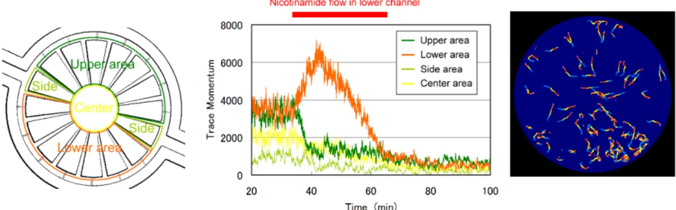
We examined the effect of nicotinamide [4] on E. graciis cells by flowing 100-mM nicotinamide solution to the lower bypass channel with a flow rate of 0.1 mL/h. Figure 3 shows the four area sectioning for TM evaluation and the temporal evolution of TM in each area. The solution was colored with brilliant blue dye for visualization. The cells were spreading in the whole area of the micro-aquarium before the flow of nicotinamide. When a small amount of the nicotinamide/BW solution was injected in the micro-aquarium, the cells moved toward the lower bypass channel, probably because they attracted to organic brilliant blue dye. The ratio of TMlower/(TMlower + TMupper)
reached 0.85 at 10 min after the injection of nicotiamide/BW. After 10 min, TMlower started decreasing together with
TMs for the other areas, indicating that the cell activity in the micro-aquarium was decayed as many cells ceased swimming. At 80 min, the total TM is decreasing, down to 20% of the original value before the injection, revealing that nicotinamide has a toxic effect, and the 80% cells cease swimming.
We previously investigated on the toxic effects of ethanol and H2O2 [3,5], i.e., small-molecule chemicals
permeable through PDMS. The E. gracilis cells exposed to ethanol showed a simple negative chemotaxis, whereas those exposed to H2O2 lost the locomotive control of swimming, fell into continuous on-site rotation, and finally
1453 Kazunari Ozasa et al. / Procedia Engineering 168 ( 2016 ) 1450 – 1453
continuous on-site rotation, but simply ceased swimming mainly at the edge part of the lower area. The cells seemed not trying to escape from the area with higher nicotinamide concentration, nor loosing locomotive control of swimming,as shown in Fig. 3(c). The observation suggests that nicotinamide has a different type of toxic effect on the cells, compared with ethanol or H2O2.
Fig. 3. (a, left) Four areas for TM measurement, upper and lower area, side area, and center area. (b, center) TM change observed when100-mM nicotinamide solution was supplied in the lower bypass channel. (c, right) Swimming traces observed at 55 min. Traces were colored in the order blue, yellow, and red to indicate swimming direction.
4. Conclusion
This study showed a high feasibility of trench structure in the combination of cell-confining micro-aquarium and bypass micro-channels, in terms of environmental toxicity monitoring and toxic effect typing. The temporal concentration gradient of test chemicals formed in the micro-aquarium was sustained for a couple of 10 min, enough to observed the chemotactic responses of the cells. The Euglena cells ceased swimming by nicotinamide exposure, showing that the chemical has a unique toxic effect to the cells, differs from ethanol or H2O2.
Acknowledgements
The authors would like to thank Dr. Kengo Suzuki and Ms. Ayaka Nakashima at Euglena Co. Ltd. (http://euglena.jp/english) for supplying the Euglena cells and culture medium, together with information on cell culture. This work was supported by the Ministry of Education, Science, Sports and Culture, Grant-in-Aid for Scientific Research (B) [grant number 25280092 (2013-2016)], and also partially by a National Research Foundation of Korea (NRF) grant funded by the Ministry of Education, Science and Technology [2013R1A2A2A01014234, 2012R1A6A1029029].
References
[1] K.Ozasa, J.Lee, S.Song, M.Hara, M.Maeda, Gas/liquid sensing via chemotaxis of Euglena cells confined in an isolated micro-aquarium, LabChip, 13 (2013) 4033–4039.
[2] T.Hirotsu, H.Sonoda, T.Uozumi, Y.Shinden, K.Mimori, Y.Maehara, N.Ueda, M.Hamakawa, A Highly Accurate Inclusive Cancer Screening Test Using Caenorhabditis elegans Scent Detectio, PLOS ONE, 10 (2015) e0118699.
[3] K.Ozasa, J.Lee, S.Song, M.Hara, M.Maeda, Gas/liquid sensing via chemotaxis of Euglena cells confined in an isolated micro-aquarium, LabChip, 13 (2013) 4033–4039.
[4] G.S.Byng, R.J.Whitaker, C.L.Shapiro, R.A.Jensen, The aromatic amino acid pathway branches at L-arogenate in Euglena gracilis, Mol. Cell Biol., 1(1981) 426–38.
[5] K.Ozasa, J.Lee, S.Song, M.Maeda, Real-time analysis of chemotactic motion of Euglena cells confined in a microchip toxicity sensor, Key Eng. Mater., 644 (2015) 185–188.