저작자표시-비영리-변경금지 2.0 대한민국 이용자는 아래의 조건을 따르는 경우에 한하여 자유롭게 l 이 저작물을 복제, 배포, 전송, 전시, 공연 및 방송할 수 있습니다. 다음과 같은 조건을 따라야 합니다: l 귀하는, 이 저작물의 재이용이나 배포의 경우, 이 저작물에 적용된 이용허락조건 을 명확하게 나타내어야 합니다. l 저작권자로부터 별도의 허가를 받으면 이러한 조건들은 적용되지 않습니다. 저작권법에 따른 이용자의 권리는 위의 내용에 의하여 영향을 받지 않습니다. 이것은 이용허락규약(Legal Code)을 이해하기 쉽게 요약한 것입니다. Disclaimer 저작자표시. 귀하는 원저작자를 표시하여야 합니다. 비영리. 귀하는 이 저작물을 영리 목적으로 이용할 수 없습니다. 변경금지. 귀하는 이 저작물을 개작, 변형 또는 가공할 수 없습니다.
A Thesis
For the Degree of Master of Science in Medicine
Galangin (3,5,7-trihydroxyflavone)
attenuates apoptosis induced by
ultraviolet B radiation in human
keratinocytes
Graduate School, Jeju National University
Department of Medicine
Madduma Hewage Susara Ruwan Kumara
인간 각질형성세포에서 자외선 B 조사로
유도되는 아포토시스에 대한 Galangin
(3,5,7-trihydroxyflavone)의 경감작용
지도교수
현 진 원
마두마
헤와게 수사라 루완 쿠마라
이
논문을 의학 석사학위 논문으로 제출함
2015년 12월
마두마
헤와게 수사라 루완 쿠마라의 의학 석사학위
논문을
인준함
심사위원장
………...
위
원………...
위
원………...
제주대학교
대학원
2015년 12월
Galangin (3,5,7-trihydroxyflavone) attenuates
apoptosis induced by ultraviolet B radiation in human
keratinocytes
Madduma Hewage Susara Ruwan Kumara
(Supervised by professor Jin Won Hyun)
A thesis submitted for the partial fulfilment of the requirement for the
degree of Master of Science in Medicine
December, 2015
This thesis has been examined and approved.
………
………
………
……….
Date
Department of Medicine
GRADUATE SCHOOL
I
Abstract
Background: Reactive oxygen species (ROS) is the root cause for most of the skin damages caused by
ultraviolet B (UVB) radiation including skin cancer and photoaging of the dermis and epidermis. Phytochemicals are known to act as antioxidants against UVB-induced oxidative stress. Galangin (3,5,7-trihydroxyflavone) is a flavonol abundantly found in plants such as Alpinia officinarum and Helichrysum aureonitens which has been proven for antiviral and anticancer properties. This study was focused to investigate the cytoprotective effects of the flavonol galangin against UVB-induced oxidative damage and subsequent induction of apoptosis in human keratinocytes.
Methods: Human skin keratinocytes (HaCaT) were cultured in RPMI 1640 containing 10% FBS. Cell
viability, ability of galangin to scavenge ROS were analyzed. Protective nature of galangin against UVB induced cellular macromolecule damage was evaluated via detecting DNA strand breaks, lipid peroxidation and protein carbonylation. Formation of apoptotic bodies, mitochondrial membrane depolarization and expression of pro-apoptotic as well as anti-apoptotic proteins were assessed to determine the degree of apoptosis.
Results: Galangin efficiently scavenged free radicals and reduced UVB-induced damage to cellular
macromolecules, such as DNA, lipids, and proteins. Furthermore, galangin rescued cells undergoing apoptosis induced by UVB radiation via recovering mitochondrial polarization and down-regulating pro-apoptotic proteins and up-regulating anti-apoptotic proteins.
Conclusion: These findings proved that galangin shields human keratinocytes against UVB
II
Content
Abstract……… I Contents……… II List of Figures……….. IV1.
Introduction……….………... 12.
Materials and Methods……….………... 32-1. Reagents
2-2. Cell culture and UVB exposure 2-3. Cell viability assay
2-4. Detection of DPPH radicals 2-5. Detection of hydroxyl radicals 2-6. Detection of intracellular ROS
2-7. Single cell gel electrophoresis (Comet assay) 2-8. 8-Isoprostane assay
2-9. Protein carbonylation assay
2-10. Nuclear staining with Hoechst 33342
2-11. Analysis of mitochondrial membrane potential (Δψm) 2-12. Western blot analysis
2-13. Statistical analysis
3.
Results……….. 8III
5.
Reference………. 246.
Abstract in Korean………. 30IV
List of Figures
Figure 1. Chemical structure of galangin………. 2
Figure 2. Galangin scavenges reactive oxygen species (ROS)……… 11
Figure 3. Galangin reduces UVB-induced cellular macromolecule damage…...…… 15
Figure 4. Galangin elevates the viability of UVB-irradiated cells and
1
1. Introduction
Solar radiation can be classified into three main types based on the wavelength which are ultraviolet (UV), visible light, and infrared (Lyons and O'Brien, 2002). Of these, UV radiation is the most responsible for photoaging and skin cancer. UV radiation is subcategorized into UVA (320-400 nm), UVB (280-320 nm), and UVC (200-280 nm). The ozone layer reflects UVC radiation, allowing only UVA and UVB to reach the earth’s surface. UVB radiation is particularly absorbed by human skin and causes erythema, burns, immune suppression, and skin cancer (Park et al., 2013). Though UVA contributes for the majority of UV radiation that reaches the earth’s surface and can get across the skin deeper than UVB, it is less carcinogenic and results aging and wrinkling of the skin (Yoshikawa et al., 1990; Donawho et al., 1996; Matsumura and Ananthaswamy, 2004).
UVB directly or indirectly damages skin cells via formation of cyclobutane pyrimidine dimers (CPDs) and pyrimidine-pyrimidone(6-4) photoproducts or via generation of reactive oxygen species (ROS), such as hydroxyl radicals (•OH), superoxide anions (•O2-), hydrogen peroxide (H2O2), and singlet oxygen (1O2) (Cunningham et al., 1985; Hattori et al., 1996; Meeran et al., 2008). UVB-exposure generates ROS by activating specific small molecules such as riboflavin, tryptophan, and porphyrin in the cells (Ikehata and Ono, 2011). The antioxidant defense system in cells equilibrates ROS production; however, when the ROS levels are raised, this antioxidant defense system is overwhelmed, resulting in oxidative stress. Uncontrolled release of ROS causes single- and double-strand DNA breaks and DNA-protein cross-linking (Caldwell et al., 2007). ROS also attack important cellular structural and functional molecules, such as proteins and lipids, causing the malfunction of cellular activities, finally leading to apoptosis, a process of programmed cell death (Tsoyi et al., 2008; Dhumrongvaraporn and Chanvorachote, 2013).
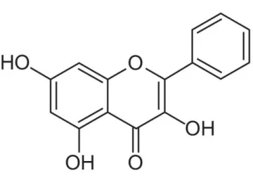
With the frequent occurrence of skin cancers and other damaging effects of UVB exposure, the protection of skin from UVB-induced oxidative damage has become a main consideration in the pharmaceutical industry. Phytochemicals are well-known for their defensive effects against oxidative stress in the skin (Sumiyoshi and Kimura, 2009). Galangin (trihydroxyflavone, IUPAC name:
2
trihydroxy-2-phenylchromen-4-one, Fig. 1) is a type of flavonoid that is commonly found in Alpinia officinarum and Helichrysum aureonitens (Afolayan and Meyer, 1997; Ciolino and Yeh, 1999). Galangin has antibacterial (Cushnie and Lamb, 2005; 2006) and antiviral (Afolayan et al., 1997) properties and represses breast tumor cell growth (So et al., 1996; Diffey, 2004). However, the cytoprotective effects of galangin against UVB-induced oxidative damage in human keratinocytes have not been studied yet. Therefore, the objective of this study was to investigate the protective effects of galangin against UVB-induced oxidative stress in human keratinocytes.
Figure 1. Chemical structure of galangin (3,5,7-trihydroxyflavone, IUPAC Name: 3,5,7-trihydroxy-2-phenylchromen-4-one).
3
2. Materials and Methods
2-1. Reagents
3,5,7-trihydroxyflavone (Galangin) was bought from Santa Cruz Biotechnology Inc. (Dallas, TX, USA). 1,1-diphenyl-2-picrylhydrazyl (DPPH), 3-(4,5-Dimethylthiazol-2-yl)-2,5-diphenyltetrazolium bromide (MTT), 5,5-dimethyl-1-pyrroline n-oxide (DMPO), 2',7'-dichlorodihydrofluorescein diacetate (DCF-DA), N-acetyl cysteine (NAC), Hoechst 33342 and primary antibody for actin were purchased from Sigma-Aldrich Inc. (St. Louis, MO, USA). 5,5',6,6'-Tetrachloro-1,1',3,3'-tetraethyl-benzimidazolylcarbocyanine iodide (JC-1) was purchased from Invitrogen (Carlsbad, CA, USA). Primary antibodies for Bax and Bcl-2 were bought from Santa Cruz Biotechnology Inc. (Dallas, TX, USA). Primary antibodies for caspase-3 and caspase-9 were bought from Cell Signaling Technology (Danvers, MA, USA). All other chemicals and reagents were of analytical grade.
2-2. Cell culture and UVB exposure
HaCaT human keratinocyte cells were purchased from Amore Pacific Company (Yongin, Korea) and maintained in a humified atmosphere conditions of 5% CO2 incubator at 37oC. RPMI 1640 culture medium with 10% heat-inactivated fetal bovine serum, streptomycin (100 μg/ml), and penicillin (100 units/ml) was used to culture cells. Cells were exposed to UVB radiation at a dose of 30 mJ/cm2. The CL-1000M UV Crosslinker (UVP, Upland, CA, USA) was used as the UVB source and delivered a UVB energy spectrum of 280-320 nm.
2-3. Cell viability assay
The influence of galangin on cell viability was examined using the MTT assay. Cells were seeded in a 96-well plate at a density of 1×10⁵ cells/ml. After 24 h, galangin was added to a final concentration of 20, 40, 80, or 100 μM and cells were incubated for 24 h. MTT stock solution (50 μl, 2 mg/ml) was added to
4
each well to yield a final reaction volume of 200 μl. The supernatant was aspirated 4 h later and formazan crystals were dissolved in 150 μl of dimethyl sulfoxide (DMSO). The absorbance at 540 nm was read using a scanning multi-well spectrophotometer (Carmichael et al., 1987).
2-4. Detection of DPPH radicals
The ability of galangin to scavenge DPPH radicals was assessed.Various concentrations of galangin (20, 40, 80, or 100 μM) or 1mM of NAC were added into a 96-well plate. DPPH dissolved in ethanol (0.1 mM) was added to each well to yield a total volume of 200 μl. After shaking for 3 h, unreacted DPPH was detected by measuring the absorbance at 520 nm using a spectrophotometer.
2-5. Detection of hydroxyl radicals
Hydroxyl radicals generated by the Fenton reaction (H2O2 + FeSO4) were reacted with DMPO. The resultant DMPO/•OH adducts were detected using an electron spin resonance (ESR) spectrometer (Li et al., 2004). The ESR spectrum was recorded 2.5 min after a phosphate buffer solution (pH 7.4) was mixed with 0.02 ml each of 0.3 M DMPO, 10 mM FeSO4, 10 mM H2O2, and 40 μM of galangin. The ESR spectrometer parameters were set as follows: central magnetic field, 336.8 mT; power, 1.00 mW; frequency, 9.4380 GHz; modulation width, 0.2 mT; amplitude, 600; sweep width, 10 mT; sweep time, 0.5 min; time constant, 0.03 sec; and temperature, 25oC.
2-6. Detection of intracellular ROS
DCF-DA fluorescence was detected to measure intracellular ROS generated by H2O2 or UVB (Rosenkranz et al., 1992). Cells were seeded at a density of 1.5×10⁵ cells/ml and incubated at 37oC for 24 h. Galangin (40 μM) or NAC (1 mM) was added to each well. After 1 h, cells were treated with H2O2 (1 mM) or exposed to UVB. After 30 min, H2O2-treated cells were treated with DCF-DA (25 μM) and incubated for another 20 min. UVB-treated cells were incubated for 24 h, treated with DCF-DA (50 μM), and incubated for a further 30 min. Fluorescence of 2',7'-dichlorofluorescein (DCF) was detected and
5
quantified using a PerkinElmer LS-5B spectrofluorometer (PerkinElmer, Waltham, MA, USA). Intracellular ROS scavenging effect of galangin (%) = ((absorbance of control cells - absorbance of galangin - or NAC-treated cells)/absorbance of control cells) × 100. Only H2O2 or UVB-treated cells were considered as controls.
2-7. Single cell gel electrophoresis (Comet assay)
DNA damage caused by oxidative stress was detected by the comet assay (Singh, 2000; Rajagopalan et al., 2003). Cells were seeded at a density of 1×10⁵ cells/ml and incubated at 37oC for 24 h. Cells were treated with galangin (40 μM) and after 1 h, exposed to UVB (30 mJ/cm2). The cell suspension was collected and mixed with 120 μl of 0.7% low melting agarose (LMA) at 37oC. The mixture was spread on a fully frosted microscopic slide pre-coated with 200 μl of 1% normal melting agarose. After this had solidified, a further 170 μl of LMA was applied to the slide. Slides were immersed in lysis solution (2.5 M NaCl, 100 mM Na-EDTA, 10 mM Tris, 1% Trion X-100, and 10% DMSO, pH 10) for 90 min at 4oC. Slides were then immersed in an unwinding buffer (300 mM NaOH and 10 mM Na-EDTA, pH 13) for 30 min at 4oC. Slides were subjected to electrophoresis in unwinding buffer solution with an electrical field of 300 mA and 25 V for 20 min at room temperature. The slides were washed three times with neutralizing buffer (0.4 M Tris, pH 7.5) for 10 min each time and then washed with 70% ethanol for 5 min. Slides were stained with 70 μl of ethidium bromide and observed under a fluorescence microscope using an image analyzer (Kinetic Imaging, Komet 5.5, UK). The tail length and the percentage of fluorescence in comet tails were recorded for 50 cells per slide.
2-8. 8-Isoprostane assay
Cells were seeded at a density of 1×10⁵ cells/ml and incubated at 37oC for 24 h. Cells were treated with galangin (40 μM) and after 1 h, exposed to UVB (30 mJ/cm2), and incubated at 37oC for another 24 h. Lipid peroxidation was assayed by colorimetric determination of the amount of 8-isoprostane secreted into the culture medium by HaCaT keratinocytes (Beauchamp et al., 2002). A commercial enzyme immunoassay
6
(Cayman Chemical, Ann Arbor, MI, USA) kit was used according to the manufacturer’s instructions.
2-9. Protein carbonylation assay
Cells were seeded at a density of 1×10⁵ cells/ml and incubated at 37oC for 24 h. Cells were treated with galangin (40 μM) and after 1 h, exposed to UVB (30 mJ/cm2), and incubated at 37oC for another 24 h. The extent of protein carbonyl formation was determined using an OxiSelectTM Protein Carbonyl ELISA Kit from Cell Biolabs (San Diego, CA, USA) according to the manufacturer’s instructions.
2-10. Nuclear staining with Hoechst 33342
Cells were seeded at a density of 1×10⁵ cells/ml and incubated at 37oC for 24 h. Cells were treated with galangin (40 μM) and after 1 h, exposed to UVB (30 mJ/cm2), and incubated at 37oC for another 24 h. The DNA-specific fluorescent dye Hoechst 33342 was added to each well and cells were incubated for 10 min at 37oC. Stained cells were visualized under a fluorescence microscope equipped with a CoolSNAP-Pro color digital camera. The degree of nuclear condensation was evaluated and apoptotic cells were counted. The ratio between apoptotic bodies and total number of cells were determined within randomly selected 0.3 mm2 area of each well.
2-11. Analysis of mitochondrial membrane potential (Δψm)
Cells were seeded in chamber slides (Nalge Nunc International) at a density of 1 × 10⁵ cells/ml and incubated for 24 h at 37°C
.
Cells were treated with galangin (40 μM) and after 1 h, exposed to UVB (30 mJ/cm2), and incubated at 37oC for another 24 h.JC-1 (1 μM of final concentration) was added to each well and cells were incubated for 30 min at 37oC. Stained cells were washed with phosphate-buffered saline (PBS), coverslips were mounted onto microscopic slides in mounting medium (DAKO, Carpinteria, CA, USA), and slides were examined using a confocal microscope. Microscopic images were collected using the Laser Scanning Microscope 5 PASCAL program (Carl Zeiss, Jena, Germany) (Cossarizza et al., 1993). In addition, mitochondrial membrane potential was analyzed by flow cytometry (Becton Dickinson,7
Mountain View, CA, USA). Cells were harvested, washed, suspended in PBS containing JC-1 (1 μM of final concentration), incubated for 30 min at 37oC, and analyzed using a flow cytometer(Troiano et al., 2007).
2-12. Western blot analysis
Harvested cells were lysed by incubation on ice for 30 min in 150 μl of lysis buffer (iNtRON Biotechnology, Republic of Korea).The resultant cell lysates were centrifuged at 13,000 rpm for 5 min. Supernatants were collected and protein concentrations were determined. Aliquots were boiled for 5 min and electrophoresed on 12% SDS-polyacrylamide gels. Protein blots of the gels were transferred onto nitrocellulose membranes. The membranes were incubated with the appropriate primary antibodies (1:1,000) followed by horseradish peroxidase conjugated anti-IgG secondary antibodies (1:5,000) (Pierce, Rockford, IL, USA). Protein bands were detected using an enhanced chemiluminescence Western blotting detection kit (Amersham, Little Chalfont, Buckinghamshire, UK).
2-13. Statistical analysis
All measurements were performed in triplicate and all values are expressed as means ± standard error. The results were subjected to an analysis of variance using Tukey’s test to analyze differences between means. In each case, a p-value of <0.05 was considered statistically significant.
8
3. Results
3-1. Galangin attenuates UVB-induced ROS generation
The MTT assay elucidated that galangin was not toxic to HaCaT cells at any concentration used. Following the treatment with each of the concentrations of galangin tested, cell viability was above than 96% of that of control cells (Fig. 2A). Galangin exhibited significant ability of DPPH radical-scavenging at the concentrations of 20-100 μM. A well-known antioxidant NAC (1 mM) was used as the positive control (Fig. 2B). Therefore, considering both cell viability and DPPH radical scavenging data, 40 μM was selected as the optimal concentration of galangin for further experiments. To assess the ability of galangin (40 μM) to scavenge hydroxyl radicals, ESR spectrometry was performed. In the Fenton reaction (Fe2+ + H2O2 → Fe3+ + •OH + OH-), DMPO/•OH adducts generated a signal of 2,915 in control cells and this signal was reduced to 1,803 in the presence of galangin (Fig. 2C). Next, the intracellular ROS scavenging activity of galangin was assessed using the DCF-DA fluorescent probe. Galangin scavenged 27% of ROS (versus 52% for NAC) in H2O2-treated cells and 27% of ROS (versus 22% for NAC) in UVB-treated cells (Fig. 2D). Taken together, these results show that galangin efficiently scavenges ROS.
10
C
11
Figure 2. Galangin scavenges reactive oxygen species (ROS). (A) Human skin keratinocytes (HaCaT
cells) were treated with various concentrations of galangin for 24 hours and cell viability was measured by the 3-(4,5-dimethylthiazol-2-yl)-2,5-diphenyltetrazolium bromide (MTT) assay. (B) Degrees of the 1-diphenyl-2-picrylhydrazyl (DPPH) radical were measured spectrophotometrically at 520 nm. N-acetyl cysteine (NAC, 1 mM) was used as the positive control. *Significantly different from the control group (p < 0.05). (C) Hydrodroxyl radical-scavenging ability was evaluated using the Fenton reaction system. *Significantly different from the control group (p < 0.05). (D) The ability of galangin to clean intracellular ROS generated by H2O2 or UVB was determined using a spectrofluorometer followed by DCF-DA staining. *,#Significantly different from cells treated with H
12
3-2. Galangin significantly attenuates UVB-induced damage of cellular macromolecules
I next investigated whether galangin can protect cellular macromolecules, such as DNA, lipids, and proteins, from UVB-induced oxidative damage. UVB-induced DNA damage and the cytoprotective effects of galangin were first studied using the comet assay. Representative microscopy images indicating the length of comet tails and the percentage of fluorescence in the tails are shown in Fig. 3A. UVB treatment increased the comet tail length and increased the percentage of fluorescence in the tail to 52%, indicating an increased level of DNA damage in keratinocytes, while galangin pretreatment significantly reduced this value to 34%. UVB-induced cellular membrane damage was measured by detecting lipid peroxidation via colorimetric determination of the level of 8-isoprostane secreted into the culture medium by HaCaT cells. The amount of 8-isoprostane was 219 pg/ml in the culture medium of UVB-exposed cells and 32 pg/ml in that of cells pretreated with galangin prior to UVB exposure (Fig. 3B). Finally, protein carbonylation was measured to assess the degree of protein damage. Oxidative stress usually modifies the amino acid side chains of proteins into carbonyl derivatives, which can be used to quantify protein damage caused by UVB-induced oxidative stress (Dalle-Donne, 2006). Protein carbonylation was markedly higher in UVB-treated cells than in control cells, whereas this increase was significantly reduced in cells pretreated with galangin prior to UVB exposure (Fig. 3C). These findings confirm that galangin can protect cellular macromolecules from UVB-induced oxidative damage.
15
Figure 3. Galangin reduces UVB-induced cellular macromolecule damage. Cells were pretreated with
40 μM of galangin and exposed to UVB (30 mJ/cm2) 1 hour later. After 24 hours, (A) the comet assay was accompanied to assess the DNA damage. Representative images and the percentage of cellular fluorescence within comet tails are given. (B) Lipid peroxidation was measured by assessing the concentration of 8-isoprostane in the conditioned medium. (C) Protein oxidation was assessed by measuring the amount of carbonyls formed. *Significantly different from control cells (p < 0.05); #significantly different from UVB only-exposed cells (p<0.05).
16 3-3. Galangin reduces UVB-induced apoptosis
The viability of HaCaT cells were assessed using the MTT assay (Fig. 4A). UVB exposure reduced the viability of keratinocytes to 60% in comparison to that of control cells. However, pretreatment with galangin increased cell viability to 76%, while pretreatment with the well-known antioxidant NAC increased cell viability to 88%. Next, cells were stained with Hoechst 33342 staining dye to visualize nuclear condensation and the formation of apoptotic bodies, which are characteristic of apoptosis. Normal nuclei were observed in control and galangin-treated cells, whereas notable nuclear condensation was found in UVB-exposed cells (Fig. 4B). The nuclear condensation was significantly lowered in galangin- and NAC-pretreated cells than in UVB only-exposed cells. Permeability of mitochondrial membrane plays a crucial role in the mitochondria-mediated apoptosis pathway. To elucidate the mechanism underlying how galangin protects keratinocytes against UVB-induced apoptosis, changes in the mitochondrial membrane potential were assessed. UVB treatment strongly increased the level of green fluorescence caused by JC-1 monomers, indicating mitochondrial depolarization (Fig. 4C). However, galangin treatment prior to UVB exposure significantly reduced the intensity of green fluorescence, showing that galangin attenuated UVB-induced mitochondrial depolarization. To confirm these data, flow cytometry analysis of JC-1 was accompanied. As expected, the green fluorescence peak was increased in UVB-treated cells. However, galangin pretreatment markedly deducted the peak of this fluorescence (Fig. 4D). Caspase-9, an initiator caspase, is cleaved to activate followed by the releasing cytochrome c from the depolarized mitochondria into the cytosol, cleaved caspase-9 propagates further activation of downstream apoptotic proteins which causes for cleavage of procaspase-3 to activate caspase-3 which execute the apoptotic process (Adrain and Martin, 2001). To assess the caspase-9 and -3 expression levels, a western blot analysis was performed. UVB exposure resulted up-regulation of cleaved caspase-9 and -3 in HaCaT cells, interestingly galangin pretreatment significantly down-regulated the levels of cleaved caspase-9 and -3. The vulnerability of cells towards apoptosis is ultimately determined by the pro-apoptotic and anti-apoptotic proteins in the B-cell leukaemia/lymphoma-2 (Bcl-2) family (Basu and Haldar, 1998). Bcl-2 itself is considered as an
17
apoptosis protein while Bcl-2-associated x protein (Bax) is known as a pro-apoptotic protein in the Bcl-2 protein family (Wei et al., 2001) Therefore levels of Bcl-2 and Bax proteins were examined after galangin treatment (Fig. 4E). Galangin treatment prior to UVB exposure resulted up-regulation of Bcl-2 proteins whereas down-regulation of Bax proteins. These results indicate that galangin inhibits apoptotic process via establishing mitochondrial membrane polarity and regulating apoptotic proteins.
20
Figure 4. Galangin elevates the viability of UVB-irradiated cells and attenuates apoptosis. HaCaT
cells were pretreated with 40 μM of galangin and exposed to UVB (30 mJ/cm2) 1 hour later. (A) After 24 hours, the MTT assay was committed to assess cell death. *Significantly different from control cells (p < 0.05). #Significantly different from UVB only-exposed cells (p < 0.05). (B) Cells were stained with Hoechst 33342 staining dye and observed by a fluorescence microscopy. Apoptotic bodies were quantified (arrows). JC-1 staining was performed to examine the mitochondrial membrane potential via (C) confocal microscopy and (D) flow cytometry. (E) Western blot analysis was accompanied to detect the expression levels of cleaved caspase- 9, -3, Bcl-2 and Bax proteins.
21
4. Discussion
Chronic solar UVB exposure, also known as photoaging, is the most well understood skin aging mechanism (Nichols and Katiyar, 2010; Svobodova and Vostalova, 2010). Notwithstanding the skin possesses a complex enzymatic and non-enzymatic defensive mechanism to protect it from adverse effects, prolonged UVB exposure can overwhelm this system. Solar UVB radiation has become one of the most common carcinogens in the environment and excessive UVB exposure leads to skin cancer. Meanwhile, due to the depletion of the stratospheric ozone layer and climate change, the earth welcome elevated levels of UV radiation (De Gruijl et al., 2003; Diffey, 2004). UVB damages DNA directly via the formation of pyrimidine dimers and indirectly via ROS generation (Ichihashi et al., 2003). DNA is the prime information molecule in the cell; therefore, nuclear DNA must exist intact for the whole life time of the cell. DNA damage can critically affect cellular functions. ROS attack not only nucleic acids but also proteins and lipids, thereby interrupting cellular metabolism. In this regard, prevention of the adverse effects caused by exposure to harmful UV radiation has become a popular theme in the cosmetic and pharmaceutical industries. Phytochemicals are well-known for their abilities to protect the skin against harmful UV radiation. Galangin is a flavone whose antioxidant ability depends on the donation of hydrogen atoms to free radicals (Sim et al., 2007). In this study, galangin scavenged DPPH radicals and hydroxyl radicals (Fig. 2B, C). DCF-DA staining showed that intracellular ROS generated via H2O2 and UVB (Fig. 2D) were removed by galangin. Flavonoid is known to possess antioxidant activity by donating hydrogen to radicals, depending on the substitution pattern of hydroxyl groups. Hydroxylated position is related with stability of the resulting phenoxyl radical by hydrogen donation or electron delocalization. The presence of 3-OH and 5-OH and C2-C3 double bond conjugated with the 4-oxo function increase antioxidant activity (Rice-Evans et al., 1996; Jung et al., 2005). In general, O-dihydroxy groups known as catechol structure in B ring increases radical scavenging activity, however it is reported that absence of catechol structure in the B ring like galangin is not always related with low antioxidant activity by compensating lack of catechol structure through combination of C2-C3 double bond with the 3-OH and 4-keto groups (Amic et al., 2003),
22
demonstrating antioxidant effect as shown in the results of my study.
UVB-induced ROS cause oxidative damage to DNA, lipids, and proteins in keratinocytes. Formation of CPDs and 6-4 photoproducts are considered as the main types of lesions caused by UVB radiation in DNA, and these adducts causes DNA strand breaks and induction of mutations (You et al., 2000). ROS especially hydroxyl radicals abstract allylic hydrogen forming carbon centered lipid radicals which will rapidly react with oxygen to form lipid peroxy radicals, interestingly these peroxy radicals are capable of abstract hydrogens from another lipid molecules generating lipid radicals initiating chain reactions (Ayala et al., 2014). ROS attack proteins and cause reversible and/or irreversible modifications, such as protein carbonylation, formation of adducts with lipid peroxidation products and protein protein cross-linking (Perluigi et al., 2010). Formation of protein carbonyls could be either by oxidative cleavage of proteins or by direct oxidation of lysine, arginine, proline, and threonine residues (Sander et al., 2002). The comet assay showed that galangin significantly suppressed DNA strand breaks induced by UVB (Fig. 3A). Detection of 8-isoprostane released by cells into the extracellular environment is an accurate means of assaying lipid peroxidation. Galangin-pretreated cells showed notably lower levels of 8-isoprostane in the culture medium than UVB alone-treated cells (Fig.3B), suggesting that galangin attenuated UVB-induced lipid peroxidation in skin cells. Proteins are oxidized by ROS and formation of protein carbonyls is a hallmark of oxidative stress. These results illustrated that the level of carbonyls was significantly attenuated in cells pretreated with galangin prior to UVB exposure (Fig. 3C).
At elevated concentration of ROS overwhelms the antioxidant defense system and interrupts cellular metabolism. In response to DNA damage, cells can undergo apoptosis (Barzilai and Yamamoto, 2004). Recent studies show that UVB induced cell death is basically governed through apoptotic pathway (Ji et al., 2012). In addition to DNA damage and cell cycle arrest by UVB induced oxidative stress, following the membrane damage via lipid peroxidation and protein carbonylation, ROS weaken the inner cellular structures especially mitochondrial membranes which causes to release cytochrome c into the cytosol and activates apoptotic proteins (Kulms et al., 2002). Galangin pretreatment significantly increased the viability of UVB-exposed cells (Fig. 4A). Hoechst 33342 staining revealed that galangin pretreatment effectively
23
reduced the formation of apoptotic bodies and DNA condensation induced by UVB exposure (Fig. 4B). Furthermore, I investigated the impact of galangin pretreatment on mitochondria-mediated apoptosis via JC-1 staining. Confocal microscopy (Fig. 4C) and flow cytometry (Fig. 4D) analyses confirmed that galangin restored mitochondrial membrane polarization. These data showed that galangin reduced apoptosis of UVB-radiated cells by establishing mitochondrial polarization. Pro-apoptotic protein Bax and anti-apoptotic protein Bcl-2 regulate the release of cytochrome c into the cytosol (Atan et al., 1999). Caspases are synthesized in their inactive forms in the cells and activated in respond to apoptotic signals (Thornberry and Lazebnik, 1998). Released cytochrome c into the cytosol binds to apoptotic protease activating factor 1, which then activates caspase-9 by cleaving procaspase-9 (Bossy-Wetzel et al., 1998). Cleaved caspase-9 activates caspase-3 and which then triggers downstream caspase cascades to execute apoptosis (Soengas et al., 1999). Results of the study elucidated that galangin suppressed the expression of Bax, cleaved caspase-9 and -3 while increased the expression of Bcl-2 protein level (Fig. 4E).
Concluding the results of this study it is justice to confirm that galangin own antioxidant properties and it intervenes the intrinsic pathway of apoptosis through down regulating apoptotic proteins and thereby galangin shields the HaCaT cells from UVB-induced oxidative damage and ultimately from the apoptosis
The thesis for Degree of Master of Science in Medicine referring to experimental data and contents are from “Galangin (3,5,7-trihydroxyflavone) shields human keratinocytes from ultraviolet
B-induced oxidative stress” which was published in the journal of “Biomolecules and Therapeutics (Seoul)” with the “DOI: 10.4062/biomolther.2014.130.” in the year 2015.
24
5. References
Adrain, C. and Martin, S. J. (2001) The mitochondrial apoptosome: a killer unleashed by the cytochrome seas. Trends Biochem. Sci. 26, 390-397.
Afolayan, A. J. and Meyer, J. J. (1997) The antimicrobial activity of 3,5,7-trihydroxyflavone isolated from the shoots of Helichrysum aureonitens. J. Ethnopharmacol. 57, 177–181.
Afolayan, A. J., Meyer, J. J., Taylor, M. B. and Erasmus, D. (1997) Antiviral activity of galangin isolated from the aerial parts of Helichrysum aureonitens. J. Ethnopharmacol. 56, 165–169.
Amic, D., Davidovic-Amic, D., Beslo, D. and Trinajstic, N. (2003) Structure-radical scavenging activity relationships of flavonoids. Croatica Chemica Acta. 76, 55-61.
Atan, G., James, M. M. and Stanley, J. K. (1999) Bcl-2 family members and the mitochondria in Apoptosis. Genes Dev. 13, 1899-1911.
Ayala, A. Muñoz, M. F. and Argüelles, S. (2014) Lipid peroxidation: production, metabolism, and signaling mechanisms of malondialdehyde and 4-hydroxy-2-nonenal. Oxid. Med. Cell. Longev. 2014, 360438-360469
Barzilai, A. and Yamamoto, K. (2004) DNA damage responses to oxidative stress. DNA Repair (Amst). 3, 1109–1115.
Basu, A. and Haldar, S. (1998) The relationship between BcI-2, Bax and p53: consequences for cell cycle progression and cell death. Mol. Hum. Reprod. 4, 1099-1109.
Beauchamp, M. C., Letendre, E. and Renier, G. (2002) Macrophage lipoprotein lipase expression is increased in patients with heterozygous familial hypercholesterolemia. J. Lipid. Res. 43, 215–222. Bossy-Wetzel, E., Newmeyer, D. D. and Green, D. R. (1998) Mitochondrial cytochrome c release in
25
apoptosis occurs upstream of DEVD-specific caspase activation and independently of mitochondrial transmembrane depolarization. EMBO J. 17, 37-49.
Caldwell, M. M., Bornman, J. F., Ballare, C. L., Flint, S. D. and Kulandaivelu, G. (2007) Terrestrial ecosystems, increased solar ultraviolet radiation and interactions with other climate change factors. Photochem. Photobiol Sci. 6, 252–266.
Carmichael, J., De Graff, W. G., Gazdar, A. F., Minna, J. D. and Mitchell, J. B. (1987) Evaluation of a tetrazolium-based semiautomated colorimetric assay assessment of chemosensitivity testing. Cancer Res.
47, 936–942.
Ciolino, H. P. and Yeh, G. C. (1999) The flavonoid galangin is an inhibitor of CYP1A1 activity and an agonist/antagonist of the aryl hydrocarbon receptor. Br. J. Cancer. 79, 1340–1346.
Cossarizza, A., Baccarani-Contri, M., Kalashnikova, G. and Franceschi, C. (1993) A new method for the cytofluorimetric analysis of mitochondrial membrane potential using the J-aggregate forming lipophilic cation 5,50,6,60-tetrachloro-1,10,3,30-tetraethylbenzimidazolcarbocyanine iodide (JC-1). Biochem. Biophys. Res. Commun. 197, 40–45.
Cunningham, M. L., Krinsky, V., Giovanazzi, S. M. and Peak, M. J. (1985) Superoxide anion is generated from cellular metabolites by solar radiation and its components. J. Free. Radic. Biol. Me. 1, 381–385. Cushnie, T. P. T. and Lamb, A. J. (2005) Detection of galangin-induced cytoplasmic membrane damage in
Staphylococcus aureus by measuring potassium loss. J. Ethnopharmacol. 101, 243–248.
Cushnie, T. P. T. and Lamb, A. J. (2006) Assessment of the antibacterial activity of galangin against 4-quinolone resistant strains of Staphylococcus aureus. Phytomedicine. 13, 187–191.
Dalle-Donne, I, Aldini, I., Carini, G., Colombo, M., Rossi, R. and Milzani, R. (2006) Protein carbonylation, cellular dysfunction, and disease progression. J. Cell. Mol. Med. 10, 389-406.
26
De Gruijl, F. R., Longstreth, J., Norval, M., Cullen, A. P., Slaper, H., Kripke, M. L., Takizawa, Y. and Van der Leun, J. C. (2003) Health effects from stratospheric ozone depletion and interactions with climate change. Photochem. Photobiol. Sci. 2, 16–28.
Dhumrongvaraporn, A. and Chanvorachote, P. (2013) Kinetics of ultraviolet B irradiation-mediated reactive oxygen species generation in human keratinocytes. J. Cosmet. Sci. 64, 207-217.
Diffey, B. (2004) Climate change, ozone depletion and the impact on ultraviolet exposure of human skin. Phys. Med. Biol. 49, R1-R11.
Donawho, C. K., Muller, H. K., Bucana, C. D. and Kripke, M. L. (1996) Enhanced growth of murine melanoma in ultraviolet-irradiated skin is associated with local inhibition of immune effector mechanisms. J. Immunol. 157, 781–786.
Hattori, Y., Nishigori, C., Tanaka, T., Uchida, K., Nikaido, O., Osawa, T., Hiai, H., Imamura, S. and Toyokuni, S. (1996) 8-hydroxy-20-deoxyguanosine is increased in epidermal cells of hairless mice after chronic ultraviolet B exposure. J. Invest. Dermatol. 107, 733–737.
Ichihashi, M., Ueda, M., Budiyanto, A., Bito, T., Oka, M. and Fukunaga, M. (2003) UV-induced skin damage. Toxicology. 189, 21–39.
Ikehata, H. and Ono, T. (2011) The mechanisms of UV mutagenesis. J. Radiat. Res. 52, 115–125.
Ji, C. Yang, B. Yang, Z. Tu, Y. Yang, Y.L. He, L. and Bi, Z.G. (2012) Ultra-violet B (UVB)-induced skin cell death occurs through a cyclophilin D intrinsic signaling pathway. Biochem. Biophys. Res. Commun.
425, 825-829.
Jung, C. H., Seog, H. M., Choi, I. W. and Cho, H. Y. (2005) Antioxidant activities of cultivated and wild Korean ginseng leaves. Food Chem. 92, 535-540.
27
Kulms, D. and Schwartz, T. (2000) Molecular mechanisms of UV-induced apoptosis. Photodermatol. Photoimmunol. Photomed. 16, 195–201.
Li, L., Abe, Y., Kanagawa, K., Usui, N., Imai, K., Mashino, T., Mochizuki, M. and Miyata, N. (2004) Distinguishing the 5,5-dimethyl-1-pyrroline N-oxide (DMPO)-OH radical quenching effect from the hydroxyl radical scavenging effect in the ESR spin-trapping method. Anal. Chim. Acta. 512, 121-124. Lyons, N. M. and O’Brien, N. M. (2002) Modulatory effects of an algal extract containing astaxanthin on
UVA-irradiated cells in culture. J. Dermatol. Sci. 30, 73–84.
Matsumura, Y. and Ananthaswamy, H. N. (2004) Toxic effects of ultraviolet radiation on the skin. J. Toxycol. Applied. Pharm. 195, 298– 308.
Meeran, S.M., Punathil, T. and Katiyar, S. K. (2008) Interleukin-12-deficiency exacerbates inflammatory responses in UV irradiated skin and skin tumors. J. Invest. Dermatol. 128, 2716–2727.
Nichols, J. A. and Katiyar, S. K. (2010) Skin photoprotection by natural polyphenols: anti-inflammatory, antioxidant and DNA repair mechanisms. Arch. Dermatol. Res. 302, 71–83.
Park, H. M., Kim, H. J., Jang, Y. P. and Kim, S. Y. (2013) Direct Analysis in Real Time Mass Spectrometry (DART-MS) Analysis of skin metabolome changes in the ultraviolet B-induced mice. Biomol Ther. 21, 470-475.
Perluigi, M. Di Domenico, F. Blarzino, C. Foppoli, C. Cini, C. Giorgi, A. Grillo, C. De Marco, F. Butterfield, D. A. Schinin , M. E. and Co ccia, R. (2010) Effects of UVB induced oxidative stress on protein expression and specific protein oxidation in normal human epithelial keratinocytes: a proteomic approach. Proteome Sci. 8, 13.
Rajagopalan, R., Ranjan, S. K. and Nair, C. K. (2003) Effect of vinblastine sulfate on gamma-radiation-induced DNA single-strand breaks in murine tissues. Mutat Res. 536, 15-25.
28
Rice-Evans, C. A., Miller, N. J. and Paganga, G. (1996) Structure-antioxidant activity relationships of flavonoids and phenolic acids. Free Radic. Biol. Med. 20, 933-956.
Rosenkranz, A. R., Schmaldienst, S., Stuhlmeier, K. M., Chen, W., Knapp, W. and Zlabinger, G. J. (1992) A microplate assay for the detection of oxidative products using 2',7' dichlorofluorescin-diacetate. J. Immunol. Methods. 156, 39-45.
Sander, C. S., Chang, H., Salzmann, S., Müller, C. S., EkanayakeMudiyanselage, S., Elsner, P. and Thiele, J. J. (2002) Photoaging is associated with protein oxidation in human skin in vivo. J. Invest. Dermatol.
118, 618-625.
Sim, G., Lee, B., Ho, S. C., Jae, W. L., Kim, J., Lee, D. H., Kim, J. H., Pyo, H. B., Moon, D. C., Oh, K. W., Yun, Y. P. and Hong, J. T. (2007) Structure activity relationship of antioxidative property of flavonoids and inhibitory effect on matrix metalloproteinase activity in UVA-irradiated human dermal fibroblast. Arch. Pharm. Res. 30, 290-298.
Singh, N. P. (2000) Microgels for estimation of DNA strand breaks, DNA protein crosslinks and apoptosis. Mutat Res. 455, 111-127.
So, F. V., Guthrie, N., Chambers, A. F., Moussa, M. and Carroll, K. K. (1996) Inhibition of human breast cancer cell proliferation and delay of mammary tumorigenesis by flavonoids and citrus juices. Nutr Cancer. 26, 167–181.
Soengas, M. S., Alarcon, R. M., Yoshida, H., Giaccia, A. J., Hakem, R., Mak, T. W. and Lowe, S. W. (1999) Apaf-1 and caspase-9 in p53-dependent apoptosis and tumor inhibition. Science 284, 156-159.
Sumiyoshi, M. and Kimura, Y. (2009) Effects of a turmeric extract (Curcuma longa) on chronic ultraviolet B irradiation-induced skin damage in melanin-possessing hairless mice. Phytomedicine. 16, 1137–1143. Svobodova, A. and Vostalova, J. (2010) Solar radiation induced skin damage: review of protective and
29
preventive options. Int. J. Radiat. Bio. 86, 999–1030.
Thornberry, N. A. and Lazebnik, Y. (1998) Caspases: Enemies within. Science. 281, 1312-1316.
Troiano, L., Ferraresi, R., Lugli, E., Nemes, E., Roat, E., Nasi, M., Pinti, M. and Cossarizza, A. (2007) Multiparametric analysis of cells with different mitochondrial membrane potential during apoptosis by polychromatic flow cytometry. Nat Protoc. 2, 2719–2727.
Tsoyi, K., Park, H. B., Kim, Y. M., Chung, J. I., Shin, S. C., Shim, H. J., Lee, W. S., Seo, H. G., Lee, J. H., Chang, K. C. and Kim, H. J. (2008) Protective effect of anthocyanins from black soybean seed coats on UVB induced apoptotic cell death in vitro and in vivo. J. Agric. Food. Chem. 56, 10600–10605. Wei, M. C., Zong, W. X., Cheng, E. H., Lindsten, T. P. V., Roth, K. A., MacGregor, G. R., Thompson, C. B.
and Korsmeyer, S. J. (2001) Proapoptotic BAX and BAK: a requisite gateway to mitochondrial dysfunction and death. Science. 292, 727-730.
Yoshikawa, T., Rae, V., Bruins-Slot, W., Van den Berg, J. W., Taylor, J. R. and Streilein, J. W. (1990) Susceptibility to effects of UVB radiation on induction of contact hypersensitivity as a risk factor for skin cancer in humans. J. Invest. Dermatol. 95, 530–536.
You, H. Y. Szabo, P. E. and Pfeifer, G. P. (2000) Cyclobutane pyrimidine dimers form preferentially at the major p53 mutational hotspot in UVB-induced mouse skin tumors. Carcinogenesis. 21, 2113-2117.
30
6. Abstract in Korean
배경: 활성 산소종 (ROS)은 자외선 B (UVB) 조사에 의한 피부 손상의 근본적인 원인이며 피부
암 또는 표피와 진피의 광 노화가 이에 포함된다. 파이토 케미컬은 UVB로 유도되는 산화적 스 트레스에 대하여 항산화제 작용을 하는 것으로 알려져 있다. Galangin은 일종의 플라보놀로서 Alpinia officinarum, Helichrysum aureonitens과 같은 식물에 풍부하게 함유되어 있으며 항바이러 스와 항암특성을 나타낸다는 것이 이미 증명되었다. 이 연구는 인간의 각질형성세포에서 UVB 로 유도되는 아포토시스에 대한 플라보놀 galangin의 세포 보호 효과를 조사하기 위해 초점을 맞추었다.
방법: 인간 피부 각질형성세포(HaCaT)는 10% 소 태아 혈청이 함유된 RPMI1640 배양액으로 배
양하였다. 세포 생존율, galangin의 ROS 소거능을 분석하였고 Galangin의 보호 특성은 UVB로 유도되는 세포 내 거대분자의 손상 즉 DNA의 사슬 붕괴, 지질 과산화작용 및 단백질의 카보닐 화의 측정을 통해 평가하였다. 아포토틱 소체의 형성, 미토콘드리아 막 탈분극 및 아포토시스 의 촉진 또는 억제 단백질의 발현을 통해 아포토시스의 정도를 측정하였다.
결과: Galangin은 효율적으로 프리 라디칼을 소거하고 UVB로 유도되는 DNA, 지질, 단백질과
같은 세포 내 거대분자의 손상을 감소시켰다. 또한, galangin은 미토콘드리아 분극화의 복구, 아 포토틱 촉진 단백질들의 하향조절과 아포토틱 억제 단백질들의 상향조절을 통하여 UVB 조사 로 유도된 아포토시스를 보호하였다.
31
결론: 이러한 발견은 galangin이 ROS 소거작용을 통하여 UVB 조사로 유도되는 산화적 스트레
32
7. Acknowladgements
Apart from the effort of mine, the success of my thesis depends largely on the encouragement and guidelines of many others. I take this opportunity to express my gratitude to the people who have been instrumental in the successful completion of this project. I would like to express my deepest and sincere gratitude to my supervisor in charge Prof. Jin Won Hyun, for providing me this valuable opportunity to commence my master degree In Jeju National University and for keen attention throughout my study, valuable suggestions and critical reviews of the reports rendered to me during my studies which made the foundation for success of my two years study in Republic of Korea.
I am also grateful to Prof. Ko Young Sang and Prof. Yoo Eun Sook, School of medicine Jeju National University, who provided valuable instructions and suggestions to complete my thesis. Further all the professors guided me in the school of Medicine are sincerely appreciated.
Especially I must mention the names who were always behind me for the every success step of my laboratory career as well as my entire stay in Republic of Korea, Dr. Mei Jing Piao and Dr. Ki Cheon Kim. Most importantly, regardless of the time as well as language barriers, the kind attention they paid for me is invaluable. I would like to express my heartiest thank for my two senior brothers Jian Zheng and Cheng Wen Yao, my senior sister Ji Won Cha and my colleague Xia Han for assisting me in various means not only on my experiments but also in my personal life since the very first day I came to the school and for fueling me to reach this benchmark. I am also thankful to Min Chang Oh, Yoon Hae Kyung and Moon You Jin for their kind help and assistance.
My special thank goes to Prasad Tharanga Jayasooriya, Dilshara Matharage and Jayasinghage Don Handun Eranga Jayasinghe for guiding, supporting and encouraging me to take this valuable opportunity and make it true. I am grateful to my classmate, friend and roommate Madushan Fernando for strengthening and supporting me at every hard time. During the period of two years, many friends were helpful to color my life. I use this opportunity to acknowledge all my colleagues in Jeju National University for making those joyful memories.
33
I owe my sincere gratitude to Dr. Kumudu Fernando, former director, Agricultural Biotechnology Centre, Faculty of Agriculture, University of Peradeniya, Sri Lanka and Prof. Susantha D Pathirana, Faculty of Engineering, University of Peradeniya, Sri Lanka for their kind instructions and guidance, especially allowing me to work at Agricultural Biotechnology Centre, Universit of Peradeniya, Sri Lanka where I got the first experience on this field. Further I owe my gratitude to my former supervisor Dr. H. M. V. G. Herath and Dr. H. A. M Wickramasinghe, Faculty of Agriculture, University of Peradeniya, Sri Lanka who showed me this path and dedicated their time and effort for me.
At last but not the least I want to thank my beloved fiancée Rashmi Keshika Siribaddana and my dear Amma and Thaththa who appreciated my work and always motivated me in many means to reach this milestone.